



BuChE-IN-2
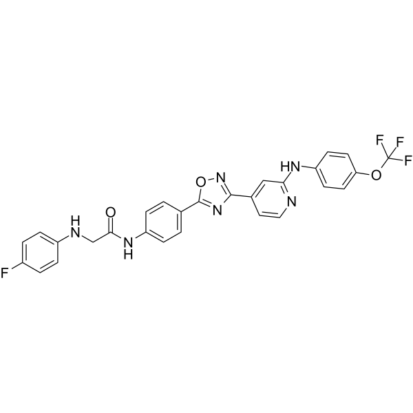
BuChE-IN-2 is an excellent butyrylcholinesterase (BuChE) inhibitor (IC50s of 1.28 μM and 0.67 μM for BuChE and NO). BuChE-IN-2 can inhibit the aggregation of Aβ, ROS formation and chelate Cu2+, exhibiting proper blood-brain barrier (BBB) penetration. BuChE-IN-2 has potential to research Alzheimer’s disease.
For research use only. We do not sell to patients.
- CAS No.: 2745118-93-6
- Formula: C28H20F4N6O3
- Molecular Weight:564.49
-
Storage:
Please store the product under the recommended conditions in the Certificate of Analysis.

To place orders, for customer services and technical support, please contact: MedChemExpress USA
Tel: 609-228-6898 E-mail: [email protected] [email protected]


-
 Biological Activity
-
Chemical Information
- Protocols
- Purity & Documentation
- References
-
 Help & FAQs




Biological Activity
Description
IC50 & Target
IC50: 1.28 μM (BuChE), 0.67 μM (NO)[1]
Cellular Effect
|
Cell Line

|
Type | Value | Description | References |
|---|---|---|---|---|
| BV-2 | IC50 |
0.67 μM
Compound: F9
|
Inhibition of NO production in LPS-induced mouse BV-2 cells pretreated for 1 hr followed by LPS stimulation and measured after 24 hrs by ELISA
Inhibition of NO production in LPS-induced mouse BV-2 cells pretreated for 1 hr followed by LPS stimulation and measured after 24 hrs by ELISA
|
[PMID: 34752955] |
| BV-2 | IC50 |
1.61 μM
Compound: F9
|
Inhibition of LPS-induced IL-1beta secretion in mouse BV-2 cells preincubated for 1 hr followed by LPS stimulation and measured after 24 hrs by ELISA
Inhibition of LPS-induced IL-1beta secretion in mouse BV-2 cells preincubated for 1 hr followed by LPS stimulation and measured after 24 hrs by ELISA
|
[PMID: 34752955] |
| BV-2 | IC50 |
4.15 μM
Compound: F9
|
Inhibition of LPS-induced TNF-alpha secretion in mouse BV-2 cells preincubated for 1 hr followed by LPS stimulation and measured after 24 hrs by ELISA
Inhibition of LPS-induced TNF-alpha secretion in mouse BV-2 cells preincubated for 1 hr followed by LPS stimulation and measured after 24 hrs by ELISA
|
[PMID: 34752955] |
In Vitro
BuChE-IN-2 (compound f9) (5-50 μM; 24 hours) shows obvious neuroprotection on H2O2-induced PC12 cells at 20 μM[1].
BuChE-IN-2 (100 μM; 48 hours) can inhibit Aβ aggregation[1].
BuChE-IN-2 (0.1-20 μM; 24 hours) has the obviously inhibitory effect on the secretion of inflammatory factors and IL-1β (IC50=1.61 μM) and TNF-α (IC50=4.15 μM) in BV2 cells[1].
BuChE-IN-2 (1-10 μM; 1 hour) can significantly reduce the expression of COX-2 and iNOS in a concentration-dependent manner[1].
BuChE-IN-2 (1-50 μM; 6 hours) has a significant inhibitory effect on ROS accumulation at 20 μM[1].
BuChE-IN-2 (10-1000 μM; 2 hours) decreases the DPPH concentration dramatically from 86.09% to 34.62% when the concentration of BuChE-IN-2 increases from 10 μM to 1000 μM[1].
BuChE-IN-2 (75 μM; 2 hours; MDCKII-MDR1 cells) exhibits proper blood-brain barrier permeability[1].
MedChemExpress (MCE) has not independently confirmed the accuracy of these methods. They are for reference only.
-
Cell Line:PC12 cells[1]
-
Concentration:5, 20, 25, 50 μM
-
Incubation Time:24 hour
-
Result:Showed obvious neuroprotection on H2O2-induced PC12 cells at 20 μM.
-
Cell Line:BV2 cells[1]
-
Concentration:1, 3, and 10 μM
-
Incubation Time:1 hour
-
Result:Significantly reduced the expression of COX-2 and iNOS in a concentration-dependent manner.
In Vivo
BuChE-IN-2 (10 and 30 mg/kg; i.g., single) can remarkably improve the cognitive impairment in scopolamine-induced mouse models according to Morris water maze experiment[1].
MedChemExpress (MCE) has not independently confirmed the accuracy of these methods. They are for reference only.
-
Animal Model:Male C57BL mice (8-week-old, 18-23 g)[1]
-
Dosage:100, 80, 64, 51.2, 40.96 mg/kg
-
Administration:i.g.; single
-
Result:The median Lethal Dose (LD50) of BuChE-IN-2 was 75.372 (62.383-101.673) mg/kg (95% confidence limit).
-
Animal Model:Male C57BL mice (8-week-old, 18-23 g)[1]
-
Dosage:30 mg/kg, 10 mg/kg
-
Administration:i.g., single
-
Result:BuChE-IN-2 could remarkably improve the cognitive impairment in scopolamine-induced mouse models according to Morris water maze experiment.


Chemical Information
-
CAS No. 2745118-93-6
-
Molecular Weight 564.49
-
Formula C28H20F4N6O3
-
SMILES
O=C(NC1=CC=C(C2=NC(C3=CC(NC4=CC=C(OC(F)(F)F)C=C4)=NC=C3)=NO2)C=C1)CNC5=CC=C(F)C=C5
-
Shipping
Room temperature in continental US; may vary elsewhere.
-
Storage
Please store the product under the recommended conditions in the Certificate of Analysis.


Purity & Documentation
References

Calculators
Concentration (start) × Volume (start) = Concentration (final) × Volume (final)


BuChE-IN-2 Related Classifications
HY-143413 Related Classifications
 Powered by Bioz