



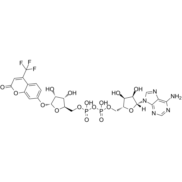
TFMU-ADPr
TFMU-ADPr is a selective reporter substrate of SARS-CoV-2 Macro1 (IC50=0.59 μM), with an excitation wavelength (λEx) of 385 nm, and an emission wavelength (λEm) of 502 nm (or 495 nm). TFMU-ADPr can also undergo enzymatic hydrolysis with Poly(ADP-ribose) Glycohydrolase (PARG) sourced from human, Tetrahymena thermophila and ADP-ribosylhydrolase 3 from human to release fluorophores, thereby directly reporting total poly (ADP-ribose) hydrolase activity. TFMU-ADPr binds to the ADPr-binding site of SARS-CoV-2 Macro1, and its TFMU moiety inserts into the narrow hydrophobic groove of this protein. TFMU-ADPr can thus be used to evaluate small-molecule inhibitors targeting PAR hydrolases under in vitro conditions, to investigate the regulatory mechanisms of ADP-ribosyl catabolic enzymes, or to detect PAR hydrolase activity in whole-cell lysate assays. TFMU-ADPr is also applicable to COVID-19-related research.
Nos produits utilisent uniquement pour la recherche. Nous ne vendons pas aux patients.
- CAS No.: 2412923-11-4
- Formule: C25H26F3N5O16P2
- Masse moléculaire:771.44
-
Stockage:
Please store the product under the recommended conditions in the Certificate of Analysis.

To place orders, for customer services and technical support, please contact: MedChemExpress USA
Tel: 609-228-6898 E-mail: [email protected] [email protected]


-
 Activité biologique
-
Chemical Information
- Pureté et documentation
- Références
-
 Help & FAQs




Activité biologique
TFMU-ADPr (0-500 μM, 0-120 μM; 5 minutes) serves as a continuous fluorescent substrate for hPARG (full-length PARG from Homo sapiens), ttPARG (PARG from Tetrahymena thermophila) and hARH3 (full-length ARH3 from H. sapiens), enabling robust enzyme kinetic assays. The corresponding KM values for the three enzymes are 66.2 μM, 210 μM and 6.3 μM, respectively[1].
TFMU-ADPr (200 μM) can report the combined hydrolase activity of PARG and ARH3 in U2OS cell lysates, and this activity is detected in both wild-type cells and ARH3 knockout cells (the latter represents PARG-only activity)[1].
TFMU-ADPr (0-50 μM) serves as an effective reporter substrate for detecting the inhibitory activity of hARH3, which is confirmed by the potent inhibitory effect of the endogenous metabolite ADPr-Arg (Ki=18 nM)[1].
TFMU-ADPr (in a dose-response manner; incubation duration 30 min) potently binds SARS-CoV-2 Macro1 (IC50=0.59 μM), human MacroD1, human MacroD2, VEEV Macro, and human PARP9 Macro2; compared with ADPr, it shows significantly improved binding potency for all tested macrodomains except CHIKV Macro[2].
TFMU-ADPr (2 mM; 5 d) binds to the conserved ADPr-binding pocket of SARS-CoV-2 Macro1, and its TFMU group forms favorable interactions with hydrophobic residues in the adjacent hydrophobic groove, thereby contributing to high binding affinity[2].
TFMU-ADPr acts as an inhibitor of the ADPr hydrolase activity of SARS-CoV-2 Macro1 and human MacroD1 in cell lysates[2].
Experimental procedure for in vitro detection using TFMU-ADPr mainly focuses on enzyme kinetics and inhibition assays, as specified below[3]:
1. Experimental Preparation:
Establish a reaction system in a 384-well plate. The system contains 50 mM Na2HPO4 (pH 7.4), 10 mM MgCl2, 5 mM DTT, TFMU-ADPr at a specified concentration as the substrate, and the enzyme to be tested (such as hARH1, hARH3, etc.). For inhibitor studies, pre-incubate the enzyme with the inhibitor at room temperature for 5 minutes first.
2. Reaction Initiation and Signal Detection:
After shaking the reaction system for 5 seconds, record the fluorescence signal using a Molecular Devices SpectraMax M3 microplate reader. Set the excitation wavelength (λEx) to 385 nm and the emission wavelength (λEm) to 502 nm (or 495 nm). Read each well 6 times in low-gain mode, and record the fluorescence value every 5 seconds for a continuous detection duration of 15 min.
3. Data Processing:
Determine the initial reaction rate by fitting the linear segment of the reaction progress curve. Fit the initial rate to the substrate concentration, use the nonlinear curve fitting algorithm in GraphPad Prism 6, and analyze enzyme kinetic parameters based on the Michaelis-Menton equation. For inhibition assays, use enzyme-free reactions and inhibitor-free reactions as positive and negative controls, respectively, calculate the inhibition percentage, and obtain relevant inhibition parameters through dose-response curve fitting.
MedChemExpress (MCE) has not independently confirmed the accuracy of these methods. They are for reference only.
502
385


Chemical Information
-
CAS No. 2412923-11-4
-
Masse moléculaire 771.44
-
Formule C25H26F3N5O16P2
-
SMILES
O[C@H]1[C@](O[C@@H]([C@H]1O)COP(OP(OC[C@H]2O[C@@H]([C@@H]([C@@H]2O)O)OC3=CC=C(C(C(F)(F)F)=CC(O4)=O)C4=C3)(O)=O)(O)=O)([H])N5C6=NC=NC(N)=C6N=C5
-
Livraison
Room temperature in continental US; may vary elsewhere.
-
Stockage
Please store the product under the recommended conditions in the Certificate of Analysis.


Pureté et documentation
Références
[1]. Rack JGM, et al. (ADP-ribosyl)hydrolases: Structural Basis for Differential Substrate Recognition and Inhibition. Cell Chem Biol. 2018 Dec 20;25(12):1533-1546.e12. [Content Brief]
[2]. Drown BS, et al. Monitoring Poly(ADP-ribosyl)glycohydrolase Activity with a Continuous Fluorescent Substrate. Cell Chem Biol. 2018;25(12):1562-1570.e19. [Content Brief]
[3]. Anmangandla A, et al. A Fluorescence Polarization Assay for Macrodomains Facilitates the Identification of Potent Inhibitors of the SARS-CoV-2 Macrodomain. ACS Chem Biol. 2023;18(5):1200-1207. [Content Brief]

Calculators
Concentration (start) × Volume (start) = Concentration (final) × Volume (final)


TFMU-ADPr Related Classifications
HY-146248 Related Classifications
 Powered by Bioz
- TFMU-ADPr
- 2412923-11-4
- SARS-CoV
- Poly(ADP-ribose) Glycohydrolase (PARG)
- Protease Activated Receptor (PAR)
- human MacroD2
- SARS-CoV-2 Macro1
- human ADP-ribosyl hydrolase 3
- Tetrahymena thermophila poly(ADP-ribose) glycohydrolase
- U2OS cell lysates
- VEEV Macro
- human MacroD1
- human PARP9 Macro2
- human poly(ADP-ribose) glycohydrolase
- covid-19
- Inhibitor
- inhibitor
- inhibit