



Chromium chloride

Based on 1 Customer Validation
Chromium chloride is a trivalent chromium compound and an essential trace mineral. Chromium chloride enhances insulin-stimulated GLUT4 translocation and glucose uptake in skeletal muscle. Chromium chloride regulates glucose and lipid metabolism, inhibits TNF-α secretion and oxidative stress in monocytes treated with high glucose or H2O2, and reverses hydrogen peroxide-induced cell growth inhibition. Chromium chloride reduces coronary and aortic lipid deposition and serum cholesterol levels in hypercholesterolemic rabbits. Chromium chloride can be used in research related to diabetes and cardiac atherosclerosis.
연구목적의 판매만을 진행합니다. 환자를 대상으로 한 판매는 하지 않습니다.
- Purity : 99.9%
- CAS No.: 10025-73-7
- 화학식: CrCl3
- 분자량:158.36
-
보관:
4°C, protect from light, stored under nitrogen
* In solvent : -80°C, 6 months; -20°C, 1 month (protect from light, stored under nitrogen)


To place orders, for customer services and technical support, please contact: MedChemExpress USA
Tel: 609-228-6898 E-mail: [email protected] [email protected]


-
 Biological Activity
-
Chemical Information
- Protocol
- 순도&문서
- References
-
 Help & FAQs


-

HY-Y0998
Potassium hexacyanoferrate(III), 99.5%Chelating Agent -

HY-W094464
Lithium sulfateInorganic Compound -
HY-W094581
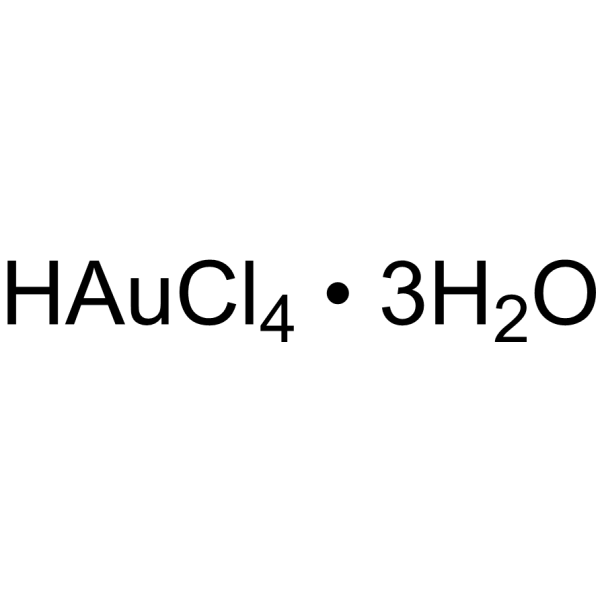
Gold(III) chloride trihydrateReducing Agent -

HY-W019894
Manganese chlorideContrast Agent -
HY-N3025
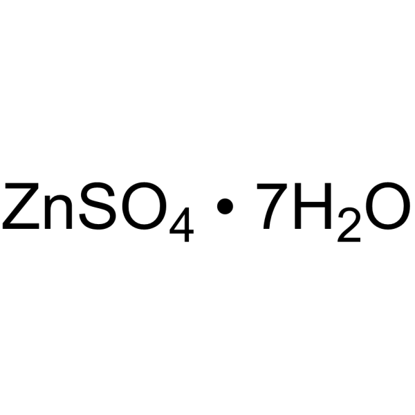
Zinc sulfate heptahydrateDietary Supplement -
HY-Y0406E
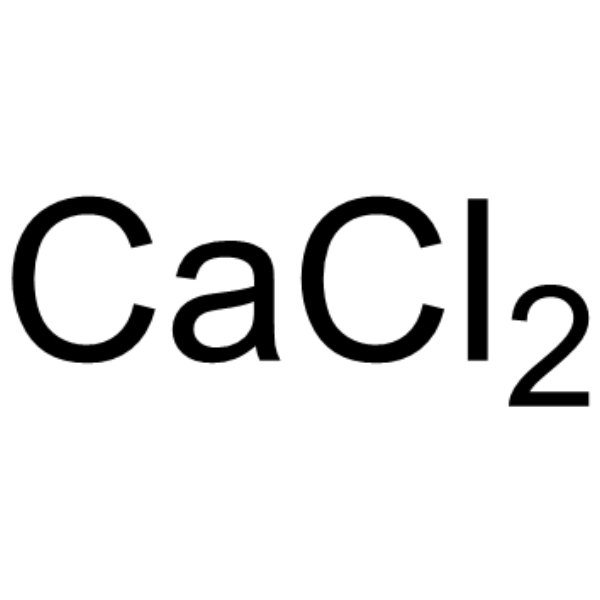
Calcium chloride anhydrous, for cell cultureCalcium Chloride


Biological Activity
제품 설명
In Vitro
Chromium chloride (1-1000 μM; 24 h) concentration-dependently inhibits elevated TNF-α secretion in high glucose-treated, PMA/lipopolysaccharide-activated U937 cells, with significant inhibitory effects observed across the concentration range after 24 h[2].
Chromium chloride (1-1000 μM; 24 h) inhibits elevated TNF-α secretion in H2O2-treated, PMA/lipopolysaccharide-activated U937 cells, with significant inhibitory effects observed across the concentration range after 24 h[2].
Chromium chloride (10-1000 μM; 24 h) prevents H2O2-induced growth inhibition in U937 cells, with significant protective effects observed across the concentration range after 24 h[2].
Chromium chloride (100 μM; 24 h) inhibits elevated lipid peroxidation levels in both high glucose-treated and H2O2-treated U937 cells after 24 h[2].
Chromium chloride (100 μM; 24 h) inhibits elevated protein oxidation levels in both high glucose-treated and H2O2-treated U937 cells after 24 h[2].
MedChemExpress (MCE) has not independently confirmed the accuracy of these methods. They are for reference only.
-
Cell Line:human promonocytic U937 cells (PMA/lipopolysaccharide-activated, high glucose-treated)
-
Concentration:1, 10, 100, 1000 μM
-
Incubation Time:24 h
-
Result:Inhibited elevated TNF-α secretion in a concentration-dependent manner, with statistically significant reductions (P < 0.02) observed at all tested concentrations compared to the high glucose-only control.
-
Cell Line:human promonocytic U937 cells (PMA/lipopolysaccharide-activated, hydrogen peroxide-treated)
-
Concentration:1, 10, 100, 1000 μM
-
Incubation Time:24 h
-
Result:Inhibited elevated TNF-α secretion at all tested concentrations, with statistically significant reductions (P < 0.02) observed compared to the H2O2-only control.
-
Cell Line:human promonocytic U937 cells (hydrogen peroxide-treated)
-
Concentration:10, 100, 1000 μM
-
Incubation Time:24 h
-
Result:Prevented H2O2-induced growth inhibition at concentrations of 10, 100, and 1000 μM, with statistically significant differences (P < 0.01) observed compared to the H2O2-only control.
In Vivo
MedChemExpress (MCE) has not independently confirmed the accuracy of these methods. They are for reference only.
-
Animal Model:New Zealand white rabbits (male adult, aged 15 weeks, initial weight 2683.6 g; hypercholesterolemic induced by 2% cholesterol diet for 30 days)[3]
-
Dosage:1 mg chromium chloride (0.33 mg chromium/kg body weight)
-
Administration:i.m.; 6 days per week; 6 weeks
-
Result:Reduced mean log10(ORO+1) value (coronary lipid deposit area) to 1.5253, which was significantly lower than untreated hypercholesterolemic rabbits.
Reduced mean area of ascending aortic lipid deposits to 255.552, which was significantly lower than untreated hypercholesterolemic rabbits.
Reduced terminal mean serum cholesterol concentration to 175.180 mg/dL, which was significantly lower than untreated hypercholesterolemic rabbits.
Increased terminal mean serum chromium concentration to 3746.4 μg/L, compared to 4.3 μg/L in untreated rabbits.
Reduced serum albumin level to mean 48.63 g/L, alkaline phosphatase to mean 35.50 U/L, triglyceride to mean 1.07 mmol/L, and HDL-C to mean 0.66 mmol/L relative to untreated hypercholesterolemic rabbits.
Showed no significant histopathological differences in liver or kidney between chromium chloride-treated and untreated hypercholesterolemic rabbits.
- ROS/oxidative-stress fluorescent staining
- Protocol for Cell Counting and Cell Density Analysis
- Research Protocol for Endocrine Diseases
- Somatic Cell Culture
- CCK-8/WST-8 Cell Proliferation Assay
- Research Protocol for Cardiovascular Diseases
- Cell Counting-Based Growth Curve Assay
- MTT Cell Proliferation Assay
- Research Protocol for Metabolic Diseases
- Cell Viability Determination by MTT Colorimetric Assay
Clinical Trial
| NCT Number | Sponsor | Condition | Start Date |
Phase

|
|---|---|---|---|---|
| NCT01329991 | 2011-05 | PHASE1 |


Chemical Information
-
CAS No. 10025-73-7
-
Appearance Solid
-
분자량 158.36
-
화학식 CrCl3
-
Color Pale purple to purple
-
SMILES
Cl[Cr](Cl)Cl
-
선적
Room temperature in continental US; may vary elsewhere.
-
보관
4°C, protect from light, stored under nitrogen
* In solvent : -80°C, 6 months; -20°C, 1 month (protect from light, stored under nitrogen)


순도&문서
-
Data Sheet (281 KB)
-
SDS (759 KB)
- English - EN (759 KB)
- Français - FR (759 KB)
- Deutsch - DE (759 KB)
- Norwegian - NO (759 KB)
- Español - ES (759 KB)
- Swedish - SV (759 KB)
- Italian - IT (759 KB)
- Korean - KR (759 KB)
- Portuguese - PT (759 KB)
-
Handling Instructions (2659 KB)
References
[1]. Doerner PG 3rd, et al. Chromium chloride increases insulin-stimulated glucose uptake in the perfused rat hindlimb. Acta Physiol (Oxf). 2014;212(3):205-213. [Content Brief]
[2]. Jain SK, et al. Chromium chloride inhibits oxidative stress and TNF-alpha secretion caused by exposure to high glucose in cultured U937 monocytes. Biochem Biophys Res Commun. 2001;289(3):687-691. [Content Brief]
[3]. Price Evans DA, et al. Chromium chloride administration causes a substantial reduction of coronary lipid deposits, aortic lipid deposits, and serum cholesterol concentration in rabbits. Biol Trace Elem Res. 2009;130(3):262-272. [Content Brief]

Calculators
Concentration (start) × Volume (start) = Concentration (final) × Volume (final)


Chromium chloride Related Classifications
HY-B1608 Related Classifications
 Powered by Bioz