



Ezurpimtrostat hydrochloride
Based on 3 publication(s) in Google Scholar
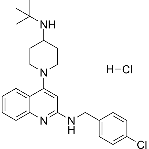
Ezurpimtrostat hydrochloride (GNS561 hydrochloride) is an orally active PPT1 inhibitor, autophagy inhibitor, immunomodulator, anti-inflammatory agent, and anticancer agent. Ezurpimtrostat hydrochloride inhibits PPT1, dysregulates lysosomal function, redistributes mTOR, and induces apoptosis. Ezurpimtrostat hydrochloride reduces IFN‑α, CRP, immune complex deposition, and SARS‑CoV‑2 viral load. Ezurpimtrostat hydrochloride can be used for the study of systemic lupus erythematosus, SARS‑CoV‑2, hepatocellular carcinoma, fibrosis, and related disorders.
For research use only. We do not sell to patients.
- Purity : 99.26%
- CAS No.: 1914148-73-4
- Formula: C25H32Cl2N4
- Molecular Weight:459.45
-
Storage:
-20°C, sealed storage, away from moisture
* In solvent : -80°C, 6 months; -20°C, 1 month (sealed storage, away from moisture)

To place orders, for customer services and technical support, please contact: MedChemExpress USA
Tel: 609-228-6898 E-mail: [email protected] [email protected]


-
 Biological Activity
-
Chemical Information
-
Solvent & Solubility
- Protocols
- Purity & Documentation
- References
-
 Help & FAQs


Publications Citing Use of MedChemExpress (MCE) Ezurpimtrostat hydrochloride
More

Biological Activity
Description
IC50 & Target
|
IFNAR1 |
In Vitro
Ezurpimtrostat (2-6 μM; 24 h) hydrochloride blocks late-stage autophagic flux in uninfected Vero E6 cells, with a dose-dependent increase in LC3-II and p62 accumulation[2].
Ezurpimtrostat (2 h pre-treatment, 24 h infection incubation) hydrochloride Ezurpimtrostat potently inhibits SARS-CoV-2 replication in Vero E6 and Calu-3 cells, with EC50 values of 0.04 μM, 0.006 μM, and 1.1 μM, and CC50 values of 8.19 μM, 2 μM, and 4.6 μM against the IHUMI-6 and USA-WA1/2020 strains[2].
Ezurpimtrostat (1 μM; 2 h pre-treatment, 48 h infection incubation) hydrochloride enhances LC3B cluster formation and signal in SARS-CoV-2-infected Vero E6 cells at 1 μM, and colocalizes with SARS-CoV-2 in LAMP2-positive lysosomes, indicating modulation of the autophagy pathway during viral infection[2].
Ezurpimtrostat (4 μM; 2 h pre-treatment, 24 h infection incubation) hydrochloride treatment at 4 μM increases autophagic vacuole volume and induces multilamellar body formation in SARS-CoV-2-infected Vero E6 cells, indicating disruption of autophagosome-lysosome fusion[2].
Ezurpimtrostat (72 h) hydrochloride potently inhibits the viability of a broad panel of human cancer cell lines and primary HCC cells[3].
Ezurpimtrostat (0-4 μM; 6-48 h) hydrochloride induces caspase-dependent apoptotic cell death in HepG2 cells in vitro in a dose- and time-dependent manner, with significant caspase activation observed after 24 hours of treatment[3].
Ezurpimtrostat (10 μM GNS561D; 90 min) hydrochloride is a lysosomotropic agent that accumulates in HepG2 cell lysosomes, and its antitumor activity in these cells is dependent on this lysosomal localization[3].
Ezurpimtrostat (0.5-10 μM; 1-24 h) hydrochloride modulates lysosomal function in HepG2 cells by inducing lysosomal enlargement, lysosomal unbound Zn2+ accumulation, impaired cathepsin maturation and activity, and blockage of autophagic flux[3].
Ezurpimtrostat (0.5-100 μM; 3-24 h) hydrochloride binds to and inhibits PPT1 in HepG2 cells, leading to MTOR displacement from the lysosomal membrane and autophagic flux inhibition; its antitumor activity is partially mediated by PPT1 inhibition, with additional contributing mechanisms[3].
Ezurpimtrostat (1-3 μM; 24-48 h) hydrochloride induces lysosomal membrane permeabilization in HepG2 cells, leading to cathepsin release and partially CTSB- and CTSD-dependent apoptotic cell death[3].
MedChemExpress (MCE) has not independently confirmed the accuracy of these methods. They are for reference only. Further protocols information, click here.
In Vivo
Ezurpimtrostat (50 mg/kg; p.o.; once daily; 24 hours pre-infection to 7 days post-infection) hydrochloride disrupts the autophagy pathway in SARS-CoV-2-infected K18-hACE2 mice and reduces lung viral load[2].
Ezurpimtrostat (50 mg/kg; p.o.) hydrochloride reduces hepatocellular carcinoma tumor volume by 37.1% and tumor weight by 34.4%, and decreases serum AFP levels significantly at days 21 and 28 post inoculation in a BALB/c nude mouse orthotopic xenograft model[3].
Ezurpimtrostat (15 mg/kg/day; p.o.; daily; 6 weeks) hydrochloride reduces hepatocellular carcinoma tumor progression by 33%, decreases mean tumor size and tumor nodule count, and lowers CCND1 and MKI67 staining in a DEN-induced cirrhotic Fischer 344 rat HCC model[3].
Ezurpimtrostat (15-50 mg/kg/day; p.o.; daily; 21-28 days) hydrochloride shows high liver tropism in rats, with the highest accumulation in liver, stomach, and lung, and limited crossing of the blood-brain and blood-testis barriers[3].
MedChemExpress (MCE) has not independently confirmed the accuracy of these methods. They are for reference only.
Clinical Trial
| NCT Number | Sponsor | Condition | Start Date |
Phase

|
|---|---|---|---|---|
| NCT01329991 | Plexxikon| | 2011-05 | PHASE1 |


Chemical Information
-
CAS No. 1914148-73-4
-
Appearance Solid
-
Molecular Weight 459.45
-
Formula C25H32Cl2N4
-
Color Off-white to light yellow
-
SMILES
CC(NC1CCN(C2=CC(NCC3=CC=C(Cl)C=C3)=NC4=CC=CC=C24)CC1)(C)C.[H]Cl
-
Synonyms
GNS561 hydrochloride
-
Shipping
Room temperature in continental US; may vary elsewhere.
-
Storage
-20°C, sealed storage, away from moisture
* In solvent : -80°C, 6 months; -20°C, 1 month (sealed storage, away from moisture)


Publications (3)
-
Journal Impact Factor
-
Most Recent
-
Research (Wash D C)
Dual Regulation of Sprouty 4 Palmitoylation by ZDHHC7 and Palmitoyl-Protein Thioesterase 1: A Potential Therapeutic Strategy for Cisplatin-Resistant Osteosarcoma. [Abstract]2025 May 23:8:0708. PMID: 40416361 -
Cell Signal
2026 Jul:143:112502. PMID: 41865945 -
Virus Res
Novel quinoline substituted autophagy inhibitors attenuate Zika virus replication in ocular cells. [Abstract]2024 Jun 18:347:199419. PMID: 38880335


Solvent & Solubility
In Vitro:
H2O : 10 mg/mL (21.77 mM; Need ultrasonic)
DMSO : 8.33 mg/mL (18.13 mM; ultrasonic and warming and heat to 60°C; Hygroscopic DMSO has a significant impact on the solubility of product, please use newly opened DMSO)
Please refer to the solubility information to select the appropriate solvent. Once prepared, please aliquot and store the solution to prevent product inactivation from repeated freeze-thaw cycles.
Storage method and period of stock solution: -80°C, 6 months; -20°C, 1 month (sealed storage, away from moisture). When stored at -80°C, please use it within 6 months. When stored at -20°C, please use it within 1 month.
* Note: If you choose water as the stock solution, please dilute it to the working solution, then filter and sterilize it with a 0.22 μm filter before use.
Please refer to the solubility information to select the appropriate solvent. Once prepared, please aliquot and store the solution to prevent product inactivation from repeated freeze-thaw cycles.
Storage method and period of stock solution: -80°C, 6 months; -20°C, 1 month (sealed storage, away from moisture). When stored at -80°C, please use it within 6 months. When stored at -20°C, please use it within 1 month.
* Note: If you choose water as the stock solution, please dilute it to the working solution, then filter and sterilize it with a 0.22 μm filter before use.
Concentration (start) × Volume (start) = Concentration (final) × Volume (final)

Protocols
-
Fibrosis/Collagen Morphometry
Fibrosis and collagen morphometry is based on the quantitative visualization of fibrillar collagen deposition in tissue sections using histochemical stains such as Sirius Red (Picrosirius Red) or Masson's trichrome, followed by image-based or polarization-enhanced analysis to estimate collagen proportional area as a surrogate of extracellular matrix accumulation during fibrotic remodeling. Sirius Red combined with polarized light microscopy enhances detection of collagen fibers due to birefringence properties, enabling more specific visualization of collagen type I and III fibrils compared to conventional bright-field histology, while whole-section or region-restricted digital morphometry reduces field-selection bias in fibrosis assessment. Alternative quantitative approaches include second harmonic generation (SHG) and two-photon excited fluorescence microscopy, which enable label-free detection of fibrillar collagen and have been validated against histological staining and biochemica
-
Collagen: Sirius Red Staining
Sirius Red or picrosirius red staining is a histochemical method for visualizing collagen-rich extracellular matrix in tissue sections, and collagen fibers are detected as red-stained structures under bright-field microscopy with enhanced birefringence under polarized light. Picrosirius red is useful for assessing total collagen organization, distribution, and fibrosis burden, but polarized color should not be interpreted as a definitive collagen type I versus type III readout because color is affected by fiber orientation, thickness, and packing.
-
Apoptosis
Apoptosis, also called programmed cell death, is generally characterized by distinct morphological characteristics.
-
TUNEL staining for apoptotic DNA fragmentation
TUNEL staining detects DNA strand breaks by using terminal deoxynucleotidyl transferase to add labeled nucleotides to exposed 3′-OH DNA termini, generating either microscopic staining in fixed cells or tissue sections, or fluorescence/cytometric signal in cell suspensions. TUNEL positivity reflects DNA fragmentation but should not be interpreted alone as definitive apoptosis, because TUNEL can also label necrotic, autolytic, mechanically damaged, or DNA-repair-associated DNA breaks.
-
Annexin V plus membrane-impermeant dye apoptosis staining
Annexin V-based apoptosis assays rely on the detection of phosphatidylserine (PS) externalization from the inner leaflet of the plasma membrane to the outer leaflet, an early biochemical hallmark of apoptosis. Fluorescently labeled Annexin V binds PS in a calcium-dependent manner, enabling identification of early apoptotic cells by flow cytometry or fluorescence microscopy. When combined with a membrane-impermeant DNA-binding dye (e. g. , propidium iodide), this approach allows discrimination between viable (Annexin V−/dye−), early apoptotic (Annexin V+/dye−), and late apoptotic or necrotic (Annexin V+/dye+) cell populations by assessing membrane integrity and PS exposure.
-
Connective Tissue: Masson's Trichrome/Collagen Trichrome Staining
Masson’s Trichrome (collagen/trichrome staining) is a histological technique that differentially stains tissue compartments using sequential acidic dyes to distinguish collagen from muscle and cytoplasmic components based on dye affinity and tissue permeability differences, enabling visualization of fibrosis and connective tissue architecture in histological sections. The classical formulation typically uses Weigert's iron hematoxylin for nuclear staining, Biebrich scarlet-acid fuchsin for cytoplasm and muscle, and aniline blue (or light green variants) for collagen, producing a characteristic blue/green collagen signal contrasted against red cytoplasm and dark nuclei. The staining principle relies on selective displacement of smaller dye molecules by larger anionic dyes in collagen-rich regions under controlled acidified conditions, which enhances collagen-specific dye retention. This property makes the method widely used for fibrosis assessment in organs such as heart, liver, lung, a
-
Autophagy
Autophagy is a process in which eukaryotic cells use lysosomes to degrade their own cytoplasmic proteins and damaged organelles under the regulation of autophagy related gene (Atg). Microtubule-associated proteins light chain 3 (LC3) is recognized as autophagy marker, which transfers from cytoplasmic LC3 (LC3-I) to membrane type (LC3-II). LC3-II/I ratio could be detected by Western Blot and fluorescence microscopy.
-
Apoptosis Solutions
Apoptosis is a regulated, generally non-lytic cell-death pathway that removes unwanted, damaged, infected, or abnormal cells through coordinated morphological changes, caspase activation, DNA fragmentation, and membrane remodeling. The intrinsic apoptosis pathway is controlled mainly by mitochondrial outer membrane permeabilization, BCL-2 family proteins, cytochrome c release, apoptosome formation, caspase-9 activation, and downstream executioner caspase-3/7 activation. The extrinsic apoptosis pathway is initiated by death receptors such as Fas, TNFR, and TRAIL receptors, which recruit adaptor proteins and activate caspase-8 before engaging executioner caspases or mitochondrial amplification through BID cleavage. Apoptosis is linked to many phenotypes, including cancer cell killing, tissue homeostasis, immune regulation, neurodegeneration, infection response, and treatment-induced cytotoxicity; unresolved questions include how apoptosis interacts with necroptosis, pyroptosis, ferroptos
-
Lysosome and acidic-vesicle live-cell staining
Lysosome and acidic-vesicle live-cell staining detects acidic intracellular compartments by using membrane-permeant acidotropic probes that accumulate in low-pH vesicles, including lysosomes, late endosomes, autolysosomes, and acidic phagosomes. LysoTracker staining is commonly used as an intensity-based readout of acidic lysosomal compartment abundance or enlargement, while acridine orange produces green fluorescence in less concentrated compartments and red fluorescence after concentration-dependent accumulation in acidic vesicular organelles. Loss or reduction of acridine-orange red signal can be used as a readout of lysosomal membrane permeabilization or reduced acidic-vesicle integrity. This protocol is designed for live cultured cells and can be adapted for fluorescence microscopy, high-content imaging, plate-reader readout, or flow cytometry when the selected literature supports the readout. Because these dyes report acidotropic accumulation rather than lysosome identity alone,
-
Research Protocol for Inflammation-related Diseases
The NLRP3 inflammasome is a cytosolic innate immune signaling platform that integrates priming signals and danger-signal activation to promote caspase-1 activation, maturation of IL-1β and IL-18, and gasdermin D-mediated pyroptotic cell death. The core experimental logic is to determine whether inflammatory disease phenotypes are driven by increased NLRP3 expression, ASC-containing inflammasome assembly, caspase-1 cleavage, GSDMD cleavage, and extracellular release of IL-1β/IL-18 rather than by nonspecific cell injury alone. The pathway is strongly linked to inflammation-related disease phenotypes because monosodium urate crystals activate NALP3/NLRP3 inflammasome signaling in gout-like crystal inflammation, cholesterol crystals activate NLRP3 inflammasomes in atherogenesis models, and DSS-induced intestinal inflammation has been reported to involve NLRP3 inflammasome activity. However, experimental colitis studies also show context-dependent protective effects of NLRP3 inflammasome co
-
Liver Cancer Modeling
Liver cancer can be classified into primary liver cancer and secondary liver cancer. Secondary liver cancer is the metastatic liver cancer. Primary liver cancer includes hepatocellular carcinoma (HCC), intrahepatic cholangiocarcinoma (ICC) and fibrolamellar HCC, of which HCC is the most common form, accounting for approximately 90% of primary liver cancers[1]. HCC mouse models include chemical agent-induced models, transplanted tumor models, and genetic engineered models.
-
Macroautophagy Solutions
Macroautophagy is a conserved lysosome-dependent degradation pathway in which cytoplasmic material is sequestered into double-membrane autophagosomes and delivered to lysosomes for degradation and recycling. The pathway supports cellular homeostasis during nutrient limitation, organelle stress, protein-aggregate accumulation, infection, differentiation, and tissue remodeling by coupling cargo sequestration, autophagosome maturation, lysosomal fusion, and degradation of cargo-derived macromolecules. The core molecular sequence includes initiation by nutrient- and stress-regulated autophagy machinery, autophagosome nucleation, LC3/ATG8-family conjugation to autophagosomal membranes, cargo selection through receptors such as SQSTM1/p62, autophagosome-lysosome fusion, and lysosomal degradation. LC3 was identified as a mammalian homolog of yeast Atg8 that localizes to autophagosomal membranes after processing, and p62/SQSTM1 was shown to connect ubiquitinated cargo with autophagic degradati


Purity & Documentation
-
Data Sheet (282 KB)
-
SDS (252 KB)
- English - EN (252 KB)
- Français - FR (252 KB)
- Deutsch - DE (252 KB)
- Norwegian - NO (252 KB)
- Español - ES (252 KB)
- Swedish - SV (252 KB)
- Italian - IT (252 KB)
- Korean - KR (252 KB)
- Portuguese - PT (252 KB)
-
Handling Instructions (2659 KB)
References
[1]. Toumi E, et al. GNS561 (ezurpimtrostat), a small basic lipophilic molecule, prevents lupus phenotype in a pristane-induced lupus mouse model. Br J Pharmacol. 2025;182(16):3786-3799. [Content Brief]
[2]. Bestion E, et al. GNS561 Exhibits Potent Antiviral Activity against SARS-CoV-2 through Autophagy Inhibition. Viruses. 2022;14(1):132. Published 2022 Jan 12. [Content Brief]
[3]. Brun S, et al. GNS561, a clinical-stage PPT1 inhibitor, is efficient against hepatocellular carcinoma via modulation of lysosomal functions. Autophagy. 2022;18(3):678-694. [Content Brief]


Complete Stock Solution Preparation Table
Please refer to the solubility information to select the appropriate solvent. Once prepared, please aliquot and store the solution to prevent product inactivation from repeated freeze-thaw cycles.
Storage method and period of stock solution: -80°C, 6 months; -20°C, 1 month (sealed storage, away from moisture). When stored at -80°C, please use it within 6 months. When stored at -20°C, please use it within 1 month.
| Optional Solvent | Concentration Solvent Mass | 1 mg | 5 mg | 10 mg | 25 mg |
|---|---|---|---|---|---|
| DMSO / H2O | 1 mM | 2.1765 mL | 10.8826 mL | 21.7652 mL | 54.4129 mL |
| 5 mM | 0.4353 mL | 2.1765 mL | 4.3530 mL | 10.8826 mL | |
| 10 mM | 0.2177 mL | 1.0883 mL | 2.1765 mL | 5.4413 mL | |
| 15 mM | 0.1451 mL | 0.7255 mL | 1.4510 mL | 3.6275 mL | |
| H2O | 20 mM | 0.1088 mL | 0.5441 mL | 1.0883 mL | 2.7206 mL |
* Note: If you choose water as the stock solution, please dilute it to the working solution, then filter and sterilize it with a 0.22 μm filter before use.


Ezurpimtrostat hydrochloride Related Classifications
HY-137978A Related Classifications
 Powered by Bioz