



Didrovaltrate

Based on 1 Customer Validation
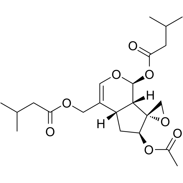
Didrovaltrate (Didrovaltratum) is an L-type calcium channel blocker, ROS scavenger, autophagy enhancer, and lipid accumulation inhibitor. Didrovaltrate blocks L-type calcium currents in a concentration-dependent manner, shifts the current-voltage curve upward, modulates steady-state inactivation kinetics, and inhibits the nuclear translocation of glucocorticoid receptors. Didrovaltrate reduces ROS levels, downregulates the expression of muscle atrophy-related genes, enhances autophagy via lipophagy, and decreases Oleic acid-induced lipid accumulation. Didrovaltrate exhibits cytotoxic activity against cancer cells. Didrovaltrate can be used in research related to skeletal muscle atrophy, non-alcoholic fatty liver disease, breast cancer, lung cancer, gastric cancer, and prostate cancer.
For research use only. We do not sell to patients.
- Purity: 92.0%
- CAS No.: 18296-45-2
- Formula: C22H32O8
- Molecular Weight:424.48
-
Storage:
-20°C, protect from light
* In solvent : -80°C, 6 months; -20°C, 1 month (protect from light)

To place orders, for customer services and technical support, please contact: MedChemExpress USA
Tel: 609-228-6898 E-mail: [email protected] [email protected]


-
 Biological Activity
-
Chemical Information
- Purity & Documentation
- References
-
 Help & FAQs


All Calcium Channel Isoforms
More

Biological Activity
|
Cell Line

|
Type | Value | Description | References |
|---|---|---|---|---|
| A549 | IC50 |
>10 μM
Compound: 20
|
Cytotoxicity against human A549 cells after 24 hrs by MTT assay
Cytotoxicity against human A549 cells after 24 hrs by MTT assay
|
[PMID: 19245261] |
| Bel-7402 | IC50 |
7.5 μM
Compound: 20
|
Cytotoxicity against human Bel7402 cells after 24 hrs by MTT assay
Cytotoxicity against human Bel7402 cells after 24 hrs by MTT assay
|
[PMID: 19245261] |
| HCT-8 | IC50 |
6.6 μM
Compound: 20
|
Cytotoxicity against human HCT8 cells after 24 hrs by MTT assay
Cytotoxicity against human HCT8 cells after 24 hrs by MTT assay
|
[PMID: 19245261] |
| PC-3M | IC50 |
3.8 μM
Compound: 20
|
Cytotoxicity against human PC3M cells after 24 hrs by MTT assay
Cytotoxicity against human PC3M cells after 24 hrs by MTT assay
|
[PMID: 19245261] |
Didrovaltrate (30-100 μg/L) reduces the peak L-type calcium current (maximum L-type calcium current), decreasing it from 6.01 pA/pF to 3.45 pA/pF and 2.16 pA/pF, with inhibition rates of 42.6% and 64.1%, respectively[1].
Didrovaltrate (2.5-5 μM; 24 h) significantly downregulates the expression of muscle atrophy marker genes *Atrogin-1*, *Murf1* and *Mstn* in atrophic C2C12 myotubes induced by Dexamethasone (DEX) (HY-14648), restores the fusion index and myotube diameter, reverses C2C12 myotube atrophy, reduces ROS production, and inhibits the nuclear translocation of glucocorticoid receptor and FOXO3a[2].
Didrovaltrate (2.5-25 μM; 24 h) reduces the viability of Huh7 hepatocytes at concentrations of 10 μM and higher[3].
Didrovaltrate (10 μM; 24 h) reduces oleic acid (HY-N1446)-induced lipid accumulation in Huh7 hepatocytes in an Atg5-dependent manner[3].
Didrovaltrate (10 μM; 0-24 h) enhances autophagic flux in Huh7 hepatocytes and increases the formation of LC3 puncta in cells[3].
Didrovaltrate (1-100 μM; 48 h) potently inhibits the viability of MCF7 human breast adenocarcinoma cells, with an IC50 of 3.9 μM; after 48 h of treatment, this compound exhibits moderate to weak cytotoxic activity against the A549, HGC27, PC3, and HUVEC cell lines[4].
MedChemExpress (MCE) has not independently confirmed the accuracy of these methods. They are for reference only.
-
Cell Line:DEX-induced atrophic C2C12 myotubes
-
Concentration:2.5, 5 μM
-
Incubation Time:24 h (co-incubated with 5 μM DEX)
-
Result:Significantly downregulated the DEX-induced upregulation of Atrogin-1, Murf1, and Mstn mRNA expression.
Reduced Atrogin-1 expression to ~1.3-fold and ~1.4-fold of the untreated control (from a DEX-induced ~2.2-fold).
Reduced Murf1 expression to ~0.7-fold and ~0.6-fold of the untreated control (from a DEX-induced ~1.9-fold).
Reduced Mstn expression to ~1-fold and ~0.5-fold of the untreated control (from a DEX-induced ~4.5-fold).
-
Cell Line:DEX-induced atrophic C2C12 myotubes
-
Concentration:2.5, 5 μM
-
Incubation Time:24 h (co-incubated with 5 μM DEX)
-
Result:Restored the fusion index from DEX-induced ~42% of the untreated control to ~55% and ~56% respectively.
Restored mean myotube diameter from DEX-induced ~10 μm (from untreated control ~12 μm) to ~11 μm and ~12 μm respectively.
Shifted the myotube diameter distribution away from small diameters (<10 μm) toward larger diameters (>14 μm), reversing the DEX-induced shift toward smaller myotubes.
-
Cell Line:DEX-induced atrophic C2C12 myotubes
-
Concentration:2.5, 5 μM
-
Incubation Time:24 h (co-incubated with 5 μM DEX)
-
Result:Reduced DEX-induced nuclear GR levels from ~2.81-fold of the untreated control to ~1.98-fold and ~1.81-fold of the untreated control respectively.
Reduced DEX-induced nuclear FOXO3a levels from ~3.75-fold of the untreated control to ~1.74-fold and ~1.53-fold of the untreated control respectively, effectively inhibiting DEX-induced nuclear translocation of both proteins.
-
Cell Line:Huh7 hepatocytes
-
Concentration:2.5, 5, 10, 25 μM
-
Incubation Time:24 h
-
Result:Reduced cell viability to ~75% of control at 10 μM.
Reduced cell viability to ~38% of control at 25 μM.
Caused no significant changes in cell viability at 2.5 and 5 μM.
-
Cell Line:Huh7 hepatocytes
-
Concentration:10 μM; 10 μM (with 25 nM BafA1 for final 1 h)
-
Incubation Time:0-24 h; 24 h (with 1 h BafA1 treatment)
-
Result:Increased LC3-II levels relative to LC3-I over time, with the highest ratio (1.5) observed at 24 hours.
Further increased LC3-II levels (ratio of 1.7) compared to BafA1 alone in the presence of BafA1.
-
Cell Line:Huh7 hepatocytes
-
Concentration:10 μM
-
Incubation Time:24 h
-
Result:Increased the formation of LC3 puncta compared to vehicle-treated cells.
-
Cell Line:human lung adenocarcinoma (A549), human breast adenocarcinoma (MCF7), human gastric carcinoma (HGC27), human prostate adenocarcinoma (PC3), human umbilical vein endothelial (HUVEC)
-
Concentration:1-100 μM
-
Incubation Time:48 h
-
Result:Inhibited cell viability of A549 cell line with an IC50 of 26.7 μM.
Inhibited cell viability of MCF7 cell line with an IC50 of 3.9 μM.
Inhibited cell viability of HGC27 cell line with an IC50 of 12.6 μM.
Inhibited cell viability of PC3 cell line with an IC50 of 69.3 μM.
Inhibited cell viability of HUVEC cell line with an IC50 of 14.8 μM.
Exhibited the most potent activity against the MCF7 breast cancer cell line.


Chemical Information
-
CAS No. 18296-45-2
-
Appearance Solid
-
Molecular Weight 424.48
-
Formula C22H32O8
-
Color White to off-white
-
SMILES
CC(O[C@@H]1[C@]2(CO2)[C@]([C@@H]3OC(CC(C)C)=O)([H])[C@](C(COC(CC(C)C)=O)=CO3)([H])C1)=O
-
Synonyms
Didrovaltratum
-
Structure Classification
-
Initial Source
-
Shipping
Room temperature in continental US; may vary elsewhere.
-
Storage
-20°C, protect from light
* In solvent : -80°C, 6 months; -20°C, 1 month (protect from light)


Purity & Documentation
-
Data Sheet (283 KB)
-
SDS (393 KB)
- English - EN (393 KB)
- Français - FR (393 KB)
- Deutsch - DE (393 KB)
- Norwegian - NO (393 KB)
- Español - ES (393 KB)
- Swedish - SV (393 KB)
- Italian - IT (393 KB)
- Korean - KR (393 KB)
- Portuguese - PT (393 KB)
-
Handling Instructions (2659 KB)
References
[1]. Xie Q, et al. Effect of didrovaltrate on I-calcium current in rabbit ventricular myocytes. J Tradit Chin Med. 2012;32(3):442-445. [Content Brief]
[2]. Kim YI, et al. Antioxidant Activity of Valeriana fauriei Protects against Dexamethasone-Induced Muscle Atrophy. Oxid Med Cell Longev. 2022 Jan 12;2022:3645431. [Content Brief]
[3]. Lee DH, et al. Iridoids of Valeriana fauriei contribute to alleviating hepatic steatosis in obese mice by lipophagy. Biomed Pharmacother. 2020;125:109950. [Content Brief]
[4]. Erdoğan M, et al. Secondary metabolites from the underground parts of Valeriana sisymbriifolia Vahl. and their in vitro cytotoxic activities. Phytochemistry. 2023;208:113590. [Content Brief]

Calculators
Concentration (start) × Volume (start) = Concentration (final) × Volume (final)


Didrovaltrate Related Classifications
HY-N3741 Related Classifications
 Powered by Bioz