



KI696 isomer

Based on 1 Customer Validation
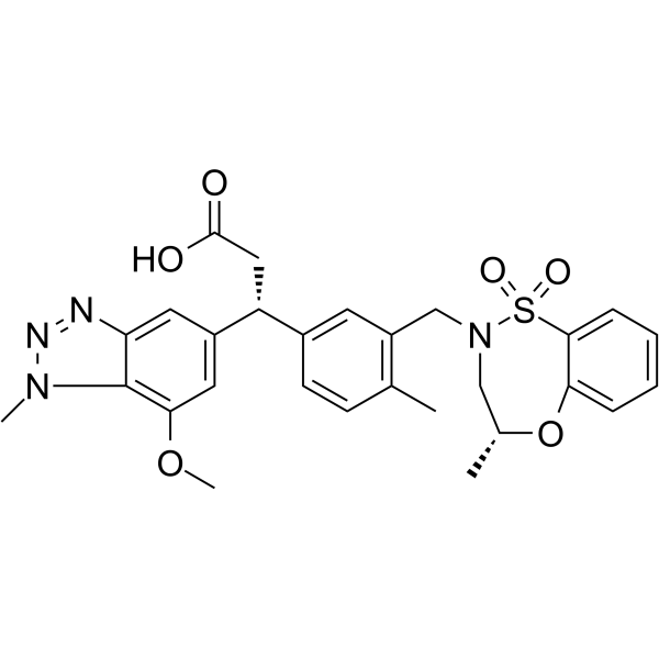
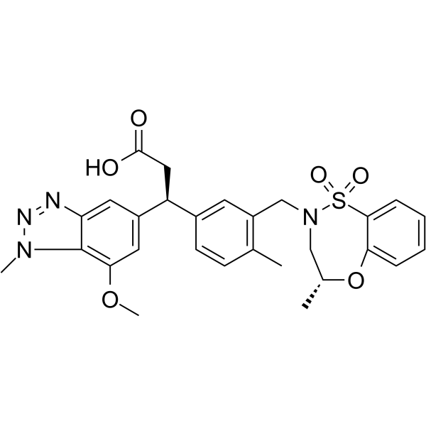
KI696 isomer is an isomer of KI696 (HY-101140). KI696 is a selective KEAP1/NRF2 protein-protein interaction inhibitor with a human Kd value of 1.3 nM. KI696 acts by competitively occupying the NRF2-binding pocket of the KEAP1 Kelch domain. KI696 blocks KEAP1-mediated ubiquitination and degradation of NRF2, promotes the translocation of NRF2 to the nucleus, activates the expression of downstream antioxidant genes, increases intracellular glutathione levels, and alleviates oxidative stress-induced cell damage and inflammatory cell infiltration in the lungs. KI696 reduces ozone-induced pulmonary oxidative damage and inflammatory cell accumulation in rats, and upregulates pulmonary antioxidant genes. KI696 can be used in research related to chronic obstructive pulmonary disease, oxidative stress and inflammation.
Nur für Forschungszwecke. Wir verkaufen nicht an Patienten.
- Reinheit : 98.37%
- CAS. Nr.: 1799974-69-8
- Formel: C28H30N4O6S
- Molecular Weight:550.63
-
Speicherung:Powder -20°C, 3 years , 4°C, 2 years ; In solvent -80°C, 2 years , -20°C, 1 year

To place orders, for customer services and technical support, please contact: MedChemExpress USA
Tel: 609-228-6898 E-mail: [email protected] [email protected]


-
 Biologische Aktivität
-
Chemical Information
-
Lösungsmittel & Löslichkeit
- Protokoll
- Reinheit & Dokumentation
- Verweise
-
 Help & FAQs


-
HY-101140
KI696KEAP1/NRF2 Protein-protein Interaction Inhibitor -
HY-101025
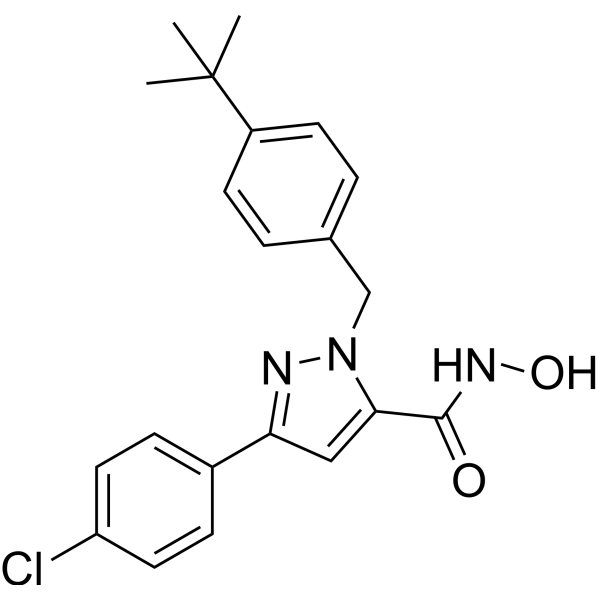
Nrf2-IN-1Nrf2 Inhibitor -
HY-12212
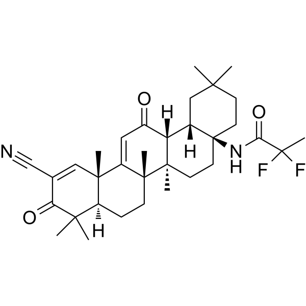
OmaveloxoloneNrf2 Activator -
HY-112675
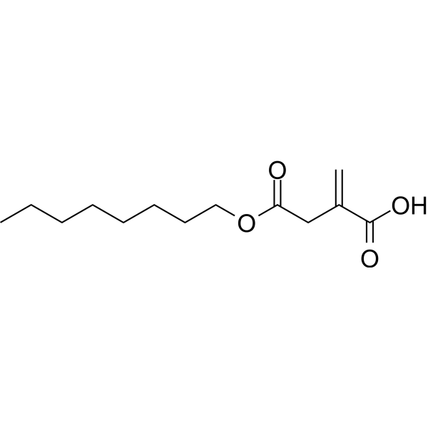
4-Octyl itaconateNrf2 Activator -
HY-13755A
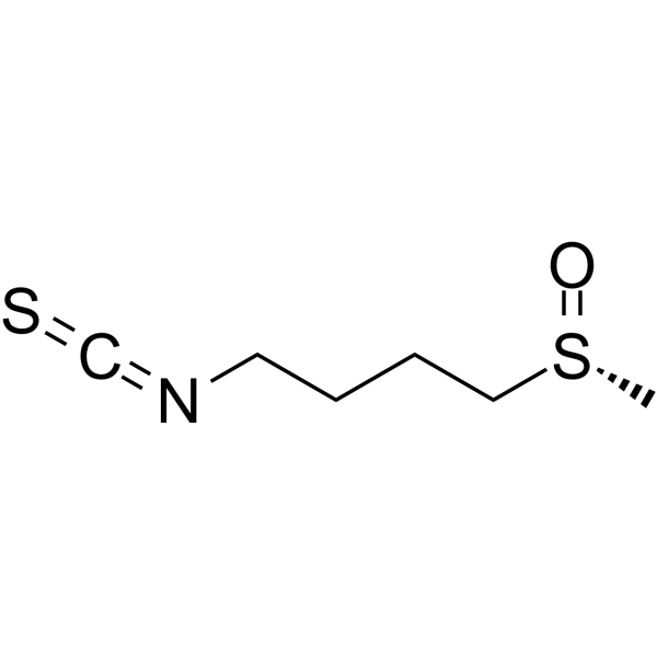
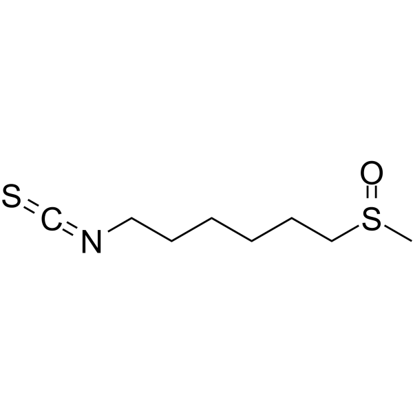
(R)-SulforaphaneKeap1/Nrf2/ARE Inducer -
HY-15725
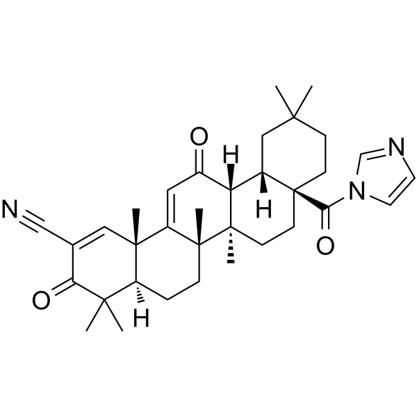
CDDO-ImNrf2 Activator -
HY-100523
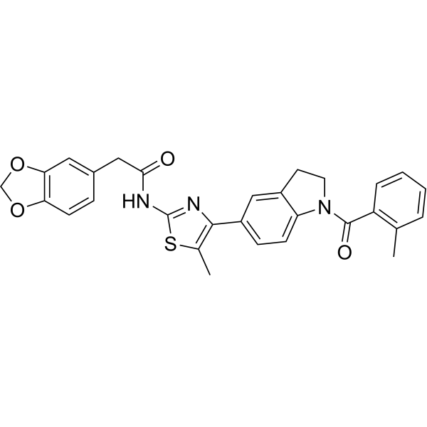
ML385Nrf2 Inhibitor -
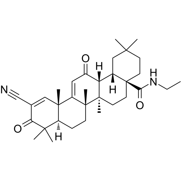
HY-12213
CDDO-EAKeap1-Nrf2 Activator -
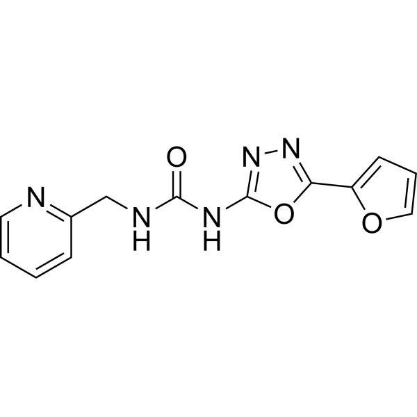
HY-19734
NK-252Nrf2 Activator -
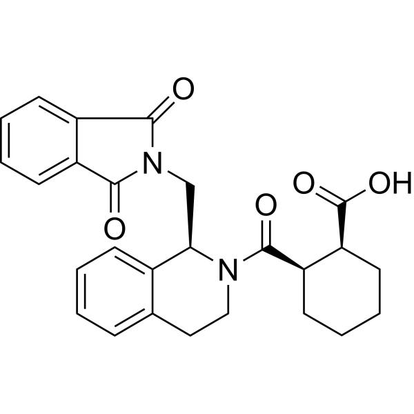
HY-110258
ML334NRF2 Activator -
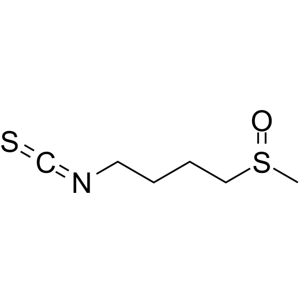
HY-13755
SulforaphaneKeap1/Nrf2/ARE Inducer -
HY-101371
HesperinNrf2 Activator


Biologische Aktivität
Beschreibung
Cellular Effect
|
Cell Line

|
Type | Value | Description | References |
|---|---|---|---|---|
| Sf9 | IC50 |
0.024 μM
Compound: 28
|
Displacement of 5'-TAMRA-labelled NRF2 peptide from human N-terminal His6-tagged KEAP1 Kelch domain (321 to 609 residues) expressed in baculovirus infected Sf9 insect cells by fluorescence polarization assay
Displacement of 5'-TAMRA-labelled NRF2 peptide from human N-terminal His6-tagged KEAP1 Kelch domain (321 to 609 residues) expressed in baculovirus infected Sf9 insect cells by fluorescence polarization assay
|
[PMID: 30973731] |
In Vitro


Chemical Information
-
CAS. Nr. 1799974-69-8
-
Appearance Solid
-
Molecular Weight 550.63
-
Formel C28H30N4O6S
-
Color White to off-white
-
SMILES
COC1=C(N2C)C(N=N2)=CC([C@@H](C3=CC(CN(C[C@H]4C)S(=O)(C5=CC=CC=C5O4)=O)=C(C)C=C3)CC(O)=O)=C1
-
Versand
Room temperature in continental US; may vary elsewhere.
-
Speicherung
Powder -20°C 3 years 4°C 2 years In solvent -80°C 2 years -20°C 1 year


Lösungsmittel & Löslichkeit
In Vitro:
DMSO : 125 mg/mL (227.01 mM; Need ultrasonic; Hygroscopic DMSO has a significant impact on the solubility of product, please use newly opened DMSO)
Please refer to the solubility information to select the appropriate solvent. Once prepared, please aliquot and store the solution to prevent product inactivation from repeated freeze-thaw cycles.
Storage method and period of stock solution: -80°C, 2 years; -20°C, 1 year. When stored at -80°C, please use it within 2 years. When stored at -20°C, please use it within 1 year.
Please refer to the solubility information to select the appropriate solvent. Once prepared, please aliquot and store the solution to prevent product inactivation from repeated freeze-thaw cycles.
Storage method and period of stock solution: -80°C, 2 years; -20°C, 1 year. When stored at -80°C, please use it within 2 years. When stored at -20°C, please use it within 1 year.
Konzentration (Stammlösung) × Volumen (Stammlösung) = Konzentration (Ziellösung) × Volumen (Ziellösung)
In Vivo:
Select the appropriate dissolution method based on your experimental animal and administration route.
- For the following dissolution methods, please ensure to first prepare a clear stock solution using an In Vitro approach and then sequentially add co-solvents:
- To ensure reliable experimental results, the clarified stock solution can be appropriately stored based on storage conditions. As for the working solution for In Vivo experiments, it is recommended to prepare freshly and use it on the same day.
- The percentages shown for the solvents indicate their volumetric ratio in the final prepared solution. If precipitation or phase separation occurs during preparation, heat and/or sonication can be used to aid dissolution.
Add each solvent one by one: 10% DMSO 40% PEG300 5% Tween-80 45% Saline
Solubility: ≥ 2.08 mg/mL (3.78 mM); Clear solution
This protocol yields a clear solution of ≥ 2.08 mg/mL (saturation unknown).
Taking 1 mL working solution as an example, add 100 μL DMSO stock solution (20.8 mg/mL) to 400 μL PEG300, and mix evenly; then add 50 μL Tween-80 and mix evenly; then add 450 μL Saline to adjust the volume to 1 mL.
Preparation of Saline: Dissolve 0.9 g sodium chloride in ddH₂O and dilute to 100 mL to obtain a clear Saline solution.
In Vivo Dissolution Calculator
Please enter the basic information of animal experiments:
-
-
-
-
Recommended: Prepare an additional quantity of animals to account for potential losses during experiments.
Please enter your animal formula composition:
-
%DMSO +
Recommended: Keep the proportion of DMSO in working solution below 2% if your animal is weak.
-
%+
-
+%Tween-80 + +
-
%Saline +
The co-solvents required include: DMSO, . All of co-solvents are available by MedChemExpress (MCE). , Tween 80. All of co-solvents are available by MedChemExpress (MCE).
Working solution concentration: 0.22 mg/mL
Method for preparing stock solution: mg drug dissolved in μL DMSO. Stock solution concentration: mg/mL.
1. Take μL DMSO stock solution;
2. Add μL .
μL , mix evenly;
3. Then add μL Tween 80, mix evenly;
4. Then add μL
Please ensure that the stock solution in the first step is dissolved to a clear state, and add co-solvents in sequence. You can use ultrasonic heating (ultrasonic cleaner, recommended frequency 20-40 kHz), vortexing, etc. to assist dissolution.


Reinheit & Dokumentation
-
Data Sheet (283 KB)
-
SDS (254 KB)
- English - EN (254 KB)
- Français - FR (254 KB)
- Deutsch - DE (254 KB)
- Norwegian - NO (254 KB)
- Español - ES (254 KB)
- Swedish - SV (254 KB)
- Italian - IT (254 KB)
- Korean - KR (254 KB)
- Portuguese - PT (254 KB)
-
Handling Instructions (2659 KB)
Verweise


Complete Stock Solution Preparation Table
Please refer to the solubility information to select the appropriate solvent. Once prepared, please aliquot and store the solution to prevent product inactivation from repeated freeze-thaw cycles.
Storage method and period of stock solution: -80°C, 2 years; -20°C, 1 year. When stored at -80°C, please use it within 2 years. When stored at -20°C, please use it within 1 year.
| Optional Solvent | Concentration Solvent Mass | 1 mg | 5 mg | 10 mg | 25 mg |
|---|---|---|---|---|---|
| DMSO | 1 mM | 1.8161 mL | 9.0805 mL | 18.1610 mL | 45.4025 mL |
| 5 mM | 0.3632 mL | 1.8161 mL | 3.6322 mL | 9.0805 mL | |
| 10 mM | 0.1816 mL | 0.9081 mL | 1.8161 mL | 4.5403 mL | |
| 15 mM | 0.1211 mL | 0.6054 mL | 1.2107 mL | 3.0268 mL | |
| 20 mM | 0.0908 mL | 0.4540 mL | 0.9081 mL | 2.2701 mL | |
| 25 mM | 0.0726 mL | 0.3632 mL | 0.7264 mL | 1.8161 mL | |
| 30 mM | 0.0605 mL | 0.3027 mL | 0.6054 mL | 1.5134 mL | |
| 40 mM | 0.0454 mL | 0.2270 mL | 0.4540 mL | 1.1351 mL | |
| 50 mM | 0.0363 mL | 0.1816 mL | 0.3632 mL | 0.9081 mL | |
| 60 mM | 0.0303 mL | 0.1513 mL | 0.3027 mL | 0.7567 mL | |
| 80 mM | 0.0227 mL | 0.1135 mL | 0.2270 mL | 0.5675 mL | |
| 100 mM | 0.0182 mL | 0.0908 mL | 0.1816 mL | 0.4540 mL |


KI696 isomer Related Classifications
HY-101140A Related Classifications
 Powered by Bioz