



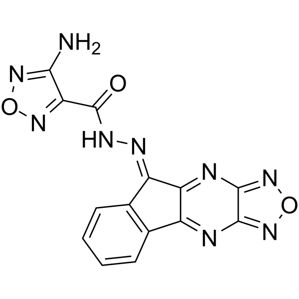
LQZ-7F

Based on 1 Customer Validation
LQZ-7F, a survivin dimerization inhibitor, induces spontaneous apoptosis and synergizes with Docetaxel in prostate cancer cells. LQZ-7F dose-dependently inhibits survival of both PC-3 and C4-2 cells with IC50s of 2.99 and 2.47 µM, respectively.
연구목적의 판매만을 진행합니다. 환자를 대상으로 한 판매는 하지 않습니다.
- Purity : 97.44%
- CAS No.: 354543-09-2
- 화학식: C14H7N9O3
- 분자량:349.26
-
보관:Powder -20°C, 3 years , 4°C, 2 years ; In solvent -80°C, 6 months , -20°C, 1 month

To place orders, for customer services and technical support, please contact: MedChemExpress USA
Tel: 609-228-6898 E-mail: [email protected] [email protected]


-
 Biological Activity
-
Chemical Information
-
용액&용해도
- Protocol
- 순도&문서
- References
-
 Help & FAQs




Biological Activity
제품 설명
Cellular Effect
|
Cell Line

|
Type | Value | Description | References |
|---|---|---|---|---|
| A549 | EC50 |
0.8 μM
Compound: LQZ-7F
|
Antiproliferative activity against human A549 cells assessed as inhibition of cell proliferation incubated for 24 hrs by MTT assay
Antiproliferative activity against human A549 cells assessed as inhibition of cell proliferation incubated for 24 hrs by MTT assay
|
[PMID: 33569941] |
| HCT-116 | EC50 |
1.6 μM
Compound: LQZ-7F
|
Antiproliferative activity against human HCT-116 cells assessed as inhibition of cell proliferation incubated for 24 hrs by MTT assay
Antiproliferative activity against human HCT-116 cells assessed as inhibition of cell proliferation incubated for 24 hrs by MTT assay
|
[PMID: 33569941] |
| HCT-116 | IC50 |
0.99 μM
Compound: 18, ZINC02092166
|
Cytotoxicity against human HCT116 cells assessed as inhibition of cell growth after 72 hrs by MTS assay
Cytotoxicity against human HCT116 cells assessed as inhibition of cell growth after 72 hrs by MTS assay
|
[PMID: 25985283] |
| HEK293 | IC50 |
0.86 μM
Compound: 18, ZINC02092166
|
Inhibition of interaction of beta-catenin (unknown origin) transfected in Wnt-activated HEK293 cells/Tcf4 assessed as decrease in transactivation of canonical Wnt signaling by TOPflash luciferase reporter assay
Inhibition of interaction of beta-catenin (unknown origin) transfected in Wnt-activated HEK293 cells/Tcf4 assessed as decrease in transactivation of canonical Wnt signaling by TOPflash luciferase reporter assay
|
[PMID: 25985283] |
| HL-60 | EC50 |
1.2 μM
Compound: LQZ-7F
|
Antiproliferative activity against human HL-60 cells assessed as inhibition of cell proliferation incubated for 24 hrs by MTT assay
Antiproliferative activity against human HL-60 cells assessed as inhibition of cell proliferation incubated for 24 hrs by MTT assay
|
[PMID: 33569941] |
| HT-29 | IC50 |
1.1 μM
Compound: 18, ZINC02092166
|
Cytotoxicity against human HT-29 cells assessed as inhibition of cell growth after 72 hrs by MTS assay
Cytotoxicity against human HT-29 cells assessed as inhibition of cell growth after 72 hrs by MTS assay
|
[PMID: 25985283] |
| LNCaP C4-2 | IC50 |
1.9 μM
Compound: 7; LQZ-7F
|
Antiproliferative activity against human LNCaP C4-2 cells assessed as cell growth inhibition
Antiproliferative activity against human LNCaP C4-2 cells assessed as cell growth inhibition
|
[PMID: 38092421] |
| LNCaP C4-2 | IC50 |
1.9 μM
Compound: 7; LQZ-7F
|
Cytotoxicity against human LNCaP C4-2 cells assessed as cell growth inhibition incubated for 24 hrs by MTT assay
Cytotoxicity against human LNCaP C4-2 cells assessed as cell growth inhibition incubated for 24 hrs by MTT assay
|
[PMID: 38092421] |
| LNCaP C4-2 | IC50 |
2.47 μM
Compound: 7F; LQZ-7F
|
Cytotoxicity against human LNCaP C4-2 cells assessed as reduction in cell viability incubated for 72 hrs by methylene blue survival assay
Cytotoxicity against human LNCaP C4-2 cells assessed as reduction in cell viability incubated for 72 hrs by methylene blue survival assay
|
[PMID: 35504208] |
| PC-3 | IC50 |
1.3 μM
Compound: 7; LQZ-7F
|
Anticancer activity against human PC-3 cells assessed as cell growth inhibition by sulforhodamine B assay
Anticancer activity against human PC-3 cells assessed as cell growth inhibition by sulforhodamine B assay
|
[PMID: 38092421] |
| PC-3 | IC50 |
2.99 μM
Compound: 7F; LQZ-7F
|
Cytotoxicity against human PC-3 cells assessed as reduction in cell viability incubated for 72 hrs by methylene blue survival assay
Cytotoxicity against human PC-3 cells assessed as reduction in cell viability incubated for 72 hrs by methylene blue survival assay
|
[PMID: 35504208] |
| PC-3 | IC50 |
4.3 μM
Compound: 7; LQZ-7F
|
Cytotoxicity against human PC-3 cells assessed as reduction in cell viability incubated for 24 hrs by MTT assay
Cytotoxicity against human PC-3 cells assessed as reduction in cell viability incubated for 24 hrs by MTT assay
|
[PMID: 38092421] |
| SW480 | IC50 |
0.71 μM
Compound: 18, ZINC02092166
|
Inhibition of beta-catenin/Tcf4 interaction in human SW480 cells assessed as decrease in transactivation of canonical Wnt signaling by TOPflash luciferase reporter assay
Inhibition of beta-catenin/Tcf4 interaction in human SW480 cells assessed as decrease in transactivation of canonical Wnt signaling by TOPflash luciferase reporter assay
|
[PMID: 25985283] |
| SW480 | IC50 |
0.85 μM
Compound: 18, ZINC02092166
|
Cytotoxicity against human SW480 cells assessed as inhibition of cell growth after 72 hrs by MTS assay
Cytotoxicity against human SW480 cells assessed as inhibition of cell growth after 72 hrs by MTS assay
|
[PMID: 25985283] |
In Vitro
LQZ-7F could be hudrolyzed under acidic conditions[1].
LQZ-7F (2.5 μM, 72 hours) displays cytotoxicity towards human cancer cells (PC-3, C4-2) with the IC50s of 2.99 μM and 2.74 μM, respectively[1].
LQZ-7F effectively inhibits survival of all cancer cell lines with IC50 values ranging between 0.4 and 4.4 mM[2].
LQZ-7F (2 μM, 24 hours) disrupts microtubule structure and cause mitotic arrest in P3 cells[2].
MedChemExpress (MCE) has not independently confirmed the accuracy of these methods. They are for reference only.
-
Cell Line:PC3, HL-60 cells
-
Concentration:0, 5, 10 μM
-
Incubation Time:
-
Result:Caused 54% to 69% apoptosis for PC3 and 66% to 98% apoptosis for HL-60 cells.
-
Cell Line:PC-3, DU145
-
Concentration:10, 20 μM
-
Incubation Time:72 hours
-
Result:Showed that the IC50 in human PC3 and DU145 cells was approximately 25 μM.
In Vivo
MedChemExpress (MCE) has not independently confirmed the accuracy of these methods. They are for reference only.
-
Animal Model:4- to 6-week-old male NSG (NOD/SCID/IL-2Rg null) mice[2].
-
Dosage:25 mg/kg
-
Administration:24 days
-
Result:Inhibited growth of xenograft tumors by inhibiting surviving.


Chemical Information
-
CAS No. 354543-09-2
-
Appearance Solid
-
분자량 349.26
-
화학식 C14H7N9O3
-
Color Yellow to orange
-
SMILES
O=C(C1=NON=C1N)N/N=C2C3=C(C4=NC5=NON=C5N=C4\2)C=CC=C3
-
선적
Room temperature in continental US; may vary elsewhere.
-
보관
Powder -20°C 3 years 4°C 2 years In solvent -80°C 6 months -20°C 1 month


용액&용해도
In Vitro:
DMSO : 5 mg/mL (14.32 mM; ultrasonic and warming and heat to 60°C; Hygroscopic DMSO has a significant impact on the solubility of product, please use newly opened DMSO)
Please refer to the solubility information to select the appropriate solvent. Once prepared, please aliquot and store the solution to prevent product inactivation from repeated freeze-thaw cycles.
Storage method and period of stock solution: -80°C, 6 months; -20°C, 1 month. When stored at -80°C, please use it within 6 months. When stored at -20°C, please use it within 1 month.
Please refer to the solubility information to select the appropriate solvent. Once prepared, please aliquot and store the solution to prevent product inactivation from repeated freeze-thaw cycles.
Storage method and period of stock solution: -80°C, 6 months; -20°C, 1 month. When stored at -80°C, please use it within 6 months. When stored at -20°C, please use it within 1 month.
Concentration (start) × Volume (start) = Concentration (final) × Volume (final)
In Vivo:
For the following dissolution methods, please prepare the working solution directly:
It is recommended to prepare fresh solutions and use them promptly within a short period of time.
The percentages shown for the solvents indicate their volumetric ratio in the final prepared solution. If precipitation or phase separation occurs during preparation, heat and/or sonication can be used to aid dissolution.
Add each solvent one by one: 50% PEG300 50% Saline
Solubility: 10 mg/mL (28.63 mM); Suspended solution; Need ultrasonic and warming and heat to 60°C


순도&문서
-
Data Sheet (279 KB)
-
SDS (393 KB)
- English - EN (393 KB)
- Français - FR (393 KB)
- Deutsch - DE (393 KB)
- Norwegian - NO (393 KB)
- Español - ES (393 KB)
- Swedish - SV (393 KB)
- Italian - IT (393 KB)
- Korean - KR (393 KB)
- Portuguese - PT (393 KB)
-
Handling Instructions (2659 KB)
References


Complete Stock Solution Preparation Table
Please refer to the solubility information to select the appropriate solvent. Once prepared, please aliquot and store the solution to prevent product inactivation from repeated freeze-thaw cycles.
Storage method and period of stock solution: -80°C, 6 months; -20°C, 1 month. When stored at -80°C, please use it within 6 months. When stored at -20°C, please use it within 1 month.
| Optional Solvent | Concentration Solvent Mass | 1 mg | 5 mg | 10 mg | 25 mg |
|---|---|---|---|---|---|
| DMSO | 1 mM | 2.8632 mL | 14.3160 mL | 28.6320 mL | 71.5799 mL |
| 5 mM | 0.5726 mL | 2.8632 mL | 5.7264 mL | 14.3160 mL | |
| 10 mM | 0.2863 mL | 1.4316 mL | 2.8632 mL | 7.1580 mL |


LQZ-7F Related Classifications
HY-122678 Related Classifications
 Powered by Bioz