



LXRβ agonist-2
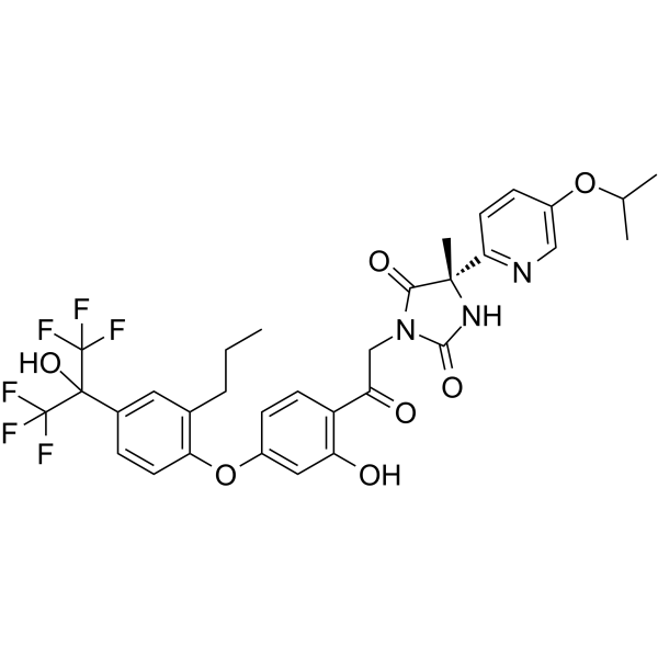
LXRβ agonist-2 is an orally active and selective LXRβ agonist. LXRβ agonist-2 increases high-density lipoprotein cholesterol levels without elevating plasma triglyceride levels. LXRβ agonist-2 decreases lipid accumulation area in the aortic arch. LXRβ agonist-2 can be used for the research of atherosclerosis.
Nos produits utilisent uniquement pour la recherche. Nous ne vendons pas aux patients.
- CAS No.: 1949801-52-8
- Formule: C32H31F6N3O7
- Masse moléculaire:683.59
-
Stockage:
Please store the product under the recommended conditions in the Certificate of Analysis.

To place orders, for customer services and technical support, please contact: MedChemExpress USA
Tel: 609-228-6898 E-mail: [email protected] [email protected]


-
 Activité biologique
-
Chemical Information
- Protocole
- Pureté et documentation
- Références
-
 Help & FAQs


-
HY-10627
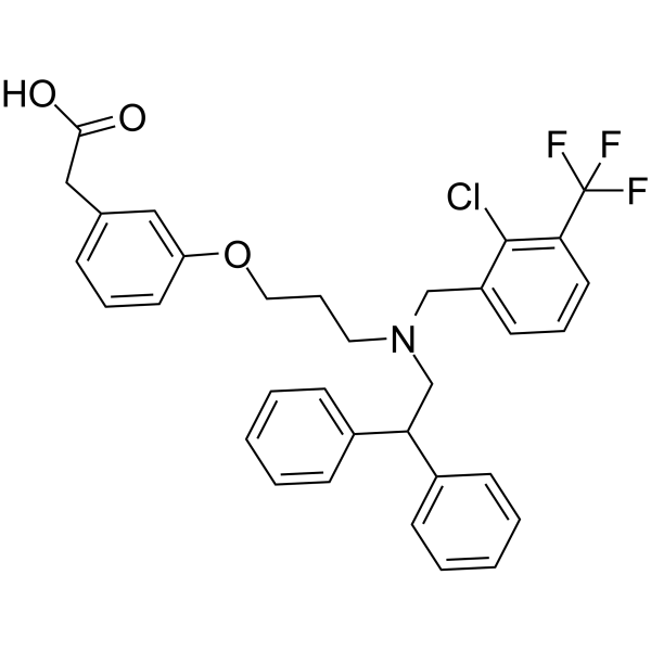
GW3965LXR Agonist -
HY-10627A
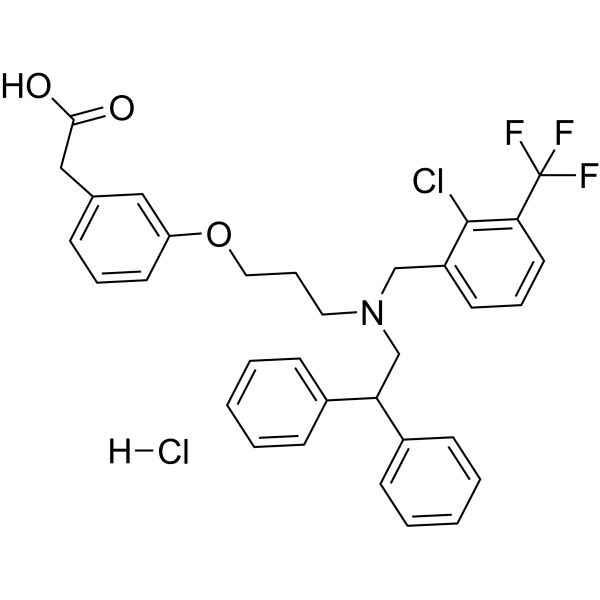
GW3965 hydrochlorideLXR Agonist -
HY-108688
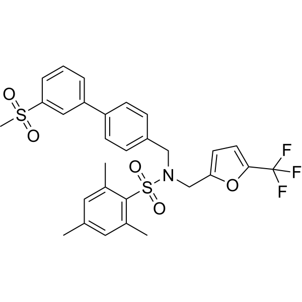
GSK2033LXR Antagonist -
HY-10629
LXR-623LXRα/β Agonist -
HY-111498A
RGX-104LXR Agonist -
HY-16972
SR9243LXR Inverse Agonist -
HY-18282
AZ876LXR Agonist -
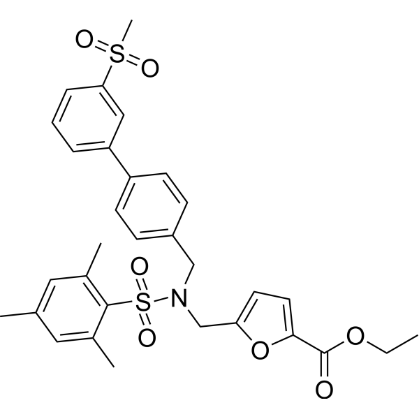
HY-101442
SR9238LXR Inverse Agonist -
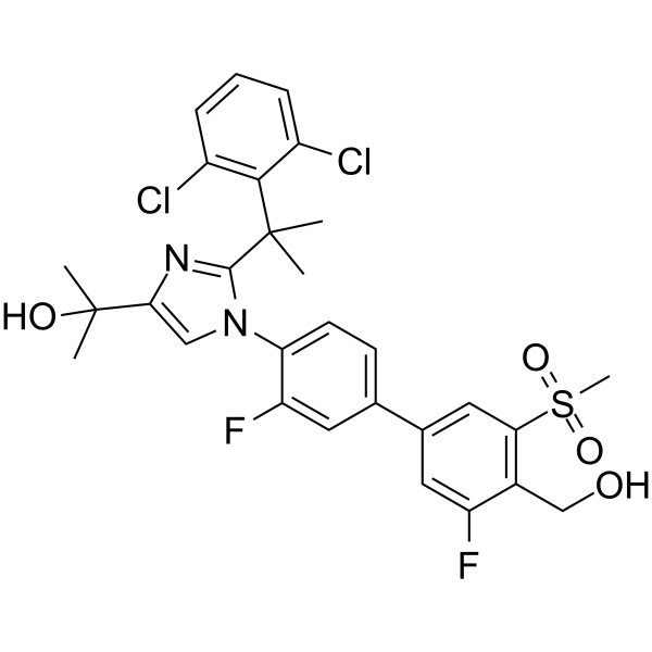
HY-101973
XL041LXRβ Agonist -
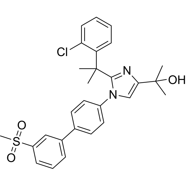
HY-19919
BMS-779788LXR Agonist -
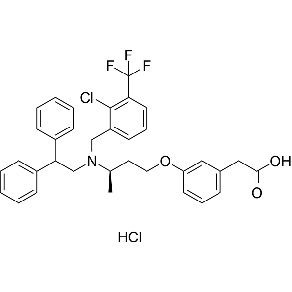
HY-111498
RGX-104 hydrochlorideLXR Agonist -
HY-109073
RovazolacLXR Modulator


Activité biologique
Description
IC50 & Target
[1]|
LXRβ |
In Vitro
LXRβ agonist-2 (Compound (-)-56) (0.01-10 μM; 24 h) potently and selectively activates LXRβ in GAL4-LXR luciferase assays using stably transfected CHO K-1 cells, with an EC50 of 0.007 μM and an LXRα/LXRβ selectivity ratio of 28.5[1].
MedChemExpress (MCE) has not independently confirmed the accuracy of these methods. They are for reference only. Further protocols information, click here.
Parmacokinetics
| Species | Dose | Route | Cmax | AUC |
|---|---|---|---|---|
| Golden hamster[1] | 10 mg/kg | p.o. | 503 ng/mL | 3027 ng·h/mL |
In Vivo
MedChemExpress (MCE) has not independently confirmed the accuracy of these methods. They are for reference only.
-
Animal Model:LDL receptor knock-out (male, 10 weeks old, fed western diet to induce atherosclerotic lesions)[1]
-
Dosage:1 mg/kg; 3 mg/kg
-
Administration:p.o.; daily; 10 weeks
-
Result:Increased HDL-C to 115% of control levels.
Maintained LDL-C at 108% of control.
Increased plasma TG to 152% of control.
Reduced aortic arch lipid accumulation area to 62% of control (p < 0.05).
Increased HDL-C to 116% of control levels.
Maintained LDL-C at 97% of control.
Increased plasma TG to 166% of control.
Reduced aortic arch lipid accumulation area to 59% of control (p < 0.05).


Chemical Information
-
CAS No. 1949801-52-8
-
Masse moléculaire 683.59
-
Formule C32H31F6N3O7
-
SMILES
OC(C(F)(F)F)(C(F)(F)F)C1=CC(CCC)=C(OC2=CC(O)=C(C(CN3C(N[C@@](C)(C4=NC=C(OC(C)C)C=C4)C3=O)=O)=O)C=C2)C=C1
-
Livraison
Room temperature in continental US; may vary elsewhere.
-
Stockage
Please store the product under the recommended conditions in the Certificate of Analysis.

Protocole
-
3T3-L1 preadipocyte-to-adipocyte differentiation
3T3-L1 preadipocytes are induced to differentiate after growth arrest using adipogenic media containing insulin, dexamethasone, and IBMX; differentiation is assessed by lipid-droplet accumulation, triglyceride increase, Oil Red O staining, and adipocyte-marker induction such as PPARγ and C/EBPα.
-
Research Protocol for Cardiovascular Diseases
Cardiovascular disease can be modeled as maladaptive cardiac remodeling, where ischemic injury or pressure overload activates inflammatory signaling, fibroblast activation, extracellular-matrix deposition, cardiomyocyte hypertrophy, vascular remodeling, and progressive ventricular dysfunction. The TGF-β/SMAD axis is a central profibrotic pathway after myocardial injury and pressure overload, while innate immune and cytokine pathways regulate leukocyte recruitment, scar formation, and adverse remodeling. Key unresolved questions include which inflammatory signals are reparative versus harmful, when fibrosis is protective versus maladaptive, and whether pathway inhibition improves function without weakening necessary infarct healing or compensatory remodeling.
-
Lipid Droplets: Oil Red O/Sudan Dye Lipid Staining
Lipid droplets are intracellular organelles with a neutral-lipid core that stores triacylglycerols and sterol esters, and Oil Red O or Sudan dyes detect these hydrophobic lipid deposits by partitioning into retained lipids in fresh or frozen specimens. Oil Red O stains neutral triglycerides and lipids in frozen tissue sections or air-dried cytologic preparations, while Sudan Black B has also been used as a histochemical fat stain for lipid-rich tissue structures.


Pureté et documentation
Références

Calculators
Concentration (start) × Volume (start) = Concentration (final) × Volume (final)


LXRβ agonist-2 Related Classifications
HY-100469 Related Classifications
 Powered by Bioz