



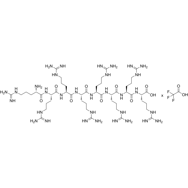
Octaarginine TFA
Octaarginine TFA is a cell-penetrating peptide and proteasome inhibitor. Octaarginine TFA exhibits mixed-type inhibition against 20S proteasome chymotrypsin-like, caspase-like, and trypsin-like activities, and inhibits 26S proteasome activity with decreased efficiency. Octaarginine TFA induces ubiquitin-conjugated protein accumulation, mediates HSPG-dependent cellular internalization via macropinocytosis, enhances liposomal cargo uptake and gene delivery. Octaarginine TFA can be used for the research of cervix carcinoma, collagen antibody-induced arthritis, and bacterial infections.
For research use only. We do not sell to patients.
- Formula: C48H98N32O9.xC2HF3O2
- Molecular Weight:1267.50 (free base)
-
Storage:
Sealed storage, away from moisture.
Powder -80°C, 2 years , -20°C, 1 year* In solvent : -80°C, 6 months; -20°C, 1 month (sealed storage, away from moisture)

To place orders, for customer services and technical support, please contact: MedChemExpress USA
Tel: 609-228-6898 E-mail: [email protected] [email protected]


-
 Biological Activity
-
Chemical Information
-
Solvent & Solubility
- Protocols
- Purity & Documentation
- References
-
 Help & FAQs




Biological Activity
Description
In Vitro
OctaarginineTFA potently inhibits the chymotrypsin-like and caspase-like activities of purified rat skeletal muscle 20S proteasomes with IC50 values of 100 nM and 200 nM, respectively, and weakly inhibits the trypsin-like activity; this inhibitory activity remains stable under physiological salt conditions[1].
Octaarginine (up to 8 μM; 5-30 min) TFA inhibits the activities of purified human erythrocyte 26S proteasomes and 20S/PA28 proteasome complexes, though with lower efficiency than for isolated 20S proteasomes[1].
Octaarginine (30-60 μM; 2 h) TFA inhibits chymotrypsin-like and caspase-like proteasome activities in cultured HeLa cells, leading to accumulation of ubiquitin-conjugated proteins, but does not significantly affect trypsin-like activity[1].
Free Octaarginine peptide efficiently delivers protein, peptide, and plasmid DNA cargos into cultured cells, with stearylation of R8 significantly enhancing plasmid DNA transfection efficiency to match Lipofectamine levels[2].
Octaarginine (3.9-7.9 nmol; 16 h) TFA inhibits the growth of E. coli and S. aureus bacterial cells, reducing survival to 60% at 7.9 nmol (16 h incubation) and 40% at 3.9 nmol (16 h incubation), respectively[4].
Octaarginine TFA causes membrane damage and permeabilization in both E. coli and S. aureus bacterial cells[4].
Octaarginine (5-30 nmol; 24 h) TFA inhibits HeLa human cancer cell growth in a concentration-dependent manner, with maximum inhibition of 49% at 15 nmol (24 h incubation)[4].
MedChemExpress (MCE) has not independently confirmed the accuracy of these methods. They are for reference only. Further protocols information, click here.
In Vivo
Rhodamine-labeled octaarginine (R8) (20 μL of 0.1 mM; intra-articular injection; single dose) accumulation is significantly decreased in the degenerative articular cartilage of CAIA mice, reflecting loss of chondroitin sulfate proteoglycans[3].
MedChemExpress (MCE) has not independently confirmed the accuracy of these methods. They are for reference only.


Chemical Information
-
Appearance Solid
-
Molecular Weight 1267.50 (free base)
-
Formula C48H98N32O9.xC2HF3O2
-
Sequence
Arg-Arg-Arg-Arg-Arg-Arg-Arg-Arg
-
Sequence Shortening
RRRRRRRR
-
Shipping
Room temperature in continental US; may vary elsewhere.
-
Storage
Sealed storage, away from moisture
Powder -80°C 2 years -20°C 1 year * In solvent : -80°C, 6 months; -20°C, 1 month (sealed storage, away from moisture)


Solvent & Solubility
In Vitro
H2O
Peptide Solubility and Storage Guidelines:
1. Calculate the length of the peptide.
2. Calculate the overall charge of the entire peptide according to the following table:
| Contents | Assign value | |
|---|---|---|
| Acidic amino acid | Asp (D), Glu (E), and the C-terminal -COOH. | -1 |
| Basic amino acid | Arg (R), Lys (K), His (H), and the N-terminal -NH2 | +1 |
| Neutral amino acid | Gly (G), Ala (A), Leu (L), Ile (I), Val (V), Cys (C), Met (M), Thr (T), Ser (S), Phe (F), Tyr (Y), Trp (W), Pro (P), Asn (N), Gln (Q) | 0 |
3. Recommended solution:
| Overall charge of peptide | Details |
|---|---|
| Negative (<0) |
1. Try to dissolve the peptide in water first. 2. If water fails, add NH4OH (<50 μL). 3. If the peptide still does not dissolve, add DMSO (50-100 μL) to solubilize the peptide. |
| Positive (>0) |
1. Try to dissolve the peptide in water first. 2. If water fails, try dissolving the peptide in a 10%-30% acetic acid solution. 3. If the peptide still does not dissolve, try dissolving the peptide in a small amount of DMSO. |
| Zero (=0) |
1. Try to dissolve the peptide in organic solvent (acetonitrile, methanol, etc.) first. 2. For very hydrophobic peptides, try dissolving the peptide in a small amount of DMSO, and then dilute the solution with water to the desired concentration. |

Protocols
-
Collagen-Induced Arthritis
Collagen-induced arthritis (CIA) is an autoimmune murine model of rheumatoid arthritis in which immunization with type II collagen (CII) emulsified in an adjuvant induces a T cell- and autoantibody-driven inflammatory arthritis characterized by synovial hyperplasia, immune cell infiltration, and joint destruction. The model typically relies on genetically susceptible mouse strains (e. g. , DBA/1) and reproduces key features of human rheumatoid arthritis, including anti-collagen immune responses and progressive joint inflammation. Disease onset generally occurs within ~3-4 weeks after immunization, depending on antigen/adjuvant combinations and protocol variation. The immunopathology is driven by adaptive immune activation against CII, leading to systemic and local joint inflammation mediated by pro-inflammatory cytokines and effector immune cells, making CIA a standard preclinical platform for evaluating immunomodulatory and anti-arthritic interventions.
-
Apoptosis Solutions
Apoptosis is a regulated, generally non-lytic cell-death pathway that removes unwanted, damaged, infected, or abnormal cells through coordinated morphological changes, caspase activation, DNA fragmentation, and membrane remodeling. The intrinsic apoptosis pathway is controlled mainly by mitochondrial outer membrane permeabilization, BCL-2 family proteins, cytochrome c release, apoptosome formation, caspase-9 activation, and downstream executioner caspase-3/7 activation. The extrinsic apoptosis pathway is initiated by death receptors such as Fas, TNFR, and TRAIL receptors, which recruit adaptor proteins and activate caspase-8 before engaging executioner caspases or mitochondrial amplification through BID cleavage. Apoptosis is linked to many phenotypes, including cancer cell killing, tissue homeostasis, immune regulation, neurodegeneration, infection response, and treatment-induced cytotoxicity; unresolved questions include how apoptosis interacts with necroptosis, pyroptosis, ferroptos


Purity & Documentation
-
Data Sheet (273 KB)
-
SDS (252 KB)
- Français - FR (252 KB)
- Deutsch - DE (252 KB)
- Norwegian - NO (252 KB)
- Español - ES (252 KB)
- Swedish - SV (252 KB)
- Italian - IT (252 KB)
- Korean - KR (252 KB)
- Portuguese - PT (252 KB)
-
Handling Instructions (2659 KB)
References
[1]. Kloss A, et al. The cell-penetrating peptide octa-arginine is a potent inhibitor of proteasome activities. Eur J Pharm Biopharm. 2009;72(1):219-225. [Content Brief]
[2]. Khalil IA, et al. Octaarginine-modified liposomes: enhanced cellular uptake and controlled intracellular trafficking. Int J Pharm. 2008;354(1-2):39-48. [Content Brief]
[3]. Inagawa K, et al. Optical imaging of mouse articular cartilage using the glycosaminoglycans binding property of fluorescent-labeled octaarginine. Osteoarthritis Cartilage. 2009;17(9):1209-1218. [Content Brief]
[4]. Ratrey P, et al. Enhancing Aqueous Solubility and Antibacterial Activity of Curcumin by Complexing with Cell-Penetrating Octaarginine. ACS Omega. 2020;5(30):19004-19013. Published 2020 Jul 23. [Content Brief]

Calculators
Concentration (start) × Volume (start) = Concentration (final) × Volume (final)


Octaarginine TFA Related Classifications
HY-P2483A Related Classifications
 Powered by Bioz