



Oritinib mesylate
Based on 1 publication(s) in Google Scholar
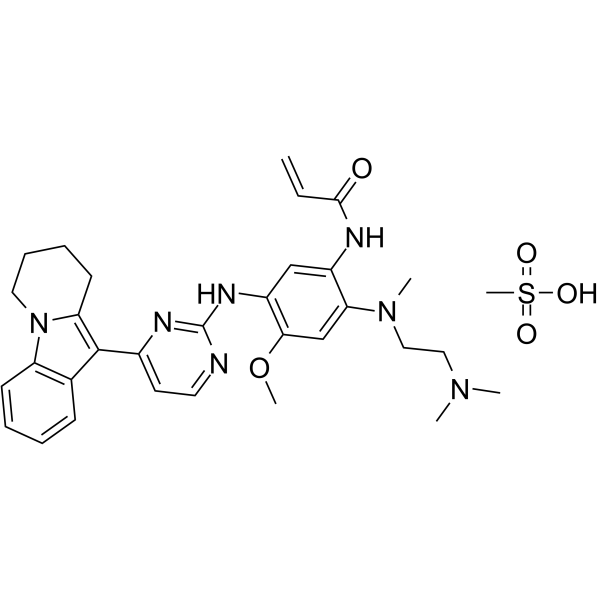
Oritinib (SH-1028) mesylate is a selective, orally active, and pyrimidine-based irreversible inhibitor of EGFR with an IC50 of 18 nM. Oritinib (SH-1028) mesylate exhibits potent activity against EGFR sensitive and resistant (T790 M) mutations. Oritinib (SH-1028) mesylate significantly inhibits proliferation of tumor cells with EGFR sensitive and resistant mutation.
For research use only. We do not sell to patients.
- CAS No.: 2180164-79-6
- Formula: C32H41N7O5S
- Molecular Weight:635.78
-
Storage:
Please store the product under the recommended conditions in the Certificate of Analysis.

To place orders, for customer services and technical support, please contact: MedChemExpress USA
Tel: 609-228-6898 E-mail: [email protected] [email protected]


-
 Biological Activity
-
Chemical Information
- Protocols
- Purity & Documentation
- References
-
 Help & FAQs


Publications Citing Use of MedChemExpress (MCE) Oritinib mesylate
MoreAll EGFR Isoforms
More

Biological Activity
Description
IC50 & Target
[1]|
EGFR (WT) 18 nM (IC50) |
EGFRL858R 0.7 nM (IC50) |
EGFRL861Q 4 nM (IC50) |
EGFRL858R/T790M 0.1 nM (IC50) |
EGFRd746-750 1.4 nM (IC50) |
EGFRd746-750/T790M 0.89 nM (IC50) |
In Vitro
Oritinib (SH-1028) (72 hours; 10 μmol/L and the 3-fold dilution; nine times) mesylate selectively inhibits EGFR-mutated NCI-H1975, H3255 and PC-9 cells, with IC50 values of 3.93, 9.39 and 7.63 nM, respectively, which is more sensitive than the inhibition of wild-type EGFR in A431 cells[1].
MedChemExpress (MCE) has not independently confirmed the accuracy of these methods. They are for reference only.
-
Cell Line:A431 (EGFRWT), H3255 (EGFRL858R), PC-9 (EGFRd746-750) and NCI-H1975 (EGFRL858R/T790M) cells
-
Concentration:0.001, 0.01, 0.1, 1, and 10 μM
-
Incubation Time:72 hours
-
Result:Selectively inhibited EGFR-mutated NCI-H1975, H3255 and PC-9 cells, with IC50s of 3.93±1.12, 9.39±0.88 and 7.63±0.18 nmol/L, respectively, which were about 198-, 83- and 102-fold more sensitive than the inhibition of wild-type EGFR in A431 cells (IC50=778.89±134.74 nM).
In Vivo
Oritinib (SH-1028) (p.o.; once daily for consecutive 14 days; 2.5-15 mg/kg) mesylate only induces a moderate tumor growth inhibition in A431 (wild-type EGFR) tumor xenografts, while causes profound and sustained tumor shrinkage in both NCI-H1975 and PC-9 xenograft models with EGFR mutations with 5 mg/kg/day[1].
Oritinib (SH-1028) (p.o.; once daily for consecutive 14 days; 2.5-15 mg/kg) mesylate shows good bioavailability, and is distributed extensively from the plasma to the tissues with Tmax of 1.5-2 h, and AUC0–t values of SH-1028 in plasma are 118, 300 and 931 ng × h/ml on Day 1, while 272, 308 and 993 ng × h/ml on Day 14[1].
MedChemExpress (MCE) has not independently confirmed the accuracy of these methods. They are for reference only.
-
Animal Model:Nu/Nu female nude mice (6-8 weeks) bearing human lung cancer cell lines[1]
-
Dosage:2.5, 5, and 15 mg/kg (SH-1028) and control group (osimertinib, 5 mg/kg)
-
Administration:p.o.; once daily for consecutive 14 days
-
Result:Only induced a moderate tumor growth inhibition in A431 (wild-type EGFR) tumor xenografts, while caused profound and sustained tumor shrinkage in both NCI-H1975 and PC-9 xenograft models with EGFR mutations with 5 mg/kg/day.


Chemical Information
-
CAS No. 2180164-79-6
-
Molecular Weight 635.78
-
Formula C32H41N7O5S
-
SMILES
CS(=O)(O)=O.O=C(NC1=CC(NC2=NC(C3=C(CCCC4)N4C5=C3C=CC=C5)=CC=N2)=C(C=C1N(CCN(C)C)C)OC)C=C
-
Synonyms
SH-1028 mesylate
-
Shipping
Room temperature in continental US; may vary elsewhere.
-
Storage
Please store the product under the recommended conditions in the Certificate of Analysis.


Publications (1)
-
Journal Impact Factor
-
Most Recent
-
J Med Chem
2025 Aug 28;68(16):17917-17932. PMID: 40801664


Purity & Documentation
References

Calculators
Concentration (start) × Volume (start) = Concentration (final) × Volume (final)


Oritinib mesylate Related Classifications
HY-139920A Related Classifications
 Powered by Bioz