



(Rac)-Nutlin-3

Based on 1 Customer Validation
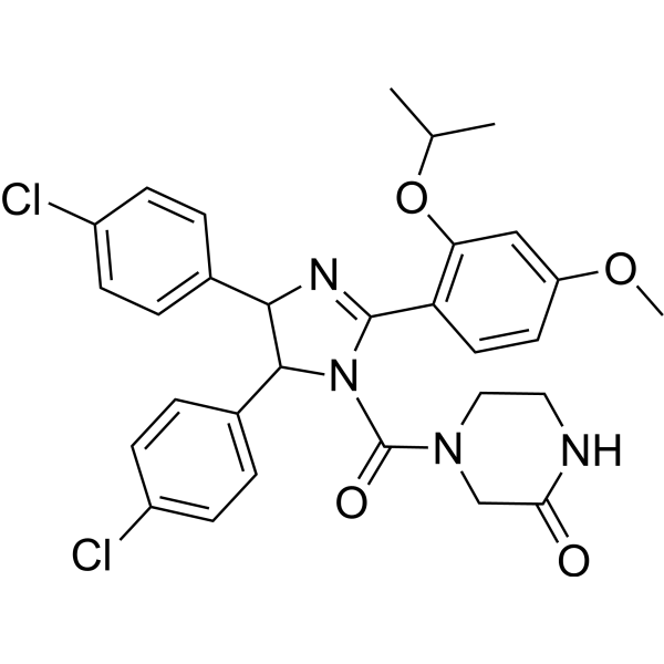
(Rac)-Nutlin-3 (Rebemadlin), an active enantiomer of Nutlin-3, is a potent murine double minute (MDM2) inhibitor (IC50=90 nM). (Rac)-Nutlin-3 inhibits MDM2-p53 interactions and stabilizes the p53 protein, and induces cell autophagy and apoptosis. (Rac)-Nutlin-3 has the potential for the study of TP53 wild-type ovarian carcinomas.
Nos produits utilisent uniquement pour la recherche. Nous ne vendons pas aux patients.
- Pureté : 98.18%
- CAS No.: 890090-75-2
- Formule: C30H30Cl2N4O4
- Masse moléculaire:581.49
-
Stockage:Powder -20°C, 3 years , 4°C, 2 years ; In solvent -80°C, 6 months , -20°C, 1 month

To place orders, for customer services and technical support, please contact: MedChemExpress USA
Tel: 609-228-6898 E-mail: [email protected] [email protected]


-
 Activité biologique
-
Chemical Information
-
Solvant et solubilité
- Protocole
- Pureté et documentation
- Références
-
 Help & FAQs


-
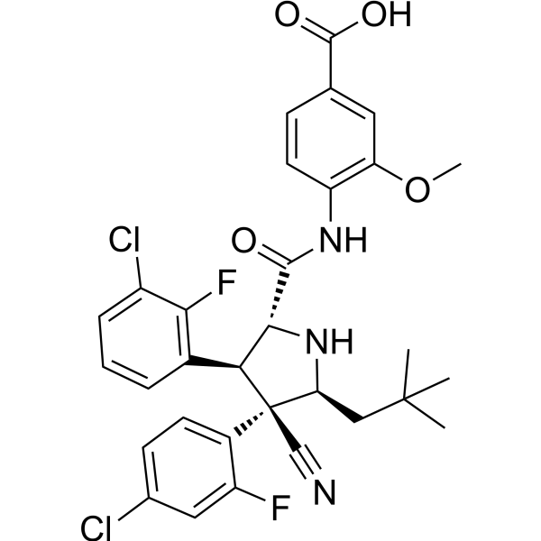
HY-15676
IdasanutlinMDM2 Inhibitor -
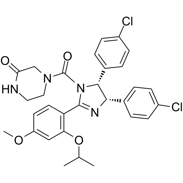
HY-10029
Nutlin-3aMDM2 Antagonist -
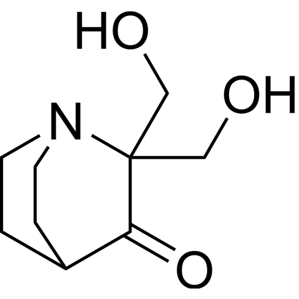
HY-19980A
PRIMA-1Mutant p53 Reactivator -
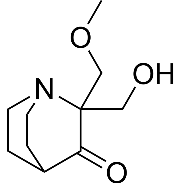
HY-19980
Eprenetapoptp53 Activator -
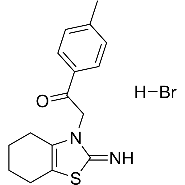
HY-15484
Pifithrin-α hydrobromidep53 Inhibitor -
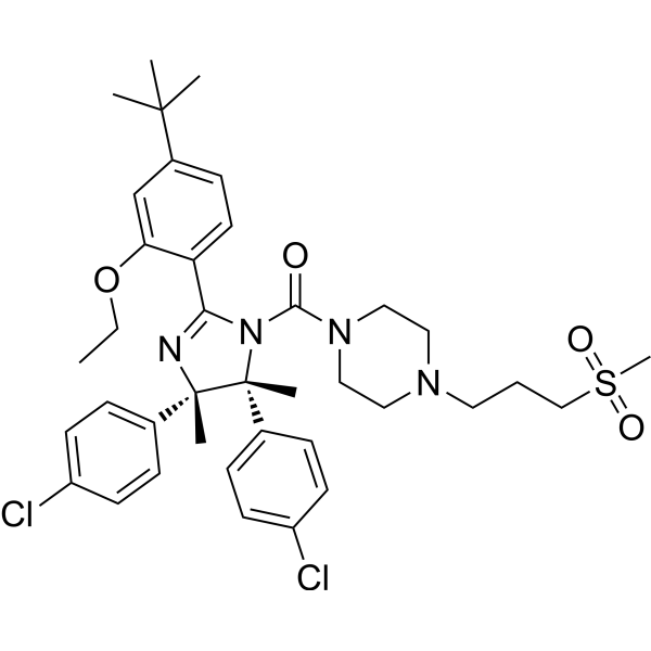
HY-10959
RG7112MDM2 Antagonist -
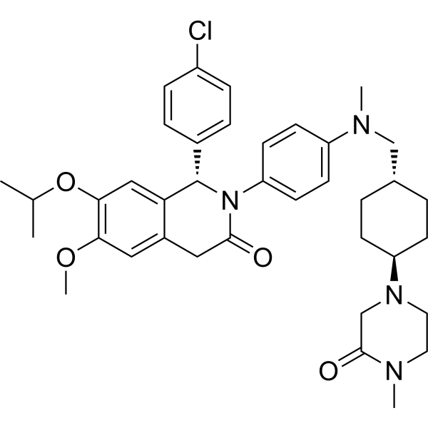
HY-15954
NVP-CGM097MDM2 Inhibitor -
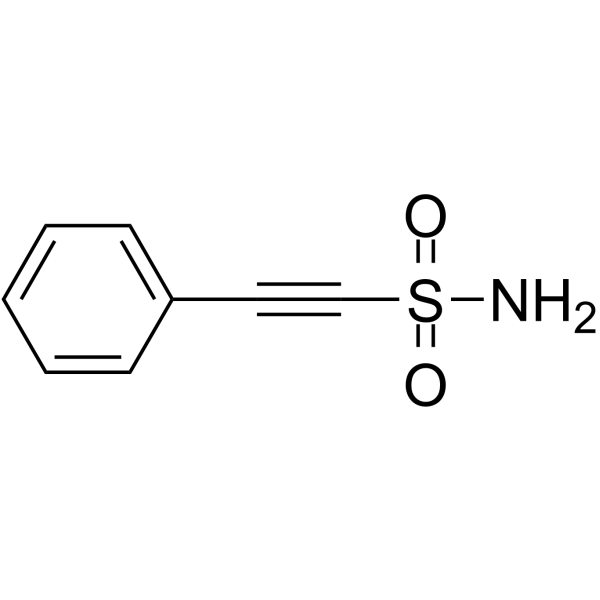
HY-10940
Pifithrin-μp53 and HSP70 Inhibitor -
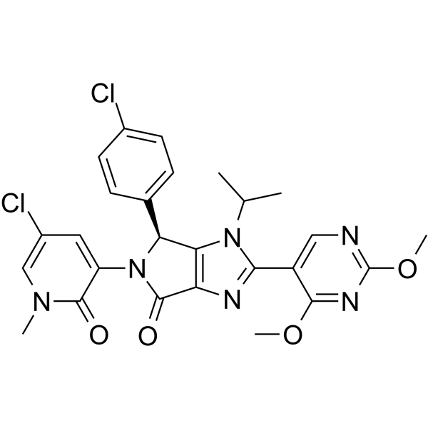
HY-18658
Siremadlinp53-MDM2 Inhibitor -
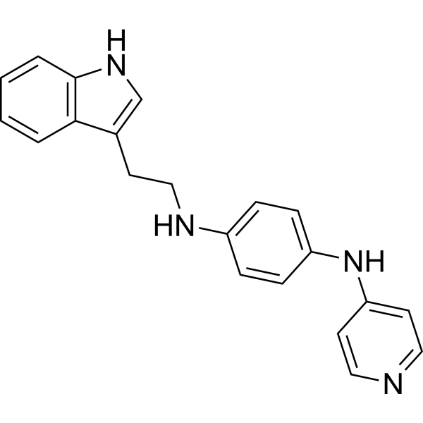
HY-12025
SerdemetanAnticancer Aagent -
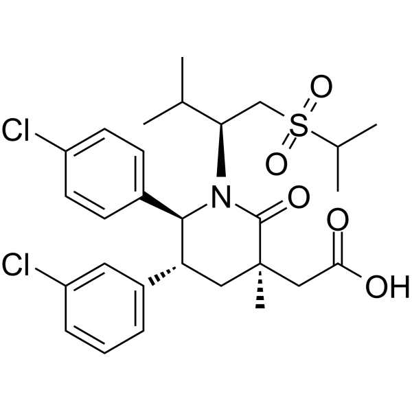
HY-12296
NavtemadlinMDM2-p53 Interaction Inhibitor -
HY-16664
SJ-172550MDM-2/p53 MDM2 Inhibitor


Activité biologique
Description
Cellular Effect
|
Cell Line

|
Type | Value | Description | References |
|---|---|---|---|---|
| A549 | IC50 |
0.41 μM
Compound: Nutlin-3
|
Cytotoxicity against human A549 cells after 72 hrs by SRB assay
Cytotoxicity against human A549 cells after 72 hrs by SRB assay
|
10.1039/C2MD20208E |
| HCT-116 | IC50 |
4.63 μM
Compound: Nutlin-3
|
Cytotoxicity against human HCT116 cells after 72 hrs by SRB assay
Cytotoxicity against human HCT116 cells after 72 hrs by SRB assay
|
10.1039/C2MD20208E |
| Huh-7 | CC50 |
26.8 μM
Compound: 43
|
Cytotoxicity against human Huh-7 cells
Cytotoxicity against human Huh-7 cells
|
[PMID: 32283298] |
| MCF7 | IC50 |
14.63 μM
Compound: Nutlin-3
|
Antiproliferative activity against human MCF7 cells assessed as reduction in cell growth incubated for 48 hrs by CCK8 assay
Antiproliferative activity against human MCF7 cells assessed as reduction in cell growth incubated for 48 hrs by CCK8 assay
|
[PMID: 33725632] |
| MCF7 | IC50 |
15.72 μM
Compound: Nutlin-3
|
Antiproliferative activity against human MCF7 cells assessed as reduction in cell growth incubated for 48 hrs in presence of Fer-1 by CCK8 assay
Antiproliferative activity against human MCF7 cells assessed as reduction in cell growth incubated for 48 hrs in presence of Fer-1 by CCK8 assay
|
[PMID: 33725632] |
| MDA-MB-231 | IC50 |
28.47 μM
Compound: nutlin-3
|
Antiproliferative activity against human MDA-MB-231 cells assessed as cell growth inhibition incubated for 24 hrs by CCK-8 assay
Antiproliferative activity against human MDA-MB-231 cells assessed as cell growth inhibition incubated for 24 hrs by CCK-8 assay
|
[PMID: 36512711] |
| PC-3 | IC50 |
6.37 μM
Compound: Nutlin-3
|
Cytotoxicity against human PC3 cells after 72 hrs by SRB assay
Cytotoxicity against human PC3 cells after 72 hrs by SRB assay
|
10.1039/C2MD20208E |
| SJSA-1 | IC50 |
1.81 μM
Compound: Nutlin-3
|
Cytotoxicity against human SJSA1 cells after 72 hrs by SRB assay
Cytotoxicity against human SJSA1 cells after 72 hrs by SRB assay
|
10.1039/C2MD20208E |
| SW-620 | IC50 |
0.38 μM
Compound: Nutlin-3
|
Cytotoxicity against human SW620 cells after 72 hrs by SRB assay
Cytotoxicity against human SW620 cells after 72 hrs by SRB assay
|
10.1039/C2MD20208E |
| U-87MG ATCC | IC50 |
104.5 nM
Compound: 2
|
Binding affinity to MDM2 in human U87MG cells assessed as inhibition of MDM2/p53 protein interaction after 10 mins by quantitative sandwich immuno assay
Binding affinity to MDM2 in human U87MG cells assessed as inhibition of MDM2/p53 protein interaction after 10 mins by quantitative sandwich immuno assay
|
[PMID: 27050782] |
| U-87MG ATCC | IC50 |
6.5 μM
Compound: 2
|
Antiproliferative activity against human U87MG cells expressing wild type p53 after 48 hrs by MTS assay
Antiproliferative activity against human U87MG cells expressing wild type p53 after 48 hrs by MTS assay
|
[PMID: 27050782] |
In Vitro
Nutlin-3a is a therapeutic which inhibits MDM2, activates wild-type p53, and induces apoptosis-as a therapeutic compound for TP53 wild-type ovarian carcinomas. Three cell lines (HOC-7, OVCA429 and A2780) with wild-type TP53 are highly sensitive to Nutlin-3a (IC50=4 to 6 μM). SKOV3 cells have an IC50 of 38 μM to Nutlin-3a. The two remaining ovarian clear cell lines (TOV21G and OVAS), both with TP53 wild-type, are relatively more sensitive to growth inhibition with Nutlin-3a (IC50=14 and 25 μm respectively) than the TP53 mutant cell lines[1]. Nutlin-3a is the active enantiomer of Nutlin-3. Nutlin-3a is a highly selective MDM2 antagonist and p53 inducer. Seven days of incubation with 10 μM Nutlin-3a leads to >90% inhibition of NIH/3T3 cells’growth but does not affect the proliferation of MEF in which both targets of the drug are eliminated. Nutlin-3a effectively arrestes cell-cycle progression in all cell lines, depleting the S-phase compartment to 0.2-2% and increasing the G1- and G2/M-phase compartments, indicating G1 and G2 arrest. The p53 targets p21 and MDM2 are elevated significantly 3 h after Nutlin-3a addition and reach maximal levels at 8 h. Nutlin-3a induces apoptosis in ≈60% of SJSA-1 and MHM cells after 40 h, which increase further after 60 h (85% and 65%, respectively)[2].
MedChemExpress (MCE) has not independently confirmed the accuracy of these methods. They are for reference only.
In Vivo
MedChemExpress (MCE) has not independently confirmed the accuracy of these methods. They are for reference only.


Chemical Information
-
CAS No. 890090-75-2
-
Appearance Solid
-
Masse moléculaire 581.49
-
Formule C30H30Cl2N4O4
-
Color White to off-white
-
SMILES
O=C(N1C(C2=C(C=C(C=C2)OC)OC(C)C)=NC(C3=CC=C(C=C3)Cl)C1C4=CC=C(C=C4)Cl)N5CC(NCC5)=O
-
Synonyms
(Rac)-Rebemadlin
-
Livraison
Room temperature in continental US; may vary elsewhere.
-
Stockage
Powder -20°C 3 years 4°C 2 years In solvent -80°C 6 months -20°C 1 month


Solvant et solubilité
In Vitro:
DMSO : 125 mg/mL (214.97 mM; Need ultrasonic; Hygroscopic DMSO has a significant impact on the solubility of product, please use newly opened DMSO)
Please refer to the solubility information to select the appropriate solvent. Once prepared, please aliquot and store the solution to prevent product inactivation from repeated freeze-thaw cycles.
Storage method and period of stock solution: -80°C, 6 months; -20°C, 1 month. When stored at -80°C, please use it within 6 months. When stored at -20°C, please use it within 1 month.
Please refer to the solubility information to select the appropriate solvent. Once prepared, please aliquot and store the solution to prevent product inactivation from repeated freeze-thaw cycles.
Storage method and period of stock solution: -80°C, 6 months; -20°C, 1 month. When stored at -80°C, please use it within 6 months. When stored at -20°C, please use it within 1 month.
Concentration (start) × Volume (start) = Concentration (final) × Volume (final)


Pureté et documentation
-
Fiche technique (282 KB)
-
SDS (392 KB)
- English - EN (392 KB)
- Français - FR (392 KB)
- Deutsch - DE (392 KB)
- Norwegian - NO (392 KB)
- Español - ES (392 KB)
- Swedish - SV (392 KB)
- Italian - IT (392 KB)
- Korean - KR (392 KB)
- Portuguese - PT (392 KB)
-
Instruction de manipulation (2659 KB)
Références
[1]. Crane EK, et al. Nutlin-3a: A Potential Therapeutic Opportunity for TP53 Wild-Type Ovarian Carcinomas. PLoS One. 2015 Aug 6;10(8):e0135101. [Content Brief]
[2]. Tovar C, et al. Small-molecule MDM2 antagonists reveal aberrant p53 signaling in cancer: implications for therapy. Proc Natl Acad Sci U S A. 2006 Feb 7;103(6):1888-93. [Content Brief]


Complete Stock Solution Preparation Table
Please refer to the solubility information to select the appropriate solvent. Once prepared, please aliquot and store the solution to prevent product inactivation from repeated freeze-thaw cycles.
Storage method and period of stock solution: -80°C, 6 months; -20°C, 1 month. When stored at -80°C, please use it within 6 months. When stored at -20°C, please use it within 1 month.
| Optional Solvent | Concentration Solvent Mass | 1 mg | 5 mg | 10 mg | 25 mg |
|---|---|---|---|---|---|
| DMSO | 1 mM | 1.7197 mL | 8.5986 mL | 17.1972 mL | 42.9930 mL |
| 5 mM | 0.3439 mL | 1.7197 mL | 3.4394 mL | 8.5986 mL | |
| 10 mM | 0.1720 mL | 0.8599 mL | 1.7197 mL | 4.2993 mL | |
| 15 mM | 0.1146 mL | 0.5732 mL | 1.1465 mL | 2.8662 mL | |
| 20 mM | 0.0860 mL | 0.4299 mL | 0.8599 mL | 2.1497 mL | |
| 25 mM | 0.0688 mL | 0.3439 mL | 0.6879 mL | 1.7197 mL | |
| 30 mM | 0.0573 mL | 0.2866 mL | 0.5732 mL | 1.4331 mL | |
| 40 mM | 0.0430 mL | 0.2150 mL | 0.4299 mL | 1.0748 mL | |
| 50 mM | 0.0344 mL | 0.1720 mL | 0.3439 mL | 0.8599 mL | |
| 60 mM | 0.0287 mL | 0.1433 mL | 0.2866 mL | 0.7166 mL | |
| 80 mM | 0.0215 mL | 0.1075 mL | 0.2150 mL | 0.5374 mL | |
| 100 mM | 0.0172 mL | 0.0860 mL | 0.1720 mL | 0.4299 mL |


(Rac)-Nutlin-3 Related Classifications
HY-10029A Related Classifications
 Powered by Bioz