



SR-4133

Based on 1 Customer Validation
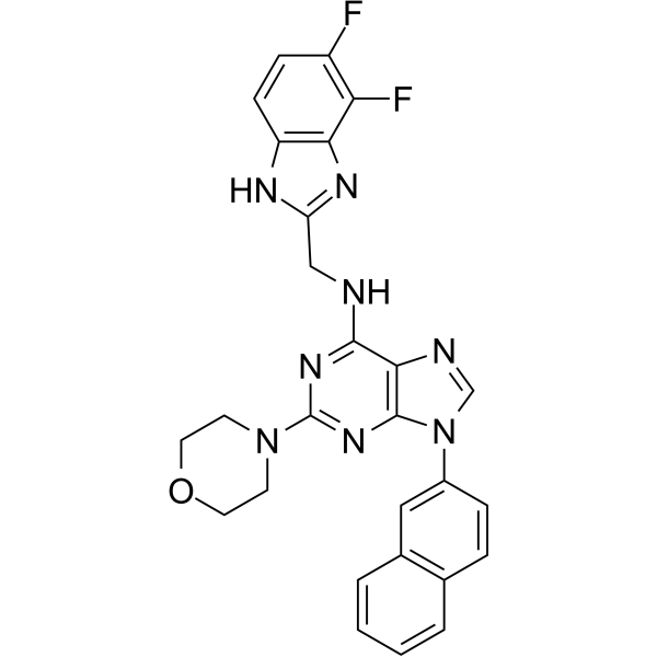
SR-4133 is a potent and highly CK1ε selective inhibitor with an IC50 of 58 nM. SR-4133 binds to the ATP-binding site of CK1ε. SR-4133 displays nanomolar growth inhibition of bladder cancer cells, and inhibits the phosphorylation of 4E-BP1.
For research use only. We do not sell to patients.
- Purity : 99.85%
- CAS No.: 2999645-23-5
- Formula: C27H22F2N8O
- Molecular Weight:512.51
-
Storage:
4°C, sealed storage, away from moisture
* In solvent : -80°C, 6 months; -20°C, 1 month (sealed storage, away from moisture)

To place orders, for customer services and technical support, please contact: MedChemExpress USA
Tel: 609-228-6898 E-mail: [email protected] [email protected]


-
 Biological Activity
-
Chemical Information
- Protocols
- Purity & Documentation
- References
-
 Help & FAQs




Biological Activity
Description
IC50 & Target
|
CK1ε 58 nM (IC50) |
CK1γ2 10 μM (IC50) |
Cellular Effect
|
Cell Line

|
Type | Value | Description | References |
|---|---|---|---|---|
| 5637 | EC50 |
314 nM
Compound: SR-4133
|
Antiproliferative activity against human 5637 cells assessed as reduction in cell proliferation incubated for 72 hrs by Cell Titer Glo assay
Antiproliferative activity against human 5637 cells assessed as reduction in cell proliferation incubated for 72 hrs by Cell Titer Glo assay
|
[PMID: 37204207] |
| T-24 | EC50 |
265 nM
Compound: SR-4133
|
Antiproliferative activity against human T24 cells assessed as reduction in cell proliferation incubated for 72 hrs by Cell Titer Glo assay
Antiproliferative activity against human T24 cells assessed as reduction in cell proliferation incubated for 72 hrs by Cell Titer Glo assay
|
[PMID: 37204207] |
| U2OS | EC50 |
373 nM
Compound: SR-4133
|
Antiproliferative activity against human U2OS cells assessed as reduction in cell proliferation incubated for 72 hrs by Cell Titer Glo assay
Antiproliferative activity against human U2OS cells assessed as reduction in cell proliferation incubated for 72 hrs by Cell Titer Glo assay
|
[PMID: 37204207] |
| UMUC3 | EC50 |
540 nM
Compound: SR-4133
|
Antiproliferative activity against human UMUC3 cells assessed as reduction in cell proliferation incubated for 72 hrs by Cell Titer Glo assay
Antiproliferative activity against human UMUC3 cells assessed as reduction in cell proliferation incubated for 72 hrs by Cell Titer Glo assay
|
[PMID: 37204207] |
In Vitro
SR-4133 (200-600 nM, 72 h) inhibits cancer cells growth significantly with EC50s of 265 nM (T24), 314 nM (5637), 540 nM (UM-UC-3 EC), 373 nM (U2-OS EC)[1].
SR-4133 (1 μM, 24 h) inhibits the phosphorylation of 4E-BP1 in T24 cells[1].
MedChemExpress (MCE) has not independently confirmed the accuracy of these methods. They are for reference only.
-
Cell Line:T24, 5637, UM-UC-3, U2-OS
-
Concentration:265 nM, 314 nM, 540 nM, 373 nM
-
Incubation Time:72 h
-
Result:Inhibited cell growth with EC50s of 265 nM (T24), 314 nM (5637), 540 nM (UM-UC-3 EC), 373 nM (U2-OS EC).
-
Cell Line:T24
-
Concentration:1 μM
-
Incubation Time:24 h
-
Result:Blocked the phosphorylation of T37/46, S65, and T70 of 4E-BP1.


Chemical Information
-
CAS No. 2999645-23-5
-
Appearance Solid
-
Molecular Weight 512.51
-
Formula C27H22F2N8O
-
Color Off-white to light yellow
-
SMILES
FC1=C(F)C2=C(NC(CNC3=NC(N4CCOCC4)=NC5=C3N=CN5C6=CC(C=CC=C7)=C7C=C6)=N2)C=C1
-
Shipping
Room temperature in continental US; may vary elsewhere.
-
Storage
4°C, sealed storage, away from moisture
* In solvent : -80°C, 6 months; -20°C, 1 month (sealed storage, away from moisture)


Purity & Documentation
-
Data Sheet (274 KB)
-
SDS (252 KB)
- English - EN (252 KB)
- Français - FR (252 KB)
- Deutsch - DE (252 KB)
- Norwegian - NO (252 KB)
- Español - ES (252 KB)
- Swedish - SV (252 KB)
- Italian - IT (252 KB)
- Korean - KR (252 KB)
- Portuguese - PT (252 KB)
-
Handling Instructions (2659 KB)
References

Calculators
Concentration (start) × Volume (start) = Concentration (final) × Volume (final)


SR-4133 Related Classifications
HY-149292 Related Classifications
 Powered by Bioz