



Withanoside V

Based on 1 Customer Validation
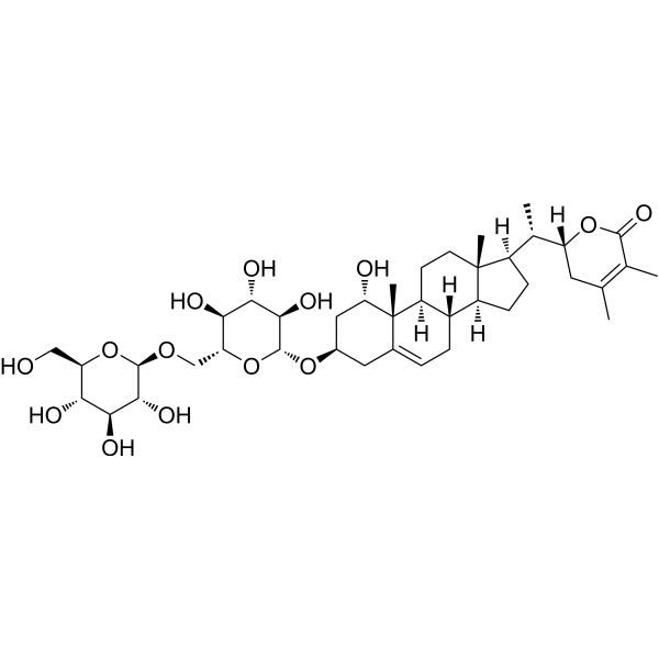
Withanoside V is a blood-brain barrier-permeable withanolide derivative. Withanoside V binds strongly to Sudlow I (domain IIA) of human serum albumin (HSA) to form a stable complex and alter the secondary structure of albumin, thereby increasing helix content and reducing β-sheet and random coil. Withanoside V binds to Aβ (1-42) to block the interaction between monomers and subsequent aggregation. Withanoside V inhibits the viability of neuroblastoma cells, reduces the number of apoptotic cells induced by Aβ (1-42), and decreases ROS production. Withanoside V inhibits SARS-CoV-2 Mpro. Withanoside V can be used for research on Alzheimer's disease and coronavirus disease 2019.
Nur für Forschungszwecke. Wir verkaufen nicht an Patienten.
- Reinheit : 99.9%
- CAS. Nr.: 256520-90-8
- Formel: C40H62O14
- Molecular Weight:766.91
-
Speicherung:Powder -20°C, 3 years ; In solvent -80°C, 6 months , -20°C, 1 month

To place orders, for customer services and technical support, please contact: MedChemExpress USA
Tel: 609-228-6898 E-mail: [email protected] [email protected]


-
 Biologische Aktivität
-
Chemical Information
- Protokoll
- Reinheit & Dokumentation
- Verweise
-
 Help & FAQs




Biologische Aktivität
Beschreibung
In Vitro
Withanoside V binds tightly to human serum albumin with a Ks of 5.33 × 10⁴ M-1, and binds to Sudlow's site I (domain IIA) of human serum albumin with a Ks of 4.8 × 10⁴ M-1, inducing conformational changes in human serum albumin[1].
Withanoside V (100 μM; 6-72 h) inhibits the aggregation of Aβ (1-42) in vitro and reduces the formation of β-sheet-rich oligomers and fibrils over an incubation period of up to 72 h[2].
Withanoside V (10-100 μM; 24-48 h) inhibits the viability of SK-N-SH cells with an IC50 of 30.14 μM, and protects SK-N-SH cells from Aβ (1-42)-induced cytotoxic damage[2].
Withanoside V (15.07 μM; 24 h) increases the proportion of early apoptotic cells in human SK-N-SH neuroblastoma cells while maintaining a high overall cell viability[2].
Compared with cells treated with Aβ (1-42), Withanoside V (15.07 μM; 24 h) reduces the level of DNA fragmentation and the number of apoptotic cells in human SK-N-SH neuroblastoma cells[2].
Withanoside V (15.07 μM; 24 h) significantly reduces Aβ (1-42)-induced intracellular ROS production in human SK-N-SH neuroblastoma cells[2].
Withanoside V is a SARS-CoV-2 Mpro inhibitor with a binding affinity of -8.96 kcal/mol. It forms stable hydrogen bonds at the active site of this enzyme, and its mechanism of action is similar to that of the natural N3 inhibitor[4].
MedChemExpress (MCE) has not independently confirmed the accuracy of these methods. They are for reference only.
-
Cell Line:human SK-N-SH neuroblastoma cells
-
Concentration:0, 20, 40, 60, 80, 100 μM/15.07 μM
-
Incubation Time:48 h/24 h
-
Result:Exhibited dose-dependent cytotoxicity against SK-N-SH cells, with an IC50 value of 30.14 μM after 48 h.
Increased neuronal cell viability significantly when cells were pre-treated with amyloid-β(1-42) then treated with half the IC50 concentration, indicating neutralization of amyloid-β(1-42)-induced cytotoxicity.
-
Cell Line:human SK-N-SH neuroblastoma cells
-
Concentration:15.07 μM (half IC50)
-
Incubation Time:24 h
-
Result:Resulted in 93.2% viable cells, 0.9% dead cells, 5.2% early apoptotic cells, and 0.7% late apoptotic cells, compared to untreated controls with 96.1% viable cells, 1.3% dead cells, 2.2% early apoptotic cells, and 0.4% late apoptotic cells.


Chemical Information
-
CAS. Nr. 256520-90-8
-
Appearance Solid
-
Molecular Weight 766.91
-
Formel C40H62O14
-
Color White to off-white
-
SMILES
C[C@]1(CC2)[C@](CC[C@]1([H])[C@@H]([C@@](OC3=O)([H])CC(C)=C3C)C)([H])[C@@](CC=C4C[C@H]5O[C@@H]([C@@H]([C@H]6O)O)O[C@@H]([C@H]6O)CO[C@@H]([C@@H]([C@H]7O)O)O[C@@H]([C@H]7O)CO)([H])[C@@]2([H])[C@]4([C@H](C5)O)C
-
Structure Classification
-
Initial Source
-
Versand
Room temperature in continental US; may vary elsewhere.
-
Speicherung
Powder -20°C 3 years In solvent -80°C 6 months -20°C 1 month


Reinheit & Dokumentation
-
Data Sheet (275 KB)
-
SDS (394 KB)
- English - EN (394 KB)
- Français - FR (394 KB)
- Deutsch - DE (394 KB)
- Norwegian - NO (394 KB)
- Español - ES (394 KB)
- Swedish - SV (394 KB)
- Italian - IT (394 KB)
- Korean - KR (394 KB)
- Portuguese - PT (394 KB)
-
Handling Instructions (2659 KB)
Verweise
[1]. Dubey S, et al. Elucidating the active interaction mechanism of phytochemicals withanolide and withanoside derivatives with human serum albumin. PLoS One. 2018;13(11):e0200053. Published 2018 Nov 7. [Content Brief]
[2]. Dubey S, et al. Improving the inhibition of β-amyloid aggregation by withanolide and withanoside derivatives. Int J Biol Macromol. 2021;173:56-65. [Content Brief]
[3]. Matsuda H, et al. Structures of withanosides I, II, III, IV, V, VI, and VII, new withanolide glycosides, from the roots of Indian Withania somnifera DUNAL. and inhibitory activity for tachyphylaxis to clonidine in isolated guinea-pig ileum. Bioorg Med Chem. 2001 Jun;9(6):1499-507. [Content Brief]
[4]. Choe J, et al. The Efficacy of Traditional Medicinal Plants in Modulating the Main Protease of SARS-CoV-2 and Cytokine Storm. Chem Biodivers. 2022;19(11):e202200655. [Content Brief]

Calculators
Konzentration (Stammlösung) × Volumen (Stammlösung) = Konzentration (Ziellösung) × Volumen (Ziellösung)


Withanoside V Related Classifications
HY-N8671 Related Classifications
 Powered by Bioz
Keywords
- Withanoside V
- 256520-90-8
- Amyloid-β
- Apoptosis
- Reactive Oxygen Species (ROS)
- SARS-CoV
- withanolide derivative
- SARS-CoV-2 main protease
- human SK-N-SH neuroblastoma cells
- human serum albumin
- Alzheimer's disease
- amyloid-β 42
- apoptosis
- blood-brain barrier
- Sudlow's drug binding site I
- reactive oxygen species
- coronavirus disease 2019
- Inhibitor
- inhibitor
- inhibit