



Cathepsin B Antibody (YA551)
(Synonyms: CPSB, CTSB, Cathepsin B, APP secretase, Cathepsin B1, APPS)
Based on 1 Customer Validation
Cathepsin B Antibody (YA551) is a Rabbit-derived and non-conjugated IgG monoclonal antibody, targeting to Cathepsin B.
-
Host:
Rabbit
-
Isotype:
IgG
-
Application:
WB, ICC/IF, IHC-P
-
Reactivity :
Human
-
Formulation:
Supplied in 1*TBS (pH7.4), 0.05% BSA and 40% Glycerol. Preservative: 0.05% Sodium Azide.
-
Conjugation:
Non-conjugated


To place orders, for customer services and technical support, please contact: MedChemExpress USA
Tel: 609-228-6898 E-mail: [email protected] [email protected]





Applications
| Application |
WB

WB: Western Blot
|
ICC/IF
ICC/IF: Immunocytochemistry/
Immunofluorescence |
IHC-P
IHC-P: Immunohistochemistry-Paraffin
|
|---|---|---|---|
| Dilution Ratio | 1:500-1:1000 | 1:50-1:200 | 1:50-1:200 |


Product Details
Cathepsin B Antibody (YA551) is a Rabbit-derived and non-conjugated IgG monoclonal antibody, targeting to Cathepsin B.
-
Host Rabbit

-
Clonality Recombinant,Monoclonal

-
Species ReactivityHuman
-
Observed Molecular WeightObserved band size: 38 kDa
Note: Due to possible protein modifications or aggregation, the molecular weight should be confirmed by actual measurement, and the predicted value is for reference only.

-
Calculated Molecular Weight Predicted band size: 38 kDa

Synthetic peptide corresponding to Human Cathepsin B.AA range:7-176.
Endogenous
Protein A affinity purified.
Non-conjugated
Unmodified
IgG


Product Properties
-
Appearance
Solution
-
Formulation
Supplied in 1*TBS (pH7.4), 0.05% BSA and 40% Glycerol. Preservative: 0.05% Sodium Azide.
-
Concentration
Batch-dependent, Please check the COA for the concentration of each lot. Check Lot Concentration
-
Storage & Stability
Stored at -20°C for 1 year. Avoid repeated freeze / thaw cycles.
-
Shipping
Shipping with blue ice.


Verification Images
-
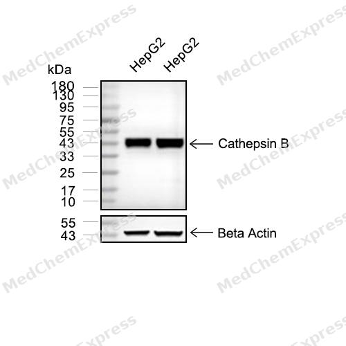 Western blot analysis of extracts from HepG2 (lane 2(20μg) and HepG2(lane 3(40μg) using Cathepsin B (HY-P80383) Rabbit mAb. Proteins were transferred to a PVDF membrane and blocked with 5% non-fat milk in TBST for 2 hour at room temperature. The primary antibody (1/1000) and Loading control antibody (Beta Actin, HY-P80438, 1/10000) was used in 5% non-fat milk in TBST at 4°C overnight. Goat Anti-Mouse/Rabbit IgG-HRP Secondary Antibody (1/10000) was used for 1 hour at room temperature.
 -
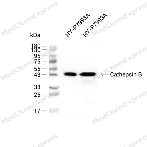 Western blot analysis of extracts from HY-P7993A (lane 2(25 ng) and HY-P7993A (lane 3(50 ng) using Cathepsin B (HY-P80383) Rabbit mAb. Proteins were transferred to a PVDF membrane and blocked with 5% non-fat milk in TBST for 2 hour at room temperature. The primary antibody (1/1000) and Loading control antibody (Beta Actin, HY-P80438, 1/10000) was used in 5% non-fat milk in TBST at 4°C overnight. Goat Anti-Mouse IgG-HRP Secondary Antibody (1/10000) was used for 1 hour at room temperature.
 -
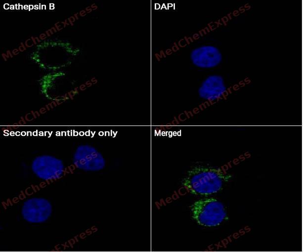 Immunocytochemistry analysis of RAW264.7 cells labeling Cathepsin B Antibody (HY-P80383) at 1/50 dilution. Cells were fixed in 4% paraformaldehyde for 15 minutes at room temperature, permeabilized with 0.1% Triton X-100 for 10 minutes at room temperature, then blocked with BSA for Immunol Staining for 10 min at room temperature. Cells were then incubated with Cathepsin B Antibody (HY-P80383) dilution in BSA for Immunol Staining at 4 ℃overnight. AF488-conjugated AffiniPure Goat Anti-Rabbit IgG H&L(HY-P8002, Green) was used as the secondary antibody at 1/1,000 dilution. PBS instead of the primary antibody was used as the secondary antibody only control. The Nuclear counterstain was DAPI (Blue).
 -
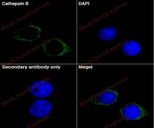 Immunocytochemistry analysis of RAW264.7 cells labeling Cathepsin B Antibody (HY-P80383) at 1/100 dilution. Cells were fixed in 4% paraformaldehyde for 15 minutes at room temperature, permeabilized with 0.1% Triton X-100 for 10 minutes at room temperature, then blocked with BSA for Immunol Staining for 10 min at room temperature. Cells were then incubated with Cathepsin B Antibody (HY-P80383) dilution in BSA for Immunol Staining at 4 ℃overnight. AF488-conjugated AffiniPure Goat Anti-Rabbit IgG H&L(HY-P8002, Green) was used as the secondary antibody at 1/1,000 dilution. PBS instead of the primary antibody was used as the secondary antibody only control. The Nuclear counterstain was DAPI (Blue).


Background
-
Function
Cathepsin B (CTSB) is a lysosomal cysteine protease predominantly localized in endosomal and lysosomal compartments, where it participates in intracellular and extracellular protein turnover and contributes to cellular proteolysis pathways[1][2]. Cathepsin B is among the most abundant lysosomal proteases and functions in protein degradation, lysosomal homeostasis, and cellular turnover, linking its activity to fundamental endo/lysosomal biological processes[1][2]. Mechanistically, Cathepsin B regulates multiple forms of programmed cell death, including apoptosis, pyroptosis, ferroptosis, necroptosis, and autophagic cell death, thereby connecting lysosomal proteolysis with cell fate determination pathways[2]. In disease models, dysregulated Cathepsin B activity contributes to neurological disorders, cardiovascular disease, inflammatory conditions, and multiple cancers, where elevated expression is frequently associated with pathological progression, tissue remodeling, invasion, and metastasis[2][3]. Compared with related cysteine cathepsins such as Cathepsin L, which is strongly involved in antigen processing, proprotein processing, and degradation of extracellular matrix proteins including collagen and elastin, Cathepsin B is distinguished by its characteristic endopeptidase and exopeptidase activities and its prominent role in programmed cell death signaling and disease-associated proteolytic networks[4][2]. For experimental applications, Cathepsin B is a widely used pharmacological target, and selective inhibitors including CA-074 and other Cathepsin B inhibitors have been extensively employed to investigate protease-dependent mechanisms and evaluate therapeutic strategies in cancer and other disease models[3][2].
-
Subcellular Localization
Lysosome; Melanosome; Secreted, extracellular space; Apical cell membrane; Peripheral membrane protein; Extracellular side
-
Expression
Tissue_specificity:Expressed in the stratum spinosum of the epidermis. Weak expression is detected in the stratum granulosum -
Subunit
Dimer of a heavy chain and a light chain cross-linked by a disulfide bond. Interacts with SRPX2. Directly interacts with SHKBP1 (PubMed:16733801)
-
SwissProt ID
-
Synonyms
CPSB, CTSB, Cathepsin B, APP secretase, Cathepsin B1, APPS
-
Research Field
Neuroscience


Documentation
-
Data Sheet (260 KB)
-
SDS (251 KB)
- English - EN (251 KB)
- Français - FR (251 KB)
- Deutsch - DE (251 KB)
- Norwegian - NO (251 KB)
- Español - ES (251 KB)
- Swedish - SV (251 KB)
- Italian - IT (251 KB)
- Korean - KR (251 KB)
- Portuguese - PT (251 KB)
-
User Guide for Antibodies (1077 KB)
[1]. Yadati T, et al. The Ins and Outs of Cathepsins: Physiological Function and Role in Disease Management. Cells. 2020 Jul 13;9(7):1679. [Content Brief]
[2]. Xie Z, et al. Cathepsin B in programmed cell death machinery: mechanisms of execution and regulatory pathways. Cell Death Dis. 2023 Apr 8;14(4):255. [Content Brief]
[3]. Frlan R, et al. Inhibitors of cathepsin B. Curr Med Chem. 2006;13(19):2309-27. [Content Brief]

 Powered by Bioz
