



IGF-2 Antibody (YA3603)
(Synonyms: IGF-II; PP9974; C11orf43; FLJ22066; FLJ44734)
Based on 1 Customer Validation
IGF-2 Antibody (YA3603) is a Mouse-derived and non-conjugated IgG1 monoclonal antibody, targeting to IGF-2.
-
Host:
Mouse
-
Isotype:
IgG
-
Application:
IHC-P, ICC/IF, FC, ELISA
-
Reactivity :
Human
-
Formulation:
Supplied in PBS with 0.05% sodium azide
-
Conjugation:
Non-conjugated


To place orders, for customer services and technical support, please contact: MedChemExpress USA
Tel: 609-228-6898 E-mail: [email protected] [email protected]





Applications
| Application |
IHC-P

IHC-P: Immunohistochemistry-Paraffin
|
ICC/IF
ICC/IF: Immunocytochemistry/
Immunofluorescence |
FC
FC: Flow Cytometry
|
ELISA
ELISA: Enzyme Linked Immunosorbent Assay
|
|---|---|---|---|---|
| Dilution Ratio | 1:200-1:1000 | 1:200-1:1000 | 1:200-1:400 | 1:10000 |


Product Details
IGF-2 Antibody (YA3603) is a Mouse-derived and non-conjugated IgG1 monoclonal antibody, targeting to IGF-2.
-
Host Mouse

-
Clonality Monoclonal

-
Species ReactivityHuman
-
Observed Molecular WeightObserved band size: 20 kDa
Note: Due to possible protein modifications or aggregation, the molecular weight should be confirmed by actual measurement, and the predicted value is for reference only.

-
Calculated Molecular Weight Predicted band size: 20 kDa

Purified recombinant fragment of human IGF2 (AA: 25-180) expressed in E. Coli.
affinity purified.
Non-conjugated
Unmodified
IgG


Product Properties
-
Appearance
Solution
-
Formulation
Supplied in PBS with 0.05% sodium azide
-
Concentration
Batch-dependent, Please check the COA for the concentration of each lot. Check Lot Concentration
-
Storage & Stability
Stored at -20°C for 1 year. Avoid repeated freeze / thaw cycles.
-
Shipping
Shipping with blue ice.


Verification Images
-
 Immunohistochemical analysis of paraffin-embedded human urothelial cancer tissue using IGF-2 Antibody (HY-P83906, 1/1000). The section was pretreated using heat mediated antigen retrieval with sodium citrate buffer (pH 6.0) for 8 minutes. The tissues were blocked with quick block buffer for 0.5 hours at room temperature, washed with PBS and PBST, and then incubated with the primary antibody overnight at 4℃. The detection was performed using an HRP conjugated compact polymer system. DAB was used as the chromogen. Tissues were counterstained with hematoxylin and mounted with DPX.
 -
 Immunohistochemical analysis of paraffin-embedded human rectum cancer tissue using IGF-2 Antibody (HY-P83906, 1/1000). The section was pretreated using heat mediated antigen retrieval with sodium citrate buffer (pH 6.0) for 8 minutes. The tissues were blocked with quick block buffer for 0.5 hours at room temperature, washed with PBS and PBST, and then incubated with the primary antibody overnight at 4℃. The detection was performed using an HRP conjugated compact polymer system. DAB was used as the chromogen. Tissues were counterstained with hematoxylin and mounted with DPX.
 -
 Immunohistochemical analysis of paraffin-embedded human pancreatic cancer tissue using IGF-2 Antibody (HY-P83906, 1/1000). The section was pretreated using heat mediated antigen retrieval with sodium citrate buffer (pH 6.0) for 8 minutes. The tissues were blocked with quick block buffer for 0.5 hours at room temperature, washed with PBS and PBST, and then incubated with the primary antibody overnight at 4℃. The detection was performed using an HRP conjugated compact polymer system. DAB was used as the chromogen. Tissues were counterstained with hematoxylin and mounted with DPX.
 -
 Immunohistochemical analysis of paraffin-embedded human placenta tissue using IGF-2 Antibody (HY-P83906, 1/1000). The section was pretreated using heat mediated antigen retrieval with sodium citrate buffer (pH 6.0) for 8 minutes. The tissues were blocked with quick block buffer for 0.5 hours at room temperature, washed with PBS and PBST, and then incubated with the primary antibody overnight at 4℃. The detection was performed using an HRP conjugated compact polymer system. DAB was used as the chromogen. Tissues were counterstained with hematoxylin and mounted with DPX.
 -
 Immunohistochemical analysis of paraffin-embedded human kidney tissue using IGF-2 Antibody (HY-P83906, 1/1000). The section was pretreated using heat mediated antigen retrieval with sodium citrate buffer (pH 6.0) for 8 minutes. The tissues were blocked with quick block buffer for 0.5 hours at room temperature, washed with PBS and PBST, and then incubated with the primary antibody overnight at 4℃. The detection was performed using an HRP conjugated compact polymer system. DAB was used as the chromogen. Tissues were counterstained with hematoxylin and mounted with DPX.
 -
 Immunohistochemical analysis of paraffin-embedded human small intestine tissue using IGF-2 Antibody (HY-P83906, 1/1000). The section was pretreated using heat mediated antigen retrieval with sodium citrate buffer (pH 6.0) for 8 minutes. The tissues were blocked with quick block buffer for 0.5 hours at room temperature, washed with PBS and PBST, and then incubated with the primary antibody overnight at 4℃. The detection was performed using an HRP conjugated compact polymer system. DAB was used as the chromogen. Tissues were counterstained with hematoxylin and mounted with DPX.
 -
 Immunohistochemical analysis of paraffin-embedded human duodenum tissue using IGF-2 Antibody (HY-P83906, 1/1000). The section was pretreated using heat mediated antigen retrieval with sodium citrate buffer (pH 6.0) for 8 minutes. The tissues were blocked with quick block buffer for 0.5 hours at room temperature, washed with PBS and PBST, and then incubated with the primary antibody overnight at 4℃. The detection was performed using an HRP conjugated compact polymer system. DAB was used as the chromogen. Tissues were counterstained with hematoxylin and mounted with DPX.
 -
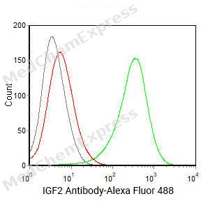
 Flow cytometric analysis of 1X10^6 HepG2 cells labeling IGF2 Antibody(HY-P83906
 -
 Flow cytometric analysis of 1X10⁶ HeLa cells labeling IGF-2 Antibody (HY-P83906, red). Cells were fixed with 4% paraformaldehyde and permeabilised with 90% methanol. Then stained with the primary antibody at 1/400 dilution for an hour at 4℃. AF488-conjugated Goat Anti-Mouse IgG H&L (HY-P8005) was used as the secondary antibody at 1/1,000 dilution for 30 minutes at 4℃. Mouse IgG Isotype Control (HY-P80757, blue) was used as the isotype control, cells without incubation with primary antibody were used as the unlabeled control (black).
 -
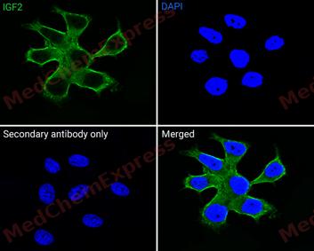
 Immunofluorescence analysis of MCF-7 cells labeling IGF2 antibody (HY-P83906) at 1/50 dilution. Cells were fixed in 4% paraformaldehyde for 15 minutes at room temperature


Background
-
Function
IGF-2 is a The insulin-like growth factors possess growth-promoting activity. Major fetal growth hormone in mammals. Plays a key role in regulating fetoplacental development. IGF2 is influenced by placental lactogen. Also involved in tissue differentiation. In adults, involved in glucose metabolism in adipose tissue, skeletal muscle and liver (Probable). Acts as a ligand for integrin which is required for IGF2 signaling. Positively regulates myogenic transcription factor MYOD1 function by facilitating the recruitment of transcriptional coactivators, thereby controlling muscle terminal differentiation. Inhibits myoblast differentiation and modulates metabolism via increasing the mitochondrial respiration rate; Preptin undergoes glucose-mediated co-secretion with insulin, and acts as a physiological amplifier of glucose-mediated insulin secretion. Exhibits osteogenic properties by increasing osteoblast mitogenic activity through phosphoactivation of MAPK1 and MAPK3[1].
-
Subcellular Localization
Secreted
-
Expression
Tissue_specificity:It is expressed in the heart, placenta, lungs, liver, muscles, kidneys, tongue, limbs, eyes, and pancreas. -
Subunit
Interacts with MYORG; this interaction is required for IGF2 secretion (By similarity). Interacts with integrins ITGAV:ITGB3 and ITGA6:ITGB4; integrin-binding is required for IGF2 signaling (PubMed:28873464). Interacts with IGFBP2 (PubMed:18563800)
-
SwissProt ID
-
Synonyms
IGF-II; PP9974; C11orf43; FLJ22066; FLJ44734


Documentation
-
Data Sheet (234 KB)
-
SDS (251 KB)
- English - EN (251 KB)
- Français - FR (251 KB)
- Deutsch - DE (251 KB)
- Norwegian - NO (251 KB)
- Español - ES (251 KB)
- Swedish - SV (251 KB)
- Italian - IT (251 KB)
- Korean - KR (251 KB)
- Portuguese - PT (251 KB)
-
User Guide for Antibodies (1077 KB)

 Powered by Bioz
