



Smad3 Antibody (YA075)
(Synonyms: MADH3, SMAD3, Mothers against decapentaplegic homolog 3, MAD homolog 3, Mad3, Mothers against DPP homolog 3, hMAD-3, JV15-2, SMAD family member 3, SMAD 3, Smad3, hSMAD3)Based on 5 publication(s) in Google Scholar
Smad3 Antibody (YA075) is a Rabbit-derived and non-conjugated IgG monoclonal antibody, targeting to Smad3.
-
Host:
Rabbit
-
Isotype:
IgG
-
Application:
WB, ICC/IF, IHC-P, FC
-
Reactivity :
Human, Mouse, Rat
-
Formulation:
Supplied in 1*TBS (pH7.4), 0.05% BSA and 40% Glycerol. Preservative: 0.05% Sodium Azide.
-
Conjugation:
Non-conjugated


To place orders, for customer services and technical support, please contact: MedChemExpress USA
Tel: 609-228-6898 E-mail: [email protected] [email protected]



-

HY-P80324
Smad2 Antibody (YA076) -
HY-P80521
TGF beta 1 Antibody (YA036) -
HY-P80484
alpha smooth muscle Actin Antibody (YA627) -
HY-P80326
Smad4 Antibody (YA074) -
HY-P80853
Phospho-Smad2 (Ser250) Antibody (YA152) -
HY-P81227
Collagen I Antibody -
HY-P8001
HRP-conjugated Goat Anti-Rabbit IgG H&L -
HY-P83739
alpha smooth muscle Actin Antibody(YA3468) -
HY-P80477
Phospho-Smad3 (Ser423/Ser425) Antibody (YA151) -
HY-P80493
Fibronectin Antibody (YA436) -
HY-P87781
Phospho-Smad3 (Ser423/Ser425) Antibody (YA7466)
Publications Citing Use of MedChemExpress (MCE) Smad3 Antibody (YA075)
More
 5 Publications Citing Use of MCE Smad3 Antibody (YA075) (1)
-
WB


Applications
| Application |
WB

WB: Western Blot
|
ICC/IF
ICC/IF: Immunocytochemistry/
Immunofluorescence |
IHC-P
IHC-P: Immunohistochemistry-Paraffin
|
FC
FC: Flow Cytometry
|
|---|---|---|---|---|
| Dilution Ratio | 1:500-1:2000 | 1:50-1:200 | 1:50-1:200 | 1:50-1:100 |


Product Details
Smad3 Antibody (YA075) is a Rabbit-derived and non-conjugated IgG monoclonal antibody, targeting to Smad3.
-
Host Rabbit

-
Clonality Recombinant,Monoclonal

-
Species ReactivityHuman, Mouse, Rat
-
Observed Molecular WeightObserved band size: 55 kDa
Note: Due to possible protein modifications or aggregation, the molecular weight should be confirmed by actual measurement, and the predicted value is for reference only.

-
Calculated Molecular Weight Predicted band size: 48 kDa

Entrez Gene: 4088 Human ; 17127 Mouse ; 25631 Rat
SwissProt: P84022 Human ; Q8BUN5 Mouse ; P84025 Rat
OMIM: 114500 Human
Synthetic peptide corresponding to Human Smad3.AA range:181-230.
Endogenous
Protein A affinity purified.
Non-conjugated
Unmodified
IgG


Product Properties
-
Appearance
Solution
-
Formulation
Supplied in 1*TBS (pH7.4), 0.05% BSA and 40% Glycerol. Preservative: 0.05% Sodium Azide.
-
Concentration
Batch-dependent, Please check the COA for the concentration of each lot. Check Lot Concentration
-
Storage & Stability
Stored at -20°C for 1 year. Avoid repeated freeze / thaw cycles.
-
Shipping
Shipping with blue ice.


Publications (5)
-
Journal Impact Factor
-
Most Recent
-
Drug Resist Updat
Dynamic immunoediting by macrophages in homologous recombination deficiency-stratified pancreatic ductal adenocarcinoma. [Abstract]2024 Sep:76:101115. PMID: 39002266 -
Anim Nutr
Proline promotes prolyl 4-hydroxylase subunit alpha 1 via transforming growth factor beta 1 to increase the synthesis and deposition of collagen in grass carp (Ctenopharyngodon idellus) muscle fibroblasts: Insights from integrated bioinformatics and functional analysis. [Abstract]2026 Jan 2:24:109-120. PMID: 41704244 -
Food Chem (Oxf)
Role of timp2 in Ctenopharyngodon idellus muscle collagen deposition and the regulatory mechanism by Cu2. [Abstract]2025 Oct 8:11:100306. PMID: 41140839
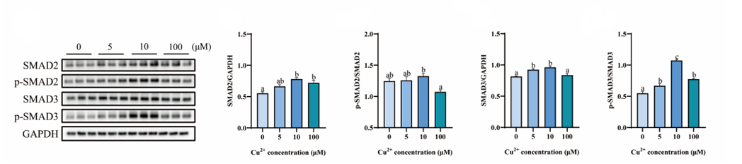
Smad3 Antibody (YA075) purchased from MedChemExpress. Usage Cited in: Food Chem (Oxf). 2025 Oct 8:11:100306. [Abstract]
Effects of CuSO₄ (24 h) on the protein levels of Smad2 (1:1,000), p-Smad2 (Ser250) (1:1,000), Smad3 (1:1,000), and p-Smad3 (Ser423/425) (1:1,000) in fibroblasts. GAPDH was used as a loading control (antibody dilution 1:5,000).
-
Eur J Cell Biol
TGF-β drives pulmonary fibrosis through dual dysregulation of TNF pathway transcription and splicing. [Abstract]2026 Feb 27;105(2):151534. PMID: 41764855


Verification Images
-
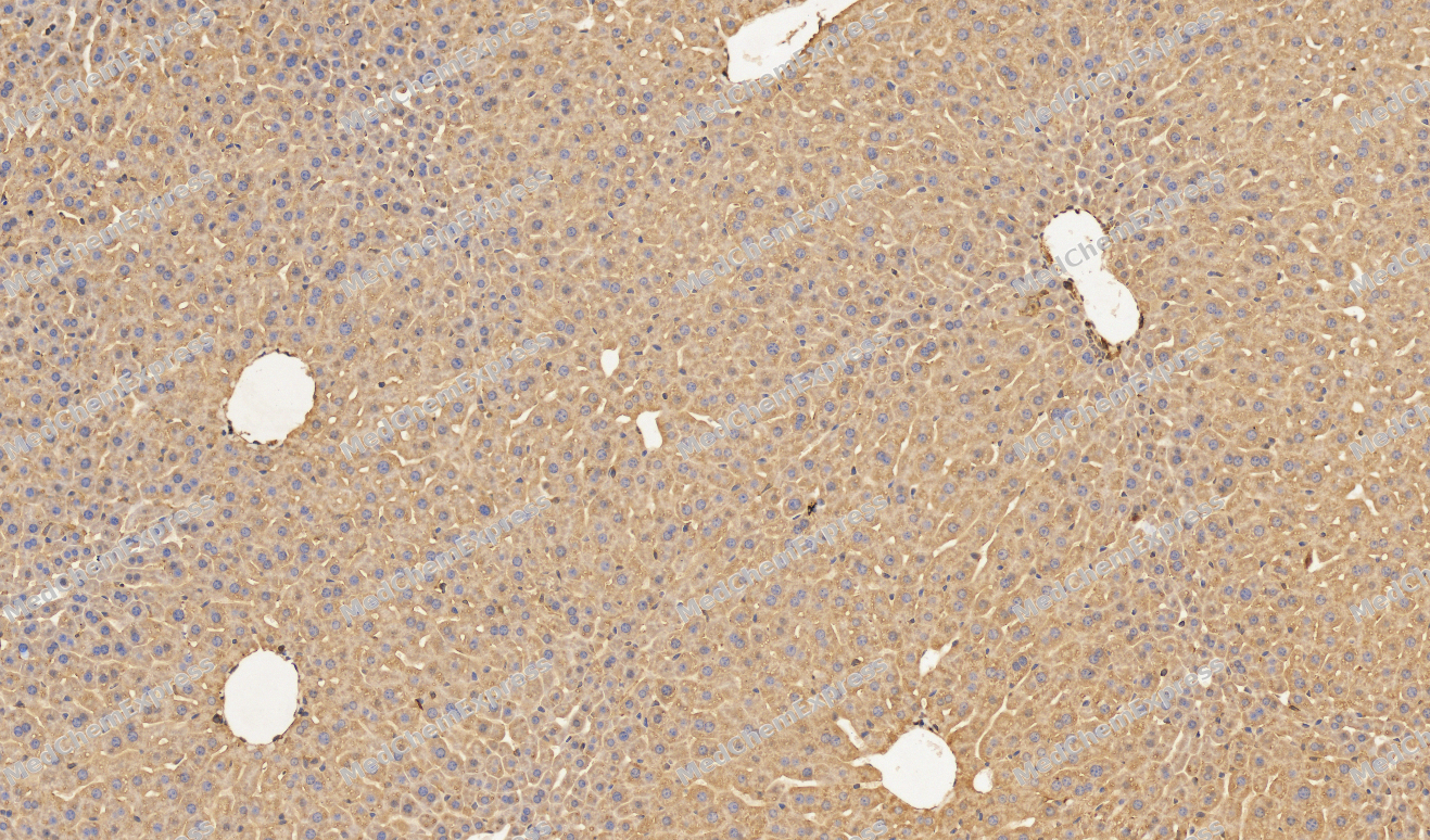 Immunohistochemical analysis of paraffin-embedded Mouse liver tissue using Smad3 Antibody. The section was pre-treated using heat mediated antigen retrieval with sodium citrate buffer (pH 6.0) for 8 minutes. The tissues were blocked in QuickBlock for 20 minutes at room temperature, washed with ddH2O and PBS, and then probed with the primary antibody (HY-P80325, 1/100) in 4℃ overnight. The detection was performed using an HRP conjugated compact polymer system. DAB was used as the chromogen. Tissues were counterstained with hematoxylin and mounted with DPX.
 -
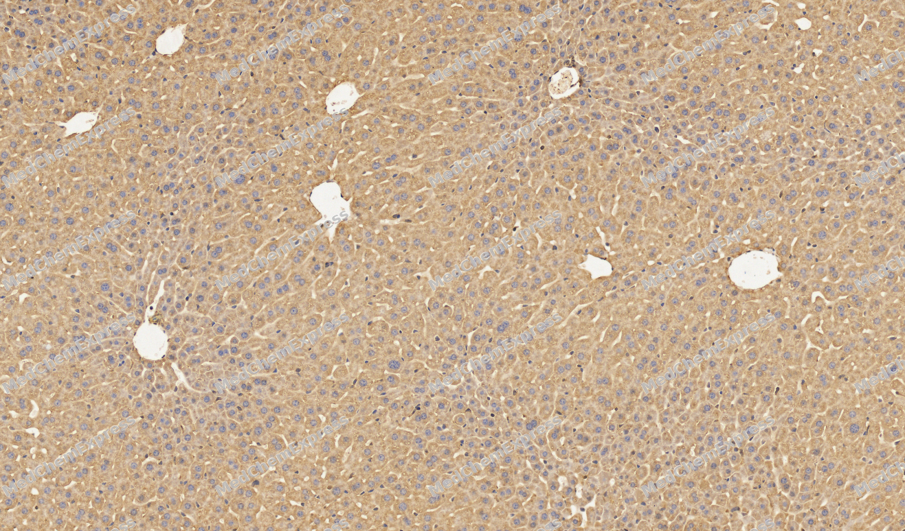 Immunohistochemical analysis of paraffin-embedded Mouse liver tissue using Smad3 Antibody. The section was pre-treated using heat mediated antigen retrieval with sodium citrate buffer (pH 6.0) for 8 minutes. The tissues were blocked in QuickBlock for 20 minutes at room temperature, washed with ddH2O and PBS, and then probed with the primary antibody (HY-P80325, 1/100) in 4℃ overnight. The detection was performed using an HRP conjugated compact polymer system. DAB was used as the chromogen. Tissues were counterstained with hematoxylin and mounted with DPX.
 -
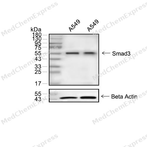 Western blot analysis of extracts from A549 (lane 2(20μg)) and A549 (lane 3(40μg)) using Smad3 (HY-P80325) Rabbit mAb. Proteins were transferred to a PVDF membrane and blocked with 5% non-fat milk in TBST for 2 hour at room temperature. The primary antibody (1/1000) and Loading control antibody (Beta Actin, HY-P80438, 1/10000) was used in 5% non-fat milk in TBST at 4°C overnight. Goat Anti-Rabbit/Mouse IgG-HRP Secondary Antibody (1/10000) was used for 1 hour at room temperature.
 -
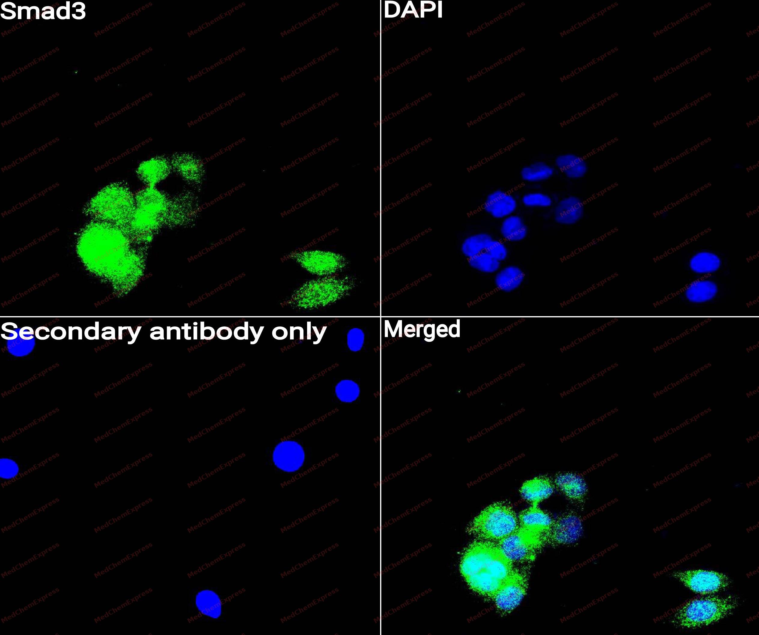 Immunocytochemistry analysis of A549 cells labeling Smad3 with Smad3antibody (HY-P80325) at 1/50 dilution. Cells were fixed in 4% paraformaldehyde for 15 minutes at room temperature, permeabilized with 0.1% Triton X-100 for 10 minutes at room temperature, then blocked with QuickBlock™ Blocking Buffer for Immunol Staining for 10 min at room temperature. Cells were then incubated with Smad3 antibody (HY-P80325) at 1/50 dilution in QuickBlock™ Blocking Buffer for Immunol Staining at 4 ℃. Alexa Fluor® 488-conjugated AffiniPure Goat Anti-Rabbit IgG H&L(HY-P8002, Green) was used as the secondary antibody at 1/1,000 dilution. PBS instead of the primary antibody was used as the secondary antibody only control. The Nuclear counterstain was DAPI (Blue).
 -
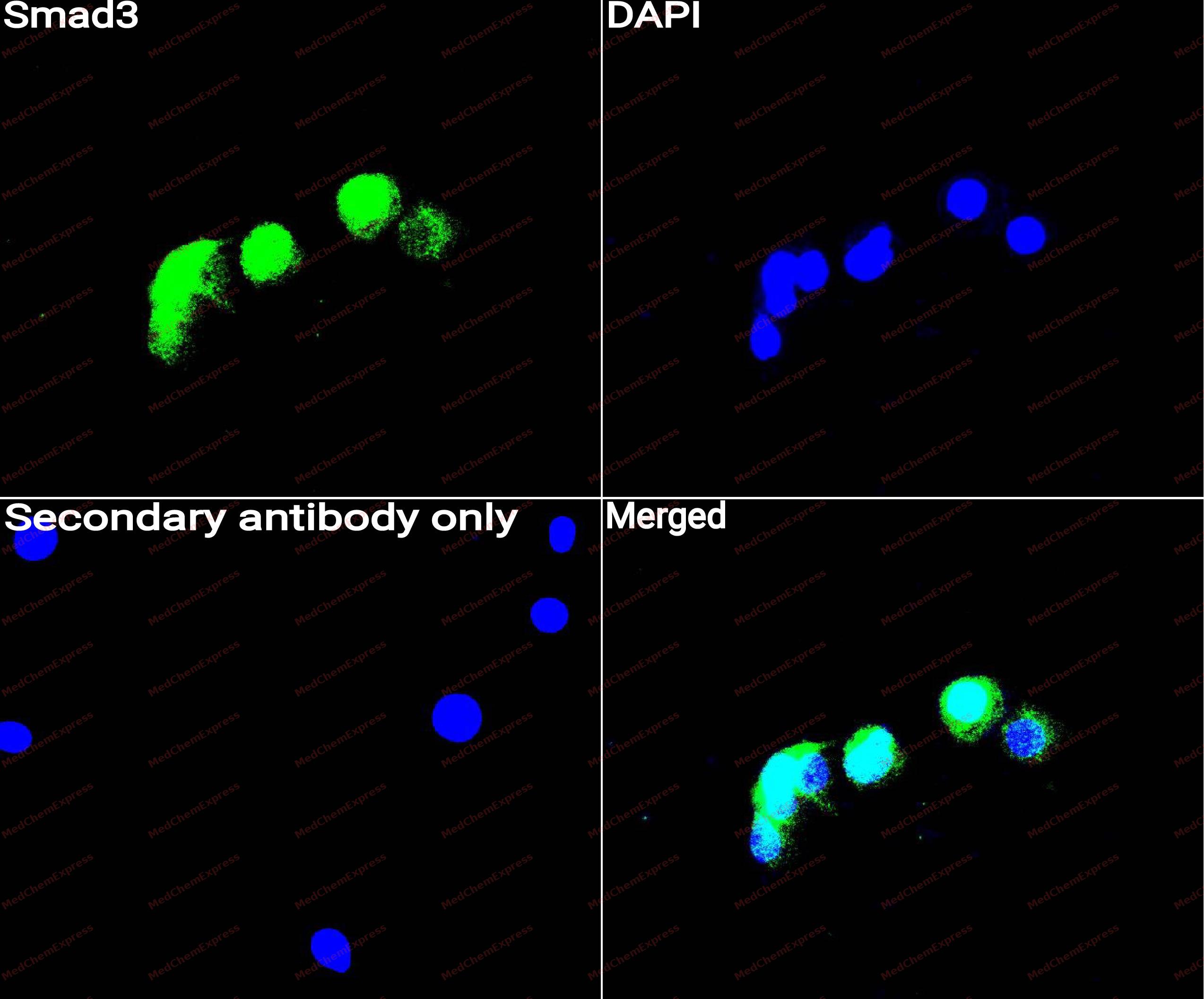 Immunocytochemistry analysis of A549 cells labeling Smad3 with Smad3 antibody (HY-P80325) at 1/100 dilution. Cells were fixed in 4% paraformaldehyde for 15 minutes at room temperature, permeabilized with 0.1% Triton X-100 for 10 minutes at room temperature, then blocked with QuickBlock™ Blocking Buffer for Immunol Staining for 10 min at room temperature. Cells were then incubated with Smad3 antibody (HY-P80325) at 1/100 dilution in QuickBlock™ Blocking Buffer for Immunol Staining at 4 ℃. Alexa Fluor® 488-conjugated AffiniPure Goat Anti-Rabbit IgG H&L(HY-P8002, Green) was used as the secondary antibody at 1/1,000 dilution. PBS instead of the primary antibody was used as the secondary antibody only control. The Nuclear counterstain was DAPI (Blue).
 -
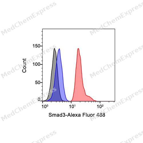 Flow cytometric analysis of 1X10^6 Hela cells labeling Smad3 Antibody (HY-P80325, red). Cells were fixed with 4% paraformaldehyde and permeabilised with 90% methanol. Then stained with the primary antibody at 1/1000 dilution for an hour at 4℃. Alexa Fluor® 488-conjugated AffiniPure Goat Anti-Rabbit IgG H&L (HY-P8002) was used as the secondary antibody at 1/1,000 dilution for 30 minutes at 4℃. Rabbit IgG Isotype Control (HY-P80879, blue) was used as the isotype control, cells without incubation with primary antibody were used as the unlabeled control (black).


Background
-
Function
SMAD3 is a receptor-regulated transcription factor in the TGF-β pathway, and activated TGF-β receptors phosphorylate SMAD3 to form SMAD complexes that regulate target-gene transcription[1]. Mechanistically, SMAD3 participates in cell proliferation, apoptosis, immune suppression, epithelial-mesenchymal transition, and extracellular matrix expression, making it useful for fibrosis, cancer progression, and inflammatory signaling studies[2][3]. In fibrosis models, SMAD3 acts as an important mediator of TGF-β profibrotic responses, and Smad3-deficient cells show reduced responses to selected chemotactic and matrix-related signals[3]. In cancer, SMAD3 can function as either a negative or positive regulator of carcinogenesis depending on cell type and tumor stage[2]. Compared with SMAD2, SMAD3 shows distinct regulation of TGF-β target genes and performs different functions in FOXH1-associated transcriptional control[4][5]. For experimental applications, SIS3 selectively inhibits TGF-β1-induced SMAD3 phosphorylation and SMAD3-SMAD4 interaction, making it a practical tool for testing SMAD3-dependent mechanisms[6].
-
Subcellular Localization
Cytoplasm; Nucleus
-
Subunit
Monomer; in the absence of TGF-beta (PubMed:9670020). Homooligomer; in the presence of TGF-beta (PubMed:9670020).
-
SwissProt ID
-
Synonyms
MADH3, SMAD3, Mothers against decapentaplegic homolog 3, MAD homolog 3, Mad3, Mothers against DPP homolog 3, hMAD-3, JV15-2, SMAD family member 3, SMAD 3, Smad3, hSMAD3
-
Research Field
Signal Transduction
Documentation
-
Data Sheet (262 KB)
-
SDS (252 KB)
- English - EN (252 KB)
- Français - FR (252 KB)
- Deutsch - DE (252 KB)
- Norwegian - NO (252 KB)
- Español - ES (252 KB)
- Swedish - SV (252 KB)
- Italian - IT (252 KB)
- Korean - KR (252 KB)
- Portuguese - PT (252 KB)
-
User Guide for Antibodies (1077 KB)
References
[1]. Hata A, et al. TGF-β signaling from receptors to Smads. Cold Spring Harb Perspect Biol. 2016;8(9):a022061. [Content Brief]
[2]. Millet C, et al. Roles of Smad3 in TGF-beta signaling during carcinogenesis. Crit Rev Eukaryot Gene Expr. 2007;17(4):281-293. [Content Brief]
[3]. Flanders KC. Smad3 as a mediator of the fibrotic response. Int J Exp Pathol. 2004;85(2):47-64. [Content Brief]
[4]. Brown KA, et al. A tale of two proteins: differential roles and regulation of Smad2 and Smad3 in TGF-beta signaling. J Cell Biochem. 2007;101(1):9-33. [Content Brief]
[5]. Aragón E, et al. Structural basis for distinct roles of SMAD2 and SMAD3 in FOXH1 pioneer-directed TGF-β signaling. Genes Dev. 2019;33(21-22):1506-1524. [Content Brief]
[6]. Jinnin M, et al. Characterization of SIS3, a novel specific inhibitor of Smad3, and its effect on transforming growth factor-beta1-induced extracellular matrix expression. Mol Pharmacol. 2006;69(2):597-607. [Content Brief]

 Powered by Bioz
