



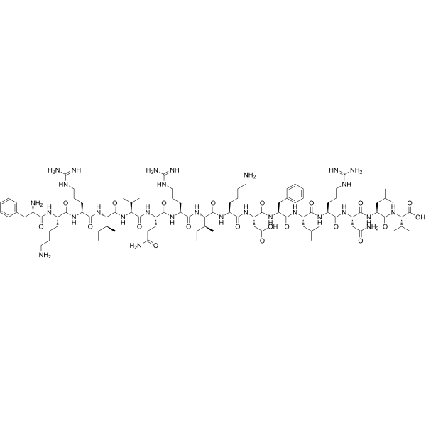
LL-37(17-32)

Based on 1 Customer Validation
LL-37 (17-32) is an ABCG2 inhibitor. LL-37 (17-32) binds to ABCG2, inhibits its transport activity in a non-competitive manner, and downregulates ABCG2 protein expression via lysosomal degradation. LL-37 (17-32) increases the accumulation of Mitoxantrone (HY-13502) in cancer cells overexpressing ABCG2, thereby reversing mitoxantrone resistance. LL-37 (17-32) can be used in the research of multidrug-resistant cancers.
For research use only. We do not sell to patients.
- Purity : 96.71%
- CAS No.: 717919-61-4
- Formula: C95H161N29O21
- Molecular Weight:2045.48
-
Storage:
Sealed storage, away from moisture.
Powder -80°C, 2 years , -20°C, 1 year* In solvent : -80°C, 6 months; -20°C, 1 month (sealed storage, away from moisture)

To place orders, for customer services and technical support, please contact: MedChemExpress USA
Tel: 609-228-6898 E-mail: [email protected] [email protected]


-
 Biological Activity
-
Chemical Information
- Protocols
- Purity & Documentation
- References
-
 Help & FAQs




Biological Activity
Description
In Vitro
LL-37 (17-32) (2-10 μM; 72 h) specifically reverses ABCG2-mediated multidrug resistance in MCF-7 FLV1000 cells and ABCG2-transfected HEK293 cells; at a concentration of 10 μM, it reduces the resistance fold of mitoxantrone to 43 and 5.4, respectively, and does not affect the cytotoxicity of non-ABCG2 substrate drugs or the viability of parental cells at effective concentrations[1].
LL-37 (17-32) (10-40 μM; 1.5 h) inhibits ABCG2-mediated pheophorbide A efflux in MCF-7 FLV1000 cells in a concentration-dependent manner and increases intracellular substrate retention[1].
LL-37 (17-32) (10-40 μM; 2 h) increases mitoxantrone accumulation in ABCG2-overexpressing MCF-7 FLV1000 cells in a concentration-dependent manner, but exerts no such effect on parental MCF-7 cells[1].
LL-37 (17-32) (10-40 μM; 55 min) enhances the binding of 5D3 antibody to ABCG2 in MCF-7 FLV1000 cells in a concentration-dependent manner, indicating a direct interaction with this transporter[1].
LL-37 (17-32) (0.4-50 μM; 45 min) inhibits vanadate-sensitive ABCG2 ATPase activity in crude membranes of ABCG2-expressing High Five insect cells in a concentration-dependent manner[1].
LL-37 (17-32) (5-20 μM; 24 h) downregulates the expression of ABCG2 protein in MCF-7 FLV1000 cells in a concentration-dependent manner via the lysosomal degradation pathway, without altering ABCG2 mRNA levels[1].
MedChemExpress (MCE) has not independently confirmed the accuracy of these methods. They are for reference only. Further protocols information, click here.
-
Cell Line:human breast cancer MCF-7 (parental), MCF-7 FLV1000 (ABCG2-overexpressing), human embryonic kidney HEK293 pcDNA3-transfected, HEK293 ABCG2-transfected
-
Concentration:2 μM, 5 μM, 10 μM
-
Incubation Time:72 h
-
Result:Reduced the IC50 of mitoxantrone from 2.116 μM to 0.821 μM in MCF-7 FLV1000 cells at 5 μM, decreasing fold-resistance from 218 to 85.
Dropped the mitoxantrone IC50 further to 0.415 μM in MCF-7 FLV1000 cells at 10 μM, with fold-resistance reduced to 43.
Reduced the mitoxantrone IC50 from 45.8 nM to 10.5 nM in HEK293 ABCG2-transfected cells at 5 μM, decreasing fold-resistance from 59 to 13.
Reduced the mitoxantrone IC50 to 4.2 nM in HEK293 ABCG2-transfected cells at 10 μM, with fold-resistance decreased to 5.4.
Did not significantly alter mitoxantrone cytotoxicity in parental MCF-7 or HEK293 pcDNA3-transfected cells.
Did not affect cisplatin cytotoxicity in any tested cell line.
-
Cell Line:human breast cancer MCF-7 FLV1000 (ABCG2-overexpressing)
-
Concentration:5 μM, 10 μM, 20 μM
-
Incubation Time:24 h
-
Result:Reduced ABCG2 protein levels in a concentration-dependent manner: 5 μM caused a small reduction, 10 μM caused a significant reduction, and 20 μM caused the greatest reduction.
Had its ABCG2 protein downregulation reversed by co-treatment with leupeptin and pepstatin.
Caused no change in ABCG2 mRNA levels at any tested concentration via quantitative real-time RT-PCR.


Chemical Information
-
CAS No. 717919-61-4
-
Appearance Solid
-
Molecular Weight 2045.48
-
Formula C95H161N29O21
-
Color White to off-white
-
Sequence
Phe-Lys-Arg-Ile-Val-Gln-Arg-Ile-Lys-Asp-Phe-Leu-Arg-Asn-Leu-Val
-
Sequence Shortening
FKRIVQRIKDFLRNLV
-
Shipping
Room temperature in continental US; may vary elsewhere.
-
Storage
Sealed storage, away from moisture
Powder -80°C 2 years -20°C 1 year * In solvent : -80°C, 6 months; -20°C, 1 month (sealed storage, away from moisture)

Protocols
-
RNA extraction experimental
By lysing cells, releasing RNA, and removing impurities such as proteins and DNA, high-purity RNA products are finally obtained. The commonly used traditional method is the guanidine isothiocyanate/phenol/chloroform method (Trizol), which is suitable for a variety of animal materials including animal tissues, microorganisms, cultured cells, etc., and most plant materials.
-
Western Blot
Western blotting (WB) is a commonly used experimental method in molecular biology, biochemistry, and immunogenetics for identifying and quantifying target proteins. It combines gel electrophoresis with immunoassay, enabling researchers to analyze protein expression, post-translational modifications, and molecular weight.
-
Lysosome and acidic-vesicle live-cell staining
Lysosome and acidic-vesicle live-cell staining detects acidic intracellular compartments by using membrane-permeant acidotropic probes that accumulate in low-pH vesicles, including lysosomes, late endosomes, autolysosomes, and acidic phagosomes. LysoTracker staining is commonly used as an intensity-based readout of acidic lysosomal compartment abundance or enlargement, while acridine orange produces green fluorescence in less concentrated compartments and red fluorescence after concentration-dependent accumulation in acidic vesicular organelles. Loss or reduction of acridine-orange red signal can be used as a readout of lysosomal membrane permeabilization or reduced acidic-vesicle integrity. This protocol is designed for live cultured cells and can be adapted for fluorescence microscopy, high-content imaging, plate-reader readout, or flow cytometry when the selected literature supports the readout. Because these dyes report acidotropic accumulation rather than lysosome identity alone,
-
Protocol For Protein Expression And Purification
Recombinant protein expression in Escherichia coli followed by purification of a His-tagged soluble protein by immobilized metal affinity chromatography (IMAC), with optional MBP fusion and TEV tag removal when the construct includes these elements. The biological readout is production of the encoded target protein, detected as an inducible band at the expected molecular mass by SDS-PAGE and quantified by total protein assay or chromatographic absorbance; the purification readout is enrichment of the target protein in elution fractions after selective binding of polyhistidine residues to immobilized Ni2+/metal-chelate resin and elution by imidazole-containing buffer. Expression is driven by an inducible bacterial expression system, commonly T7/lac-based, in which IPTG or lactose/auto-induction activates transcription and translation of the cloned gene; lower induction temperature, lower inducer concentration, induction timing, and solubility-enhancing fusion tags can influence the frac


Purity & Documentation
-
Data Sheet (263 KB)
-
SDS (254 KB)
- English - EN (254 KB)
- Français - FR (254 KB)
- Deutsch - DE (254 KB)
- Norwegian - NO (254 KB)
- Español - ES (254 KB)
- Swedish - SV (254 KB)
- Italian - IT (254 KB)
- Korean - KR (254 KB)
- Portuguese - PT (254 KB)
-
Handling Instructions (2659 KB)
References

Calculators
Concentration (start) × Volume (start) = Concentration (final) × Volume (final)


LL-37(17-32) Related Classifications
HY-P5391 Related Classifications
 Powered by Bioz