



MBM-55
Based on 1 publication(s) in Google Scholar
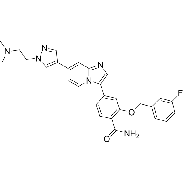
MBM-55 is a potent NIMA-related kinase 2 (Nek2) inhibitor with an IC50 of 1 nM. MBM-55 shows a 20-fold or greater selectivity in most kinases with the exception of RSK1 (IC50=5.4 nM) and DYRK1a (IC50=6.5 nM). MBM-55 effectively inhibits the proliferation of cancer cells by inducing cell cycle arrest and apoptosis. MBM-55 shows antitumor activities, and no obvious toxicity to mice.
For research use only. We do not sell to patients.
- Purity : 99.47%
- CAS No.: 2083622-09-5
- Formula: C28H27FN6O2
- Molecular Weight:498.55
-
Storage:
4°C, protect from light
* In solvent : -80°C, 6 months; -20°C, 1 month (protect from light)

To place orders, for customer services and technical support, please contact: MedChemExpress USA
Tel: 609-228-6898 E-mail: [email protected] [email protected]


-
 Biological Activity
-
Chemical Information
-
Solvent & Solubility
- Protocols
- Purity & Documentation
- References
-
 Help & FAQs



Publications Citing Use of MedChemExpress (MCE) MBM-55
More

Biological Activity
Description
IC50 & Target
|
NEK2 1 nM (IC50) |
Cellular Effect
|
Cell Line

|
Type | Value | Description | References |
|---|---|---|---|---|
| Bel-7402 | IC50 |
6.37 μM
Compound: MBM-55
|
Antitumor activity against human Bel-7402 cells assessed as inhibition of cell proliferation incubated for 72 hrs by MTT assay
Antitumor activity against human Bel-7402 cells assessed as inhibition of cell proliferation incubated for 72 hrs by MTT assay
|
[PMID: 32992252] |
| Bel-7402 | IC50 |
7.13 μM
Compound: 42g; MBM-55
|
Cytotoxicity against human Bel7402 cells measured after 72 hrs by MTT assay
Cytotoxicity against human Bel7402 cells measured after 72 hrs by MTT assay
|
[PMID: 28039836] |
| HCT-116 | IC50 |
0.7 μM
Compound: MBM-55
|
Antitumor activity against human HCT-116 cells assessed as inhibition of cell proliferation incubated for 72 hrs by MTT assay
Antitumor activity against human HCT-116 cells assessed as inhibition of cell proliferation incubated for 72 hrs by MTT assay
|
[PMID: 32992252] |
| HCT-116 | IC50 |
0.84 μM
Compound: 42g; MBM-55
|
Cytotoxicity against human HCT116 cells measured after 72 hrs by MTT assay
Cytotoxicity against human HCT116 cells measured after 72 hrs by MTT assay
|
[PMID: 28039836] |
| Hep 3B2 | IC50 |
1.07 μM
Compound: MBM-55
|
Antitumor activity against human Hep3B cells assessed as inhibition of cell proliferation incubated for 72 hrs by MTT assay
Antitumor activity against human Hep3B cells assessed as inhibition of cell proliferation incubated for 72 hrs by MTT assay
|
[PMID: 32992252] |
| MGC-803 | IC50 |
0.53 μM
Compound: 42g; MBM-55
|
Cytotoxicity against human MGC803 cells measured after 72 hrs by MTT assay
Cytotoxicity against human MGC803 cells measured after 72 hrs by MTT assay
|
[PMID: 28039836] |
| MGC-803 | IC50 |
0.65 μM
Compound: MBM-55
|
Antitumor activity against human MGC-803 cells assessed as inhibition of cell proliferation incubated for 72 hrs by MTT assay
Antitumor activity against human MGC-803 cells assessed as inhibition of cell proliferation incubated for 72 hrs by MTT assay
|
[PMID: 32992252] |
In Vitro
MBM-55 (compound 42g) inhibits MGC-803, HCT-116, Bel-7402 cells proliferation with IC50s of 0.53, 0.84, 7.13 μM, respectively[1].
MBM-55 (0.5-1 μM; 24 hours) induces G2/M phase arrest and accumulation of cells with >4N content in HCT-116 cells[1].
MBM-55 (0.5-1 μM; 24 hours) causes cell apoptosis in a concentration-dependent manner in HCT-116 cells[1].
MedChemExpress (MCE) has not independently confirmed the accuracy of these methods. They are for reference only.
-
Cell Line:HCT-116 cells
-
Concentration:0.5, 1 μM
-
Incubation Time:24 hours
-
Result:Induced G2/M phase arrest and accumulation of cells with >4N content.
-
Cell Line:HCT-116 cells
-
Concentration:0.5, 1 μM
-
Incubation Time:24 hours
-
Result:Caused cell apoptosis in a concentration-dependent manner.
In Vivo
MBM-55 (1.0 mg/kg; i.v.) treatment shows the CL, Vss, T1/2, AUC0-t, and AUC0-∞ values of 33.3 mL/min/kg, 2.53 L/kg, 1.72 hours, 495 ng/h/mL and 507 ng/h/mL, respectively.
MedChemExpress (MCE) has not independently confirmed the accuracy of these methods. They are for reference only.
-
Animal Model:Female BALB/c nu/nu mice (5-6 weeks, bearing HCT-116 xenografts)[1]
-
Dosage:20 mg/kg
-
Administration:Intraperitoneal injection; twice a day for 21 days
-
Result:Significantly suppressed tumor growth.
-
Animal Model:Male Sprague Dawley (SD) rats[1]
-
Dosage:1.0 mg/kg
-
Administration:IV injection (Pharmacokinetic Analysis)
-
Result:The CL, Vss, T1/2, AUC0-t, and AUC0-∞ values were 33.3 mL/min/kg, 2.53 L/kg, 1.72 hours, 495 ng/h/mL and 507 ng/h/mL, respectively.


Chemical Information
-
CAS No. 2083622-09-5
-
Appearance Solid
-
Molecular Weight 498.55
-
Formula C28H27FN6O2
-
Color White to off-white
-
SMILES
NC(C(C=C1)=C(OCC2=CC=CC(F)=C2)C=C1C3=CN=C4N3C=CC(C5=CN(CCN(C)C)N=C5)=C4)=O
-
Shipping
Room temperature in continental US; may vary elsewhere.
-
Storage
4°C, protect from light
* In solvent : -80°C, 6 months; -20°C, 1 month (protect from light)


Publications (1)
-
Journal Impact Factor
-
Most Recent


Solvent & Solubility
In Vitro:
DMSO : 125 mg/mL (250.73 mM; Need ultrasonic; Hygroscopic DMSO has a significant impact on the solubility of product, please use newly opened DMSO)
Please refer to the solubility information to select the appropriate solvent. Once prepared, please aliquot and store the solution to prevent product inactivation from repeated freeze-thaw cycles.
Storage method and period of stock solution: -80°C, 6 months; -20°C, 1 month (protect from light). When stored at -80°C, please use it within 6 months. When stored at -20°C, please use it within 1 month.
Please refer to the solubility information to select the appropriate solvent. Once prepared, please aliquot and store the solution to prevent product inactivation from repeated freeze-thaw cycles.
Storage method and period of stock solution: -80°C, 6 months; -20°C, 1 month (protect from light). When stored at -80°C, please use it within 6 months. When stored at -20°C, please use it within 1 month.
Concentration (start) × Volume (start) = Concentration (final) × Volume (final)


Purity & Documentation
-
Data Sheet (275 KB)
-
SDS (252 KB)
- English - EN (252 KB)
- Français - FR (252 KB)
- Deutsch - DE (252 KB)
- Norwegian - NO (252 KB)
- Español - ES (252 KB)
- Swedish - SV (252 KB)
- Italian - IT (252 KB)
- Korean - KR (252 KB)
- Portuguese - PT (252 KB)
-
Handling Instructions (2659 KB)
References


Complete Stock Solution Preparation Table
Please refer to the solubility information to select the appropriate solvent. Once prepared, please aliquot and store the solution to prevent product inactivation from repeated freeze-thaw cycles.
Storage method and period of stock solution: -80°C, 6 months; -20°C, 1 month (protect from light). When stored at -80°C, please use it within 6 months. When stored at -20°C, please use it within 1 month.
| Optional Solvent | Concentration Solvent Mass | 1 mg | 5 mg | 10 mg | 25 mg |
|---|---|---|---|---|---|
| DMSO | 1 mM | 2.0058 mL | 10.0291 mL | 20.0582 mL | 50.1454 mL |
| 5 mM | 0.4012 mL | 2.0058 mL | 4.0116 mL | 10.0291 mL | |
| 10 mM | 0.2006 mL | 1.0029 mL | 2.0058 mL | 5.0145 mL | |
| 15 mM | 0.1337 mL | 0.6686 mL | 1.3372 mL | 3.3430 mL | |
| 20 mM | 0.1003 mL | 0.5015 mL | 1.0029 mL | 2.5073 mL | |
| 25 mM | 0.0802 mL | 0.4012 mL | 0.8023 mL | 2.0058 mL | |
| 30 mM | 0.0669 mL | 0.3343 mL | 0.6686 mL | 1.6715 mL | |
| 40 mM | 0.0501 mL | 0.2507 mL | 0.5015 mL | 1.2536 mL | |
| 50 mM | 0.0401 mL | 0.2006 mL | 0.4012 mL | 1.0029 mL | |
| 60 mM | 0.0334 mL | 0.1672 mL | 0.3343 mL | 0.8358 mL | |
| 80 mM | 0.0251 mL | 0.1254 mL | 0.2507 mL | 0.6268 mL | |
| 100 mM | 0.0201 mL | 0.1003 mL | 0.2006 mL | 0.5015 mL |


MBM-55 Related Classifications
HY-101029 Related Classifications
 Powered by Bioz