



Based on 1 publication(s) in Google Scholar
MG-132 (GMP) (Z-Leu-Leu-Leu-al (GMP)) is MG-132 (HY-13259) produced by using GMP guidelines. GMP small molecules works appropriately as an auxiliary reagent for cell therapy manufacture. MG-132 (Z-Leu-Leu-Leu-al) is a potent proteasome and calpain inhibitor with IC50s of 100 nM and 1.2 μM, respectively. MG-132 effectively blocks the proteolytic activity of the 26S proteasome complex. MG-132, a peptide aldehyde, also is an autophagy activator. MG-132 also induces apoptosis.
商品は「研究用試薬」です。人や動物の医療用・臨床診断用・食品用の製品ではありません。
研究用途以外に使用した場合、当社は一切の責任を負いかねます。
- CAS 番号: 133407-82-6
- 分子式: C26H41N3O5
- 分子量:475.62
-
保管条件:
Please store the product under the recommended conditions in the Certificate of Analysis.


To place orders, for customer services and technical support, please contact: MedChemExpress USA
Tel: 609-228-6898 E-mail: [email protected] [email protected]


-
 生物活性
生物活性
-
化学情報
- プロトコル
- 純度とドキュメンテーション
- 参考文献
-
 Help & FAQs


-

HY-13259
MG-132Proteasome Inhibitor -

HY-10227
BortezomibProteasome Inhibitor -
HY-10455
CarfilzomibProteasome Inhibitor -
HY-10453
IxazomibProteasome Inhibitor -
HY-18964
MG-101Cysteine Protease Inhibitor -
HY-15282
E-64Cysteine Peptidase Inhibitor -
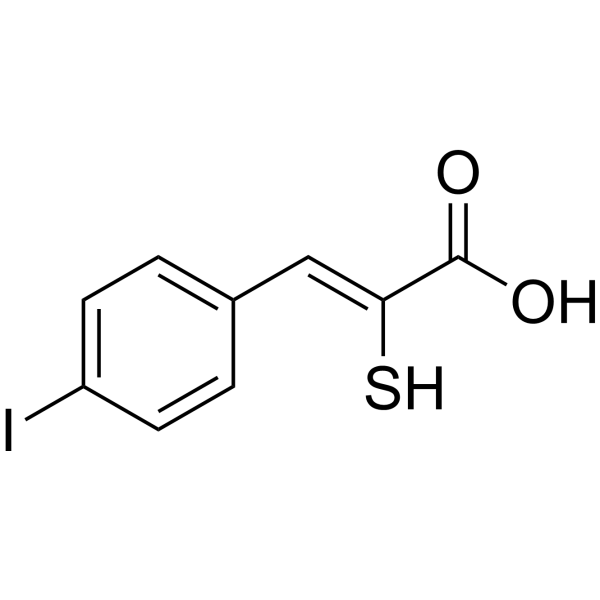
HY-100529
PD150606Calpain Inhibitor -
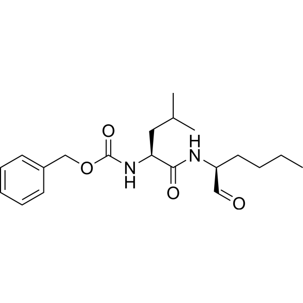
HY-100223
CalpeptinCalpain Inhibitor -
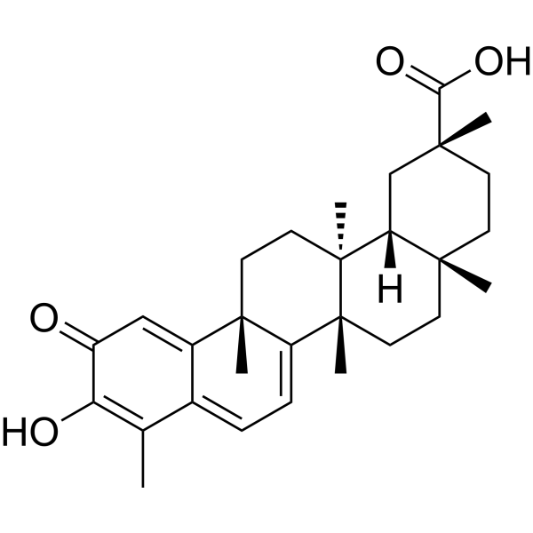
HY-13067
CelastrolAutophagy Inducer -
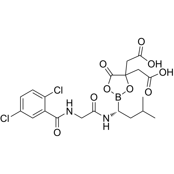
HY-10452
Ixazomib citrateProteasome Inhibitor -

HY-10227R
Bortezomib (Standard)Proteasome Inhibitor -

HY-10455R
Carfilzomib (Standard)Proteasome Inhibitor
MedChemExpress(MCE)の使用を引用している文献 MG-132 (GMP)
More

生物活性
製品説明
体外実験
MG-132 (GMP) (Z-Leu-Leu-Leu-al) initiates neurite outgrowth in PC12 cells at a low concentration (30 nM) and is a very strong inhibitor of 20S proteasome[3].
MG-132 (GMP) (10 μM; 1 hour) reverses the effects of TNF- α on I κ B degradation and NF-κ B activation in A549 cells[4].
MG-132 (GMP) (0.75-5 μM; 24 hours) potently induces p53-dependent apoptosis in KIM-2 cells by 26S proteasome inhibition[5].
MG-132 (GMP) (10-40 μM; 24 hours) significantly reduces the viability of C6 glioma cells in both time- and concentration-dependent manners and shows the IC50 of 18.5 μM at 24 hours[6].
MG-132 (GMP) (18.5 μM; 24 hours) induces down-regulation of anti-apoptotic proteins Bcl-2 and XIAP and up-regulates expression of pro-apoptotic protein Bax and caspase-3[6].
MedChemExpress (MCE) has not independently confirmed the accuracy of these methods. They are for reference only.
-
Cell Line:C6 glioma cells
-
Concentration:10, 20, 30, 40 μM
-
Incubation Time:24 hours
-
Result:Significantly reduced the viability of C6 glioma cells beginning at 6 h in both time- and concentration-dependent manners and showed the IC50 of 18.5 μM at 24 hours.
-
Cell Line:A549 cells
-
Concentration:10 μM
-
Incubation Time:1 hour
-
Result:Reversed the effects of TNF-α on IκB degradation and resulted in a reversal of TNF-α-induced NF-κB activation.
体内実験
MG-132 (GMP) (1 mg/kg; i.v.; twice a week for 4 weeks) shows potent tumor inhibitory effect against mice bearing HeLa tumors[8].
MG-132 (GMP) (1-10 μg/kg/24 hours; subcutaneously implanted osmotic pumps; for 8 days) greatly increases the expression levels of β-dystroglycan, α-dystroglycan, α-sarcoglycan, and dystrophin in skeletal muscle lysates in mice (six-month-old male C57BL/10ScSn DMD mdx mice)[9].
MedChemExpress (MCE) has not independently confirmed the accuracy of these methods. They are for reference only.
-
Animal Model:5- to 6-weeks old female athymic nude mice (EC9706 xenograft)[7]
-
Dosage:10 mg/kg
-
Administration:I.p.; daily for 25 days starting 5 days after EC9706 cells injection
-
Result:Significantly inhibited tumor growth of the EC9706 xenograft without causing toxicity to the mice.
-
Animal Model:Five-week-old female C.B-17/lcr-scid/scidJcl mice (bearing HeLa cells)[8]
-
Dosage:1 mg/kg
-
Administration:Intravenous injection; twice a week for 4 weeks
-
Result:The growth inhibition rates in HeLa tumors was 49% compared to the control.


化学情報
-
CAS 番号 133407-82-6
-
分子量 475.62
-
分子式 C26H41N3O5
-
SMILES
O=C(OCC1=CC=CC=C1)N[C@H](C(N[C@@H](CC(C)C)C(N[C@H](C([H])=O)CC(C)C)=O)=O)CC(C)C
-
別名
Z-Leu-Leu-Leu-al (GMP)
-
輸送条件
Room temperature in continental US; may vary elsewhere.
-
保管条件
Please store the product under the recommended conditions in the Certificate of Analysis.


Publications (1)
-
Journal Impact Factor
-
Most Recent
-
Cancer Cell
Targeting tumor-intrinsic STK40 induces immune vulnerability and drives T cell reinvigoration. [Abstract]2026 Jul 13;44(7):1496-1508.e10. PMID: 42208540


純度とドキュメンテーション
参考文献
[1]. Harhouri K, et al. MG132-induced progerin clearance is mediated by autophagy activation and splicing regulation. EMBO Mol Med. 2017 Sep;9(9):1294-1313. [Content Brief]
[2]. Fan WH, et al. Proteasome inhibitor MG-132 induces C6 glioma cell apoptosis via oxidative stress. Acta Pharmacol Sin. 2011 May;32(5):619-25. [Content Brief]
[3]. Tsubuki S, et al. Differential inhibition of calpain and proteasome activities by peptidyl aldehydes of di-leucine and tri-leucine. J Biochem. 1996 Mar;119(3):572-6. [Content Brief]
[4]. Fiedler MA, et al. Inhibition of TNF-alpha-induced NF-kappaB activation and IL-8 release in A549 cells with the proteasome inhibitor MG-132. Am J Respir Cell Mol Biol. 1998 Aug;19(2):259-68. [Content Brief]
[5]. MacLaren AP, et al. p53-dependent apoptosis induced by proteasome inhibition in mammary epithelial cells. Cell Death Differ. 2001 Mar;8(3):210-8. [Content Brief]
[6]. Han YH, et al. The effect of MG132, a proteasome inhibitor on HeLa cells in relation to cell growth, reactive oxygen species and GSH. Oncol Rep. 2009 Jul;22(1):215-21. [Content Brief]
[7]. Dang L, et al. Proteasome inhibitor MG132 inhibits the proliferation and promotes the cisplatin-inducedapoptosis of human esophageal squamous cell carcinoma cells. Int J Mol Med. 2014 May;33(5):1083-8. [Content Brief]
[8]. Matsumoto Y, et al. Enhanced efficacy against cervical carcinomas through polymeric micelles physically incorporating theproteasome inhibitor MG132. Cancer Sci. 2016 Jun;107(6):773-81. [Content Brief]
[9]. Bonuccelli G, et al. Proteasome inhibitor (MG-132) treatment of mdx mice rescues the expression and membrane localization of dystrophin and dystrophin-associated proteins. Am J Pathol. 2003 Oct;163(4):1663-75. [Content Brief]

Calculators
濃度 (開始) × 体積 (開始) = 濃度 (終了) × 体積 (終了)


MG-132 (GMP) 関連分類
HY-13259G 関連分類
 Powered by Bioz