



Nivatrotamab

Based on 1 Customer Validation
Nivatrotamab (Hu3F8-BsAb) is a bispecific antibody targeting GD2 and CD3, with KD values of 5.8 nM and 194 nM, respectively. Nivatrotamab redirects T cells to tumor sites and activates immune synapses, thereby mediating antibody-dependent T cell-mediated cytotoxicity. Nivatrotamab induces specific tumor cytotoxicity, stimulates the release of Th1 cytokines and promotes immune cell infiltration into tumors. In addition, its Fc segment deglycosylation design effectively prevents complement activation and cytokine storm. Nivatrotamab can be widely used in research related to various solid tumors, including melanoma, neuroblastoma, osteosarcoma, small cell lung cancer, breast cancer, Ewing's sarcoma, colon cancer, ovarian cancer and rhabdomyosarcoma.
For research use only. We do not sell to patients.
- Purity : 99.00%
- CAS No.: 2278244-14-5
- Molecular Weight:200.87 kDa
-
Storage:
Please store the product under the recommended conditions in the Certificate of Analysis.

To place orders, for customer services and technical support, please contact: MedChemExpress USA
Tel: 609-228-6898 E-mail: [email protected] [email protected]


-
 Biological Activity
-
Chemical Information
- Protocols
- Purity & Documentation
- References
-
 Help & FAQs




Biological Activity
Description
Isotype
[H-gamma1_L-kappa-scFvheavy-kappa]-dimer
Recommend Isotype Controls
Species Reactivity
Human
IC50 & Target
CD3E & ganglioside GD2
In Vitro
Nivatrotamab (Hu3F8-BsAb) (0.05-5 µg/106 T-cells; 20 min arming, 48-72 h post-arming monitoring) binds to ex vivo expanded and activated human T-cells, with surface retention lasting up to 48 hours post-arming before becoming undetectable by 72 hours[1].
Nivatrotamab (Hu3F8-BsAb) (0.05-5 µg/106 T-cells; 20 min arming, 4 h cytotoxicity assay) armed activated human T-cells potently kill GD2-positive M14 Luc melanoma cells in vitro, with maximal cytotoxicity achieved at an arming concentration of 0.5 µg/106 T-cells[1].
Nivatrotamab (0.01 μg/mL; 72 h, 4 h) has its mediated cytotoxicity against GD2+ M14 human melanoma cells enhanced across multiple E:T ratios when human PBMCs are primed with 1 nM WT-FL IL15/IL15Rα-Fc complex for 72 hours, though less effectively than priming with MUT-FL[2].
Nivatrotamab induces GD2-specific formation of mature immunological synapses and calcium flux in primary human T cells when co-cultured with GD2-positive IMR-32 neuroblastoma cells or GD2-containing supported lipid bilayers[3].
Nivatrotamab (24 hours) triggers human PBMCs and freshly isolated peripheral T cells to release pro-tumoricidal Th1 cytokines (TNFα, IFNγ, IL2) exclusively in the presence of GD2-positive tumor cells[3].
Nivatrotamab-induced Th1 cytokine (TNFα, IFNγ) release from human PBMCs co-cultured with M14 melanoma cells is dependent on the presence of monocytes[3].
MedChemExpress (MCE) has not independently confirmed the accuracy of these methods. They are for reference only.
-
Cell Line:Activated human T cells, GD2-positive human tumor cell lines (LAN-1 neuroblastoma, SW620 colon carcinoma), normal human tissue cells (cardiac myocytes, hepatocytes, adrenal cortical cells, renal mesangial cells, pulmonary alveolar epithelial cells)
-
Concentration:up to 1 μg/mL
-
Incubation Time:4 hours
-
Result:Mediated potent T-cell cytotoxicity against GD2-positive tumor cell lines, with EC50 values ranging from 4.5 fM to 3.5 nM.
Exhibited EC50 values against normal human tissue cells >105-fold higher than those against tumor cells.
In Vivo
Nivatrotamab (0.05-0.5 µg/106 T-cells; ex vivo arming for 20 minutes at room temperature; i.v.)-armed T cells suppress neuroblastoma tumor growth and significantly prolong survival in BRG mice without causing significant toxicities[1].
Nivatrotamab (10 µg; i.v.; 2x/week; 3 weeks) in combination with human PBMCs and WT-FL IL15/IL15Rα-Fc complex completely eradicates metastatic neuroblastoma growth in DKO mice[2].
Nivatrotamab (10 µg; i.v.; 2x/week; 3 weeks) in combination with human PBMCs and WT-FL IL15/IL15Rα-Fc complex completely eradicates subcutaneous melanoma growth in DKO mice[2].
Nivatrotamab (5 μg; i.v.; twice weekly; 2 weeks) plus human PBMCs exerts curative antitumor activity against subcutaneous neuroblastoma xenografts in DKO mice[3].
Nivatrotamab (40 μg; i.v.; twice weekly; 3 weeks) plus human PBMCs ex vivo exerts curative antitumor activity against subcutaneous melanoma xenografts in DKO mice[3].
Nivatrotamab (40 μg; i.v.; twice weekly; 3 weeks) plus activated human T cells suppresses metastatic neuroblastoma progression in DKO mice[3].
Nivatrotamab (5 μg; i.v.; twice weekly; 2 weeks) combined with human PBMCs or activated T cells suppresses metastatic melanoma progression and improves survival in DKO mice[3].
Nivatrotamab (40 μg; i.v.; two doses total) promotes infiltration of T cells and monocytes into subcutaneous melanoma xenograft stroma in DKO mice[3].
Monocytes are critical to the antitumor activity of Nivatrotamab (5 μg; i.v.; scheduled per experimental design) plus human PBMCs, supporting T cell infiltration and tumor suppression against subcutaneous neuroblastoma xenografts in DKO mice[3].
MedChemExpress (MCE) has not independently confirmed the accuracy of these methods. They are for reference only.
-
Animal Model:BALB-Rag2−/−IL-2R-γc-KO (BRG) (6-10 weeks old, 5 mice per group, subcutaneous flank implantation of 2 million M14 Luc cells in Matrigel)[1]
-
Dosage:0.05 µg/106 T-cells; 0.5 µg/106 T-cells; 5 µg/106 T-cells (arming doses); 5 million per dose; 10 million per dose; 20 million per dose; 40 million per dose (armed T-cell doses)
-
Administration:i.v. (armed T cells); ex vivo arming for 20 minutes at room temperature; once weekly or twice weekly
-
Result:Produced equivalent anti-tumor responses across all tested arming doses.
Improved anti-tumor responses when frequency of armed T-cell administration increased from once weekly to twice weekly.
Improved anti-tumor responses when dose of injected armed T-cells increased from 5 to 40 million per dose.
Observed significant infiltration of human CD45(+) and CD3(+) T-cells in tumors; no tumor-infiltrating lymphocytes seen with unarmed T cells.
-
Animal Model:BALB-Rag2−/−IL-2R-γc-KO (BRG) (6-10 weeks old, 5 mice per group, subcutaneous flank implantation of 2 million IMR-32-Luc cells in Matrigel)[1]
-
Dosage:0.05 µg/106 T-cells; 0.5 µg/106 T-cells (arming doses); 2×10⁷ per dose (armed T-cell dose)
-
Administration:i.v. (armed T cells); ex vivo arming for 20 minutes at room temperature
-
Result:Suppressed IMR-32 neuroblastoma tumor growth; T cells armed with control bispecific antibodies did not.
Observed no significant weight loss (>10%) or clinical toxicities during treatment or follow-up.
Significantly prolonged overall survival compared to control groups.
-
Animal Model:BALB-Rag2−/−IL-2R-γc-KO (BRG) (6-10 weeks old, 5 mice per group, subcutaneous flank implantation of Piro20-lung neuroblastoma patient-derived xenografts)[1]
-
Dosage:0.05 µg/106 T-cells; 0.5 µg/106 T-cells (arming doses); 2×10⁷ per dose (armed T-cell dose)
-
Administration:i.v. (armed T cells); ex vivo arming for 20 minutes at room temperature
-
Result:Suppressed Piro20-lung neuroblastoma PDX tumor growth, with durable responses observed for 70-80 days; T cells armed with control bispecific antibodies did not.
Observed no significant weight loss (>10%) or clinical toxicities during treatment or follow-up.
Significantly prolonged overall survival compared to control groups.
-
Animal Model:BALB-Rag2-/-IL2R-γc-KO (DKO) (metastatic neuroblastoma model via intravenous injection of 0.5×10⁶ GD2(+) IMR32 neuroblastoma cells)[2]
-
Dosage:10 µg
-
Administration:i.v.; 2x/week; 3 weeks
-
Result:Completely eradicated tumor growth when combined with human PBMCs and WT-FL IL15/IL15Rα-Fc complex.
Marginally impacted tumor growth when used alone or in combination with SU-based IL15 complexes.
-
Animal Model:BALB-Rag2-/-IL2R-γc-KO (DKO) (subcutaneous melanoma model via implantation of 4×10⁶ GD2(+) M14 melanoma cells)[2]
-
Dosage:10 µg
-
Administration:i.v.; 2x/week; 3 weeks
-
Result:Completely eradicated tumor growth when combined with human PBMCs and WT-FL IL15/IL15Rα-Fc complex.
Slowed tumor growth when used alone or in combination with SU-based IL15 complexes.
- Multiplex immunofluorescence IHC
- Cell Cytotoxicity Assay
- Mammalian live/dead viability and cytotoxicity staining
- Naïve CD4+ T-cell subset differentiation/polarization
- Multiplex immunohistochemistry
- Research Protocol for Inflammation-related Diseases
- Breast Cancer Modeling
- Research Protocol for Cancer Immunology
- Cell Viability Determination by MTT Colorimetric Assay
Clinical Trial
| NCT Number | Sponsor | Condition | Start Date |
Phase

|
|---|---|---|---|---|
| NCT01329991 | Plexxikon| | 2011-05 | PHASE1 |
Conjugated
Unconjugated
Reconsititution
The product can be reconstituted/diluted with sterile PBS or saline.
Format
-
[H-gamma1_L-kappa-scFv heavy-kappa]-dimer
.png)
Application
ELISA, FACS, Functional assay
Verified Bioactivity
-
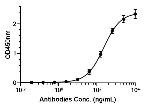 Immobilized HY-165740 Ganglioside GD2 can bind Nivatrotamab. The EC50 for this effect is 226.3 ng/mL.
 -
 Flow cytometric analysis of 1×10⁶ Jurkat cells labelling CD3E (red) with Nivatrotamab (HY-P99757). Cells were fixed with 4% paraformaldehyde and permeabilised with 90% methanol. Then stained with the primary antibody at 1/200 dilution for an hour at 4℃. Alexa Goat Anti-Human IgG H&L (AF488) (HY-P83776) was used as the secondary antibody at 1/1,000 dilution for 30 minutes at 4℃. Human IgG1 kappa Isotype Control (HY-P99001, blue) was used as the isotype control.


Chemical Information
-
CAS No. 2278244-14-5
-
Appearance Liquid
-
Molecular Weight 200.87 kDa
-
Color Colorless to light yellow
-
SMILES
[Nivatrotamab]
-
Synonyms
Hu3F8-BsAb
-
Shipping
Shipping with dry ice.
-
Formulation
Please refer to the lot-specific COA for specific buffer information.
-
Storage
Please store the product under the recommended conditions in the Certificate of Analysis.


Purity & Documentation
-
Data Sheet (272 KB)
-
SDS (251 KB)
- English - EN (251 KB)
- Français - FR (251 KB)
- Deutsch - DE (251 KB)
- Norwegian - NO (251 KB)
- Español - ES (251 KB)
- Swedish - SV (251 KB)
- Italian - IT (251 KB)
- Korean - KR (251 KB)
- Portuguese - PT (251 KB)
-
Inhibitory Antibodies User Guide (603 KB)
References
[1]. Nakajima M, et al. Potent antitumor effect of T cells armed with anti-GD2 bispecific antibody. Pediatric blood & cancer. 2021 Jul;68(7):e28971. [Content Brief]
[3]. Xu H, et al. Retargeting T cells to GD2 pentasaccharide on human tumors using Bispecific humanized antibody. Cancer immunology research. 2015 Mar;3(3):266-77. [Content Brief]
[4]. Park JA, et al. Targets and Antibody Formats for Immunotherapy of Neuroblastoma. Journal of clinical oncology : official journal of the American Society of Clinical Oncology. 2020 Jun 01;38(16):1836-1848. [Content Brief]

Calculators
Concentration (start) × Volume (start) = Concentration (final) × Volume (final)


Nivatrotamab Related Classifications
HY-P99757 Related Classifications
 Powered by Bioz