



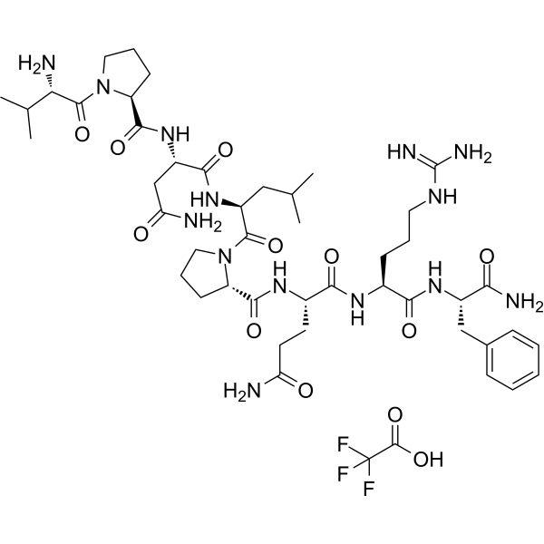
RFRP-3(human) TFA
RFRP-3 (Neuropeptide VF(124-131))(human) TFA, a human GnIH peptide homolog, is a potent inhibitor of gonadotropin secretion by inhibiting Ca2+ mobilization. RFRP-3(human) is a NPFF1 receptor agonist, it inhibits forskolin-induced production of cAMP with an IC50 of 0.7 nM.
For research use only. We do not sell to patients.
- Formula: C47H73F3N14O12
- Molecular Weight:1083.16
-
Storage:
Please store the product under the recommended conditions in the Certificate of Analysis.

To place orders, for customer services and technical support, please contact: MedChemExpress USA
Tel: 609-228-6898 E-mail: [email protected] [email protected]


-
 Biological Activity
-
Chemical Information
- Protocols
- Purity & Documentation
- References
-
 Help & FAQs


All Neuropeptide Y Receptor Isoforms
More

Biological Activity
Description
In Vitro
RFRP-3 TFA efficiently inhibits forskolin-induced production of cAMP with an IC50 of 0.7 nM[1]. Scatchard-plot analysis shows that 125I-labelled hRFRP-3 has a single class of high-affinity binding sites for the membrane fractions of CHO cells expressing rat OT7T022, the Kd value and the Bmax values are 0.19 nM and 1.3 pM, respectively.RFRP-3 TFA specifically stimulate cells transfected with a new orphan 7TMR, OT7T022, it binds to OT7T022 as a specific ligand with high affinity (Kd= 0.19 nM)[1].RFRP-3 (10-8 to 10-14M) has no effect on LH and FSH levels alone, but when it combines with GnRH, LH and FSH secretion is significantly reduced by the combination[1].
MedChemExpress (MCE) has not independently confirmed the accuracy of these methods. They are for reference only.


Chemical Information
-
Molecular Weight 1083.16
-
Formula C47H73F3N14O12
-
Synonyms
Neuropeptide VF(124-131)(human) TFA; Neuropeptide NPVF (human) TFA
-
Sequence Shortening
VPNLPQRF-NH2
-
Shipping
Room temperature in continental US; may vary elsewhere.
-
Storage
Please store the product under the recommended conditions in the Certificate of Analysis.


Purity & Documentation
References

Calculators
Concentration (start) × Volume (start) = Concentration (final) × Volume (final)


RFRP-3(human) TFA Related Classifications
HY-P1250A Related Classifications
 Powered by Bioz