



STL127705
Based on 8 publication(s) in Google Scholar
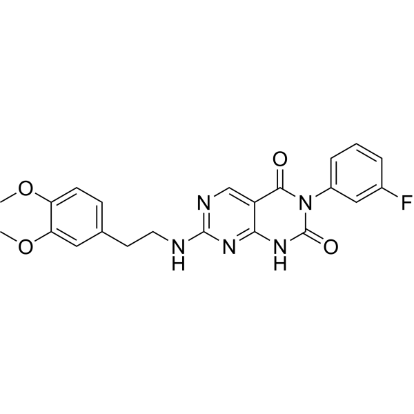
STL127705 (Compound L) is a potent Ku 70/80 heterodimer protein inhibitor with an IC50 of 3.5 μM. STL127705 interferes the binding of Ku70/80 to DNA and by inhibits the activation of the DNA-PKCS kinase. STL127705 shows antiproliferative and anticancer activity. STL127705 induces apoptosis.
For research use only. We do not sell to patients.
- Purity : 98.97%
- CAS No.: 1326852-06-5
- Formula: C22H20FN5O4
- Molecular Weight:437.42
-
Storage:Powder -20°C, 3 years , 4°C, 2 years ; In solvent -80°C, 2 years , -20°C, 1 year

To place orders, for customer services and technical support, please contact: MedChemExpress USA
Tel: 609-228-6898 E-mail: [email protected] [email protected]


-
 Biological Activity
-
Chemical Information
-
Solvent & Solubility
- Protocols
- Purity & Documentation
- References
-
 Help & FAQs


-
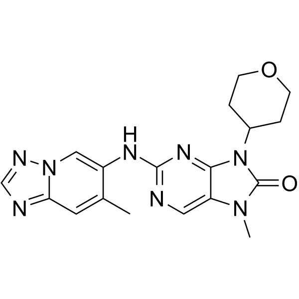
HY-111783
AZD-7648DNA-PK Inhibitor -
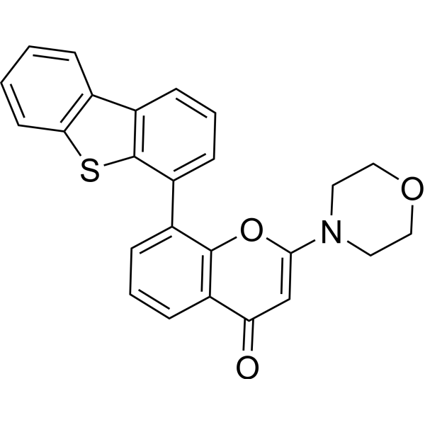
HY-11006
KU-57788DNA-PK Inhibitor -
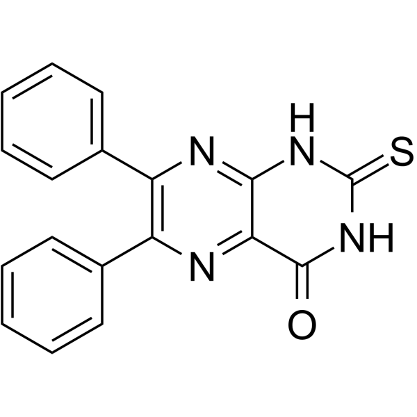
HY-107845
SCR7 pyrazineLigase IV Inhibitor/HDR Enhancer -
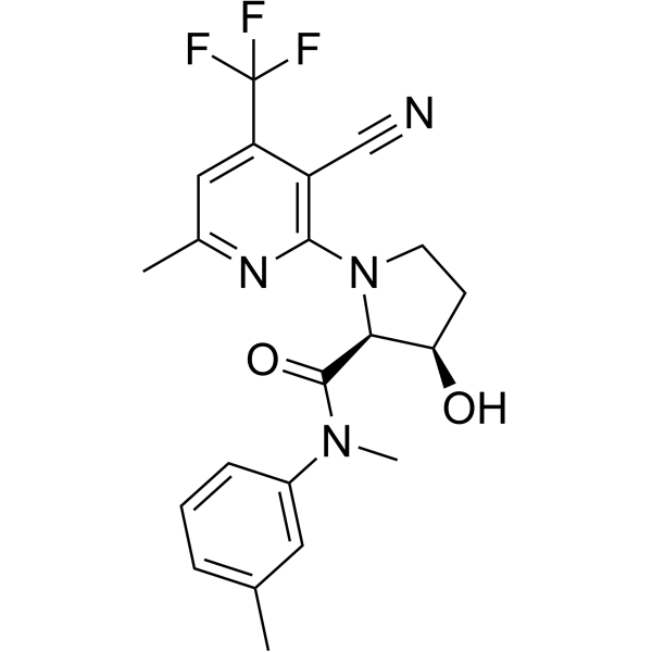
HY-141520
ART558Polθ Inhibitor -
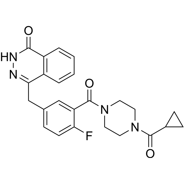
HY-10162
OlaparibPARP Inhibitor -

HY-10071
Y-27632ROCK Inhibitor -

HY-B0015
PaclitaxelAntitumor Agent -
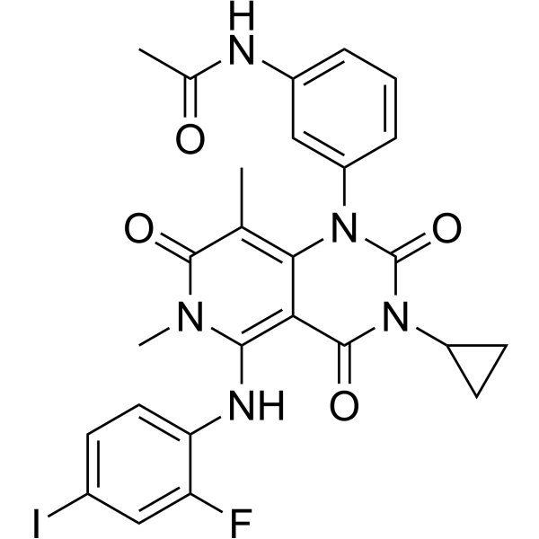
HY-10999
TrametinibMEK Inhibitor -
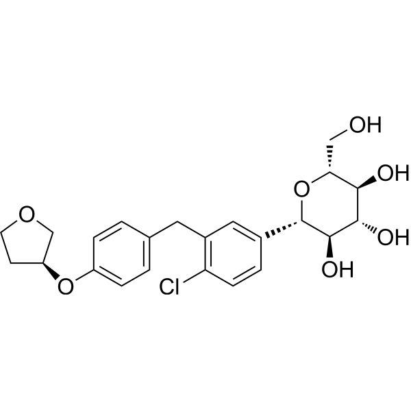
HY-15409
EmpagliflozinSGLT2 Inhibitor -
HY-13520
NocodazoleMicrotubule Inhibitor -
HY-101462
RAD51 Inhibitor B02RAD51 Inhibitor -
HY-N0005
Curcuminp300 Histone Acetylatransferase Inhibitor
Publications Citing Use of MedChemExpress (MCE) STL127705
More- Cell Res. 2026 Jan 7. [Abstract]
- Immunity. 2021 Apr 13;54(4):632-647.e9. [Abstract]
- Nucleic Acids Res. 2023 Dec 11;51(22):12242-12260. [Abstract]
- Adv Sci (Weinh). 2025 Jul;12(28):e2412698. [Abstract]
- Research (Wash D C). 2024 Mar 26:7:0331. [Abstract]
- Cell Rep. 2025 Jun 13;44(6):115851. [Abstract]
- Eur J Med Res. 2025 Sep 26;30(1):859. [Abstract]
- Biotechnol Bioeng. 2023 Sep;120(9):2419-2440. [Abstract]
 Customer Validation & Images

-
WB
-
WB

-
WB

-
Flow Cytometry

-
Cell Proliferation/Viability Assay


Biological Activity
Description
IC50 & Target
IC50: 3.5 μM (Ku 70/80), 2.5 μM (DNA-PKCS)[1]
In Vitro
STL127705 (Compound L) (0-100 μM) inhibits binding of Ku70/80 to a DNA substrate and inhibits Ku-dependent activation of the DNA-PKCS kinase[1].
STL127705 (0-100 μM; 6h) decreases the expression of DNA-PKCS auto-phosphorylation in SF-767 cells[1].
STL127705 (0-40 μM; 6h) shows antiproliferative activity in a dose dependent manner[1].
TL127705 (1 μM; 48 h) significantly promotes apoptotic when combination with gemcitabine[2].
MedChemExpress (MCE) has not independently confirmed the accuracy of these methods. They are for reference only.
-
Cell Line:SF-767, PrEC cells
-
Concentration:0-40 µM
-
Incubation Time:6 h
-
Result:Showed cytotoxicity in a dose dependent manner.
-
Cell Line:SF-767 cells
-
Concentration:0-100 µM
-
Incubation Time:pre-treated for 2 h and then co-incubation 4 h
-
Result:Decreased the DNA-PKCS autophosphorylation but total DNA-PKCS was not suppressed by STL127705.
-
Cell Line:H1299 cells
-
Concentration:1 µM
-
Incubation Time:48 h
-
Result:Induced apoptosis with apoptosis rate significantly increased to 76% when treated with STL127705 in combination with gemcitabine.


Chemical Information
-
CAS No. 1326852-06-5
-
Appearance Solid
-
Molecular Weight 437.42
-
Formula C22H20FN5O4
-
Color Off-white to light yellow
-
SMILES
O=C1NC2=NC(NCCC3=CC=C(OC)C(OC)=C3)=NC=C2C(N1C4=CC=CC(F)=C4)=O
-
Shipping
Room temperature in continental US; may vary elsewhere.
-
Storage
Powder -20°C 3 years 4°C 2 years In solvent -80°C 2 years -20°C 1 year


Publications (8)
-
Journal Impact Factor
-
Most Recent
-
Cell Res
Sensing of DNA double-strand breaks by the NHEJ system stabilizes RORγt transcriptional activity and shapes Th17 pathogenicity in autoimmunity. [Abstract]2026 Jan 7. PMID: 41495483
STL127705 purchased from MedChemExpress. Usage Cited in: Cell Res. 2026 Jan 7. [Abstract]
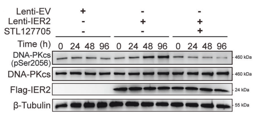
Immunoblot for pSer2056-DNA-PKcs expression in HP Th17 transfected with FLAG-IER2, with or without STL127705 (1 μmol/L).
-
Immunity
Cytoplasmic DNA sensing by KU complex in aged CD4+ T cell potentiates T cell activation and aging-related autoimmune inflammation. [Abstract]2021 Apr 13;54(4):632-647.e9. PMID: 33667382 -
Nucleic Acids Res
The RNA-binding motif protein 14 regulates telomere integrity at the interface of TERRA and telomeric R-loops. [Abstract]2023 Dec 11;51(22):12242-12260. PMID: 37930826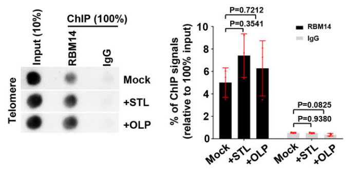
STL127705 purchased from MedChemExpress. Usage Cited in: Nucleic Acids Res. 2023 Dec 11;51(22):12242-12260. [Abstract]
Dot blot analysis of telomere DNA contents in the indicated ChIP and input from U2OS cells treated with either 1 μM Olaparib (OLP) or 2 μM STL127705 (STL) for 24 h. IgG ChIP served as a negative control. The telomeric repeat signal was detected using the Telo-C-Biotin (CCCTAA)4 probe.
-
Adv Sci (Weinh)
DNA-PKcs-Driven YAP1 Phosphorylation and Nuclear Translocation: a Key Regulator of Ferroptosis in Hyperglycemia-Induced Cardiac Dysfunction in Type 1 Diabetes. [Abstract]2025 Jul;12(28):e2412698. PMID: 40279648 -
Research (Wash D C)
DNA-PKcs Phosphorylates Cofilin2 to Induce Endothelial Dysfunction and Microcirculatory Disorder in Endotoxemic Cardiomyopathy. [Abstract]2024 Mar 26:7:0331. PMID: 38550779 -
Cell Rep
ENPP1 governs the metabolic regulation of effector T cells in autoimmunity by detecting cytosolic mitochondrial DNA. [Abstract]2025 Jun 13;44(6):115851. PMID: 40516055 -
Eur J Med Res
RHBDD1 induces cell cycle arrest in TP53-mutant NSCLC cells by promoting endoplasmic reticulum-associated degradation of p53 and DNA-PKcs. [Abstract]2025 Sep 26;30(1):859. PMID: 41013641
STL127705 purchased from MedChemExpress. Usage Cited in: Eur J Med Res. 2025 Sep 26;30(1):859. [Abstract]
Levels of RHBDD1, DNA-PKcs, and p53 proteins in sh-RHBDD1-transfected A549 cells treated with the DNA-PKcs inhibitor STL127705 (3.5 μM).
STL127705 purchased from MedChemExpress. Usage Cited in: Eur J Med Res. 2025 Sep 26;30(1):859. [Abstract]
Levels of RHBDD1, DNA-PKcs and p53 in sh-RHBDD1 transfected NCI-H596 cells treated with STL127705 (3.5 μM).
STL127705 purchased from MedChemExpress. Usage Cited in: Eur J Med Res. 2025 Sep 26;30(1):859. [Abstract]
Cell cycle analysis of transfected NCI-H596 cells co-treated with STL127705 (3.5 μM) or Nutlin-3a for 48 h by flow cytometry.
STL127705 purchased from MedChemExpress. Usage Cited in: Eur J Med Res. 2025 Sep 26;30(1):859. [Abstract]
Viability of transfected NCI-H596 cells co-treated with STL127705 (3.5 μM) or Nutlin-3a for 48 h.

STL127705 purchased from MedChemExpress. Usage Cited in: Eur J Med Res. 2025 Sep 26;30(1):859. [Abstract]
ERAD marker analysis in sh-RHBDD1- or pcDNA-RHBDD1-transfected cells treated with STL127705 (3.5 μM) or Nutlin-3a, assessed by western blotting.
-
Biotechnol Bioeng
High-efficiency and multilocus targeted integration in CHO cells using CRISPR-mediated donor nicking and DNA repair inhibitors. [Abstract]2023 Sep;120(9):2419-2440. PMID: 37039773


Solvent & Solubility
In Vitro:
H2O : < 0.1 mg/mL (insoluble)
DMSO : < 1 mg/mL (insoluble or slightly soluble)
* STL127705 is usually formulated as a suspension.


Purity & Documentation
-
Data Sheet (273 KB)
-
SDS (393 KB)
- English - EN (393 KB)
- Français - FR (393 KB)
- Deutsch - DE (393 KB)
- Norwegian - NO (393 KB)
- Español - ES (393 KB)
- Swedish - SV (393 KB)
- Italian - IT (393 KB)
- Korean - KR (393 KB)
- Portuguese - PT (393 KB)
-
Handling Instructions (2659 KB)
References
[1]. Weterings E, et al. A novel small molecule inhibitor of the DNA repair protein Ku70/80. DNA Repair (Amst). 2016 Jul;43:98-106. [Content Brief]
[2]. Guo N, et al. Inhibiting nonhomologous end-joining repair would promote the antitumor activity of gemcitabine in nonsmall cell lung cancer cell lines. Anticancer Drugs. 2022 Jun 1;33(5):502-508. [Content Brief]

Calculators
Concentration (start) × Volume (start) = Concentration (final) × Volume (final)


STL127705 Related Classifications
HY-122727 Related Classifications
 Powered by Bioz