



EL-102

Based on 1 Customer Validation
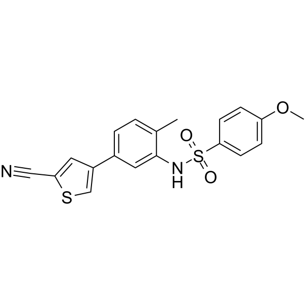
EL-102 is a hypoxia-induced factor 1 (Hif1α) inhibitor. EL-102 induces apoptosis, inhibits tubulin polymerisation and shows activities against prostate cancer. EL-102 can be used for the research of cancer.
For research use only. We do not sell to patients.
- Purity : 99.44%
- CAS No.: 1233948-61-2
- Formula: C19H16N2O3S2
- Molecular Weight:384.47
-
Storage:Powder -20°C, 3 years , 4°C, 2 years ; In solvent -80°C, 2 years , -20°C, 1 year

To place orders, for customer services and technical support, please contact: MedChemExpress USA
Tel: 609-228-6898 E-mail: [email protected] [email protected]


-
 Biological Activity
-
Chemical Information
-
Solvent & Solubility
- Protocols
- Purity & Documentation
- References
-
 Help & FAQs


-
HY-13671
LW6Apoptosis Inducer -
HY-15836
BAY 87-2243HIF-1 Inhibitor -
HY-10231
PX-478HIF-1α Inhibitor -
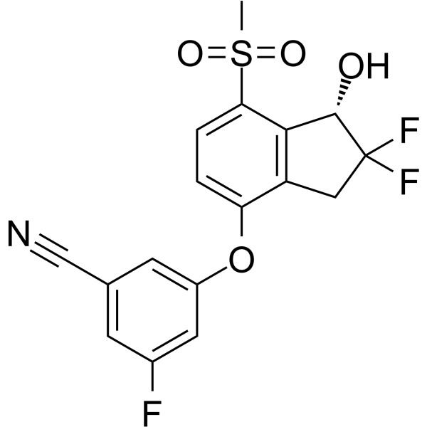
HY-12867
PT-2385HIF-2α Inhibitor -
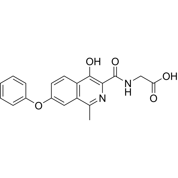
HY-13426
RoxadustatHIF-PH Inhibitor -
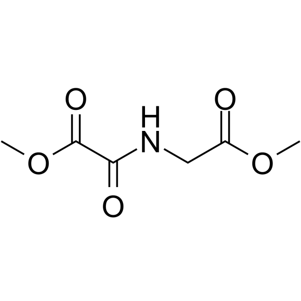
HY-15893
DMOGHIF-PH Inhibitor -
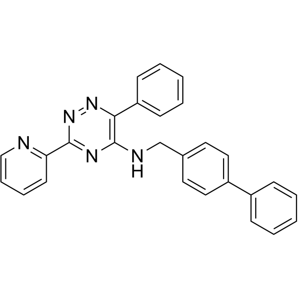
HY-12754
ML228HIF Activator -
HY-18371
TC-S 7009HIF-2α Inhibitor -
HY-17608
DaprodustatHIF-PH Inhibitor -
HY-N0055
Chlorogenic acidAntioxidant -
HY-N0560
Oroxylin AAutophagy Inducer -

HY-15893R
DMOG (Standard)HIF-PH Inhibitor


Biological Activity
Description
IC50 & Target
IC50: 24 nM (CWR22), 21.7 nM (22Rv1), 40.3 nM (DU145), 37.0 nM (PC-3), 14.4 nM (DLKP), 16.3 nM (DLKPA)[1]
In Vitro
EL-102 (0-120 nM; 72 h) inhibits prostate cancer cells proliferation in vitro[1]. EL-102 (0-100 nM; 72 h) shows cytotoxicity to prostate cancer cell lines[1]. EL-102 (10-100 nM; 24-72 h) induces cellular apoptosis and affects cell cycle[1]. EL-102 (10-100 nM; 24-48 h) affects PARP cleavage in DU145 cells[1]. EL-102 (5 nM; 0-60 min) inhibits tubulin polymerisation activity[1]. EL-102 (0-100 nM; 1 hour) inhibits Hif1α protein expression[1].
MedChemExpress (MCE) has not independently confirmed the accuracy of these methods. They are for reference only.
-
Cell Line:CWR22, 22Rv1, DU145, PC-3, DLKP and DLKPA cell lines
-
Concentration:0-120 nM
-
Incubation Time:72 hours
-
Result:Inhibited proliferation of CWR22, 22Rv1, DU145, PC-3, DLKP and doxorubicin-selected variant DLKPA cells with IC50s of 24, 21.7, 40.3, 37.0, 14.4 and 16.3 nM, respectively.
-
Cell Line:CWR22, 22Rv1, DU145 and PC-3 cell lines
-
Concentration:0-100 nM
-
Incubation Time:72 hours
-
Result:Exibited cytotoxicity to prostate cancer cell lines, and showed no additive effect on the inhibition of cell viability with docetaxel.
-
Cell Line:CWR22, 22Rv1, DU145, PC-3, DLKP and DLKPA cell lines
-
Concentration:10 and 100 nM
-
Incubation Time:24, 48 and 72 hours
-
Result:Induced cell apoptosis to inhibits cell viability with a dose of 100 nM.
-
Cell Line:DU145 cell line
-
Concentration:10 and 100 nM
-
Incubation Time:24 and 48 hours
-
Result:Increased PARP cleavage in DU145 cells and showed a more dramatic effect with docetaxel adding.
-
Cell Line:DU145 cell line
-
Concentration:10 and 100 nM
-
Incubation Time:24, 48 and 72 hours
-
Result:Increased loss of cells from G1 phase and accumulated cells in G2/M phase.
-
Cell Line:Prostate cancer cells
-
Concentration:10 , 50 and 100 nM
-
Incubation Time:24 and 48 hours
-
Result:Modestly inhibited Hif1α expression at doses of 50 and 100 nM in normoxia.
In Vivo
MedChemExpress (MCE) has not independently confirmed the accuracy of these methods. They are for reference only.
-
Animal Model:Nude mice with CWR22 xenografts[1]
-
Dosage:12 and 15 mg/kg
-
Administration:Oral gavage; 12 and 15 mg/kg 5-day on and 2-day off; from 13 to 37 days after tumour transplantation
-
Result:Showed no effect on tumor growth, but enhanced the effect of docetaxel on tumor .


Chemical Information
-
CAS No. 1233948-61-2
-
Appearance Solid
-
Molecular Weight 384.47
-
Formula C19H16N2O3S2
-
Color Light yellow to yellow
-
SMILES
O=S(C1=CC=C(OC)C=C1)(NC2=CC(C3=CSC(C#N)=C3)=CC=C2C)=O
-
Shipping
Room temperature in continental US; may vary elsewhere.
-
Storage
Powder -20°C 3 years 4°C 2 years In solvent -80°C 2 years -20°C 1 year


Solvent & Solubility
In Vitro:
DMSO : ≥ 36 mg/mL (93.64 mM; Hygroscopic DMSO has a significant impact on the solubility of product, please use newly opened DMSO)
* "≥" means soluble, but saturation unknown.
Please refer to the solubility information to select the appropriate solvent. Once prepared, please aliquot and store the solution to prevent product inactivation from repeated freeze-thaw cycles.
Storage method and period of stock solution: -80°C, 2 years; -20°C, 1 year. When stored at -80°C, please use it within 2 years. When stored at -20°C, please use it within 1 year.
Please refer to the solubility information to select the appropriate solvent. Once prepared, please aliquot and store the solution to prevent product inactivation from repeated freeze-thaw cycles.
Storage method and period of stock solution: -80°C, 2 years; -20°C, 1 year. When stored at -80°C, please use it within 2 years. When stored at -20°C, please use it within 1 year.
Concentration (start) × Volume (start) = Concentration (final) × Volume (final)


Purity & Documentation
-
Data Sheet (276 KB)
-
SDS (251 KB)
- English - EN (251 KB)
- Français - FR (251 KB)
- Deutsch - DE (251 KB)
- Norwegian - NO (251 KB)
- Español - ES (251 KB)
- Swedish - SV (251 KB)
- Italian - IT (251 KB)
- Korean - KR (251 KB)
- Portuguese - PT (251 KB)
-
Handling Instructions (2659 KB)
References


Complete Stock Solution Preparation Table
Please refer to the solubility information to select the appropriate solvent. Once prepared, please aliquot and store the solution to prevent product inactivation from repeated freeze-thaw cycles.
Storage method and period of stock solution: -80°C, 2 years; -20°C, 1 year. When stored at -80°C, please use it within 2 years. When stored at -20°C, please use it within 1 year.
| Optional Solvent | Concentration Solvent Mass | 1 mg | 5 mg | 10 mg | 25 mg |
|---|---|---|---|---|---|
| DMSO | 1 mM | 2.6010 mL | 13.0049 mL | 26.0098 mL | 65.0246 mL |
| 5 mM | 0.5202 mL | 2.6010 mL | 5.2020 mL | 13.0049 mL | |
| 10 mM | 0.2601 mL | 1.3005 mL | 2.6010 mL | 6.5025 mL | |
| 15 mM | 0.1734 mL | 0.8670 mL | 1.7340 mL | 4.3350 mL | |
| 20 mM | 0.1300 mL | 0.6502 mL | 1.3005 mL | 3.2512 mL | |
| 25 mM | 0.1040 mL | 0.5202 mL | 1.0404 mL | 2.6010 mL | |
| 30 mM | 0.0867 mL | 0.4335 mL | 0.8670 mL | 2.1675 mL | |
| 40 mM | 0.0650 mL | 0.3251 mL | 0.6502 mL | 1.6256 mL | |
| 50 mM | 0.0520 mL | 0.2601 mL | 0.5202 mL | 1.3005 mL | |
| 60 mM | 0.0433 mL | 0.2167 mL | 0.4335 mL | 1.0837 mL | |
| 80 mM | 0.0325 mL | 0.1626 mL | 0.3251 mL | 0.8128 mL |


EL-102 Related Classifications
HY-16187 Related Classifications
 Powered by Bioz