


Bcl-2 Antibody (YA590)
(Synonyms: BCL2; Apoptosis regulator Bcl-2)Based on 10 publication(s) in Google Scholar
Bcl-2 Antibody (YA590) is a Rabbit-derived and non-conjugated IgG monoclonal antibody, targeting to Bcl-2.
-
Host:
Rabbit
-
Isotype:
IgG
-
Application:
WB, IHC-P
-
Reactivity :
Human, Mouse
-
Formulation:
Supplied in 50 mM Tris-Glycine (pH 7.4), 0.15 M NaCl, 40% Glycerol and 0.05% BSA. Preservative: 0.01% Sodium azide
-
Conjugation:
Non-conjugated


 Product Image")


Publications Citing Use of MedChemExpress (MCE) Bcl-2 Antibody (YA590)
More- Rep Phys Sci. 2024 July 01.
- J Med Chem. 2026 Feb 26;69(4):5024-5054. [Abstract]
- J Med Chem. 2025 Aug 14;68(15):16138-16171. [Abstract]
- J Mater Chem B. 2025 Oct 22;13(41):13405-13422. [Abstract]
- Colloids Surf A Physicochem Eng Asp. 2025 Aug 20.
- Respir Res. 2024 Nov 7;25(1):399. [Abstract]
- Sci Rep. 2025 Apr 5;15(1):11722. [Abstract]
- Int Dent J. 2025 Oct 8;75(6):103897. [Abstract]
- Naunyn Schmiedebergs Arch Pharmacol. 2025 Oct 3. [Abstract]
- Res Sq. 2025 Jan 19.

 10 Publications Citing Use of MCE Bcl-2 Antibody (YA590) (5)
-
WB
-
WB
-
WB
-
WB
-
WB


Applications
| Application |
WB
WB: Western Blot
|
IHC-P
IHC-P: Immunohistochemistry-Paraffin
|
|---|---|---|
| Dilution Ratio | 1:500-1:1000 | 1:50-1:100 |


Product Details
Bcl-2 Antibody (YA590) is a Rabbit-derived and non-conjugated IgG monoclonal antibody, targeting to Bcl-2.
-
Host Rabbit

-
Clonality Recombinant,Monoclonal

-
Species ReactivityHuman, Mouse
-
Observed Molecular WeightObserved band size: 26 kDa
 Note: Due to possible protein modifications or aggregation, the molecular weight should be confirmed by actual measurement, and the predicted value is for reference only.
-
Calculated Molecular Weight Predicted band size: 26 kDa

Synthetic peptide corresponding to Human Bcl-2 aa1-211.
Endogenous
affinity purified
Non-conjugated
Unmodified
IgG


Product Properties
-
Appearance
Solution
-
Formulation
Supplied in 50 mM Tris-Glycine (pH 7.4), 0.15 M NaCl, 40% Glycerol and 0.05% BSA. Preservative: 0.01% Sodium azide
-
Storage & Stability
Stored at -20°C for 1 year. Avoid repeated freeze / thaw cycles.
-
Shipping
Shipping with blue ice.


Publications (10)
-
Journal Impact Factor
-
Most Recent
-
Bcl-2 Antibody (YA590) purchased from MedChemExpress. Usage Cited in: Rep Phys Sci. 2024 July 01.
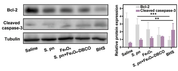
Western blotting analysis of the apoptosis-related protein expression (cleaved caspase-3 and Bcl-2) in tumor tissues after different treatments (n = 3).
-
J Med Chem
Discovery of Potent HDAC6-Selective Inhibitors Based on Artemisinin: Design, Synthesis, and Antitumor Evaluation. [Abstract]2026 Feb 26;69(4):5024-5054. PMID: 41701111 -
J Med Chem
Structure-Based Discovery of Quinazolin-2-amine Derivatives as Potent ROR1 Pseudokinase Inhibitors with In Vitro and In Vivo Efficacy against Triple-Negative Breast Cancer. [Abstract]2025 Aug 14;68(15):16138-16171. PMID: 40736991
Bcl-2 Antibody (YA590) purchased from MedChemExpress. Usage Cited in: J Med Chem. 2025 Aug 14;68(15):16138-16171. [Abstract]
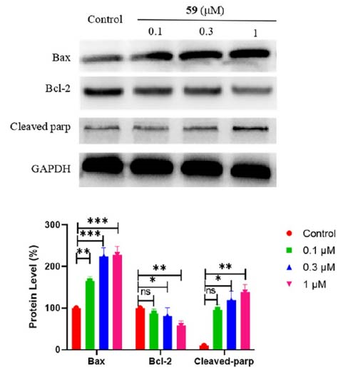
MDA-MB-231 cells were incubated with either 59 or DMSO for a period of 48h. After the treatment was completed, the cells were collected and washed three times with 1 mL of cold PBS. Subsequently, they were lysed in 50 μL of ice-cold RIPA buffer for 10 min. The cell lysates were then subjected to centrifugation at 12,000 rpm for 15 min at 4 °C. The protein concentration within the lysates was determined using the BCA assay. After lysis, the proteins were separated by SDS-PAGE and then transferred onto PVDF membranes. These membranes were then individually treated with a series of antibodies, including ROR1 antibody , Phosphorylated ROR1 antibody, cleaved PARP antibody (HY-P80448), Bax antibody (HY-P80028), BCL-2 antibody (HY-P80566) and GAPDH antibody. Afterward, the membranes were probed with a secondary antibody conjugated with horseradish peroxidase (obtained from Cell Signaling Technology). Finally, the blots were visualized using an enhanced chemiluminescence system.
-
J Mater Chem B
A metal-organic framework-based co-delivery system for atherosclerosis therapy via macrophage regulation. [Abstract]2025 Oct 22;13(41):13405-13422. PMID: 41002166 -
Bcl-2 Antibody (YA590) purchased from MedChemExpress. Usage Cited in: Colloids Surf A Physicochem Eng Asp. 2025 Aug 20.
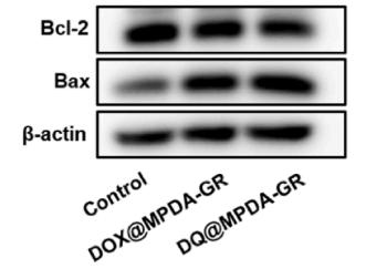
Western blot analysis of the expressions of Bcl-2 and Bax, in C5WN1 cells after treated with Control, DOX@MPDA-GR, and DQ@MPDA-GR.
-
Respir Res
Activation of LXR signaling ameliorates apoptosis of alveolar epithelial cells in Bronchopulmonary dysplasia. [Abstract]2024 Nov 7;25(1):399. PMID: 39511537
Bcl-2 Antibody (YA590) purchased from MedChemExpress. Usage Cited in: Respir Res. 2024 Nov 7;25(1):399. [Abstract]
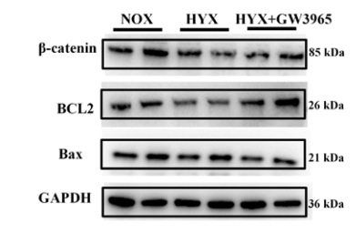
The relative protein levels of BCL2, BAX, and β-catenin were determined by western blot analysis and quantified.
-
Sci Rep
Sodium propionate protects against bronchopulmonary dysplasia by inhibiting IL-17-mediated apoptosis of alveolar epithelial cells. [Abstract]2025 Apr 5;15(1):11722. PMID: 40188136 -
Int Dent J
High-Glucose Microenvironment Accelerates Malignant Progression Via O-GlcNAcylation in Oral Squamous Cell Carcinoma. [Abstract]2025 Oct 8;75(6):103897. PMID: 41067097
Bcl-2 Antibody (YA590) purchased from MedChemExpress. Usage Cited in: Int Dent J. 2025 Oct 8;75(6):103897. [Abstract]
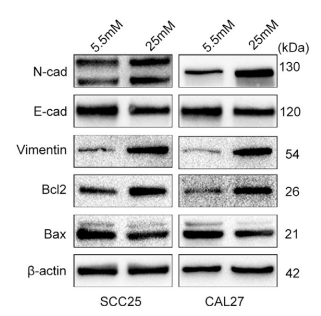
OSCC cells were treated with different concentrations of glucose for 48 hours, and Western blots were performed to detect the expression of N-cadherin, E-cadherin, vimentin, Bax, and Bcl-2.
-
Naunyn Schmiedebergs Arch Pharmacol
Dehydrocostus lactone induces apoptosis and mitophagy in gastric cancer cells through the ROS-mediated mitochondrial pathway. [Abstract]2025 Oct 3. PMID: 41039061


Verification Images
-
 Western blot analysis of extracts from HeLa (lane 1(20μg)) 、SH-SY5Y (lane 2(20μg)) 、HepG2 (lane 3(20μg)) 、HEK293 (lane 4(20μg)) 、Jurkat (lane 5(20μg)) 、HL-60 (lane 6(20μg)) 、U937 (lane 7(20μg)) and MCF7 (lane 8(20μg)) using Bcl-2 Antibody. Proteins were transferred to a PVDF membrane and blocked with 5% nonfat dry milk in TBST for 1.5 hour at room temperature. The primary antibody (HY-P80566, 1/1000) , competitor's antibody (1/1000) and Loading control antibody (Hsp90, 1/10000) was used in 5% nonfat dry milk in TBST at 4℃ overnight. Goat Anti-Rabbit IgG-HRP Secondary Antibody (1/10,000) was used for 1 hour at room temperature.
 -
 Western blot analysis of extracts from NIH3T3 (lane 2(20μg), K562 (lane 3(20μg) and Hela(lane 4(20μg) using Bcl2 (HY-P80566) Rabbit mAb. Proteins were transferred to a PVDF membrane and blocked with 5% non-fat milk in TBST for 2 hour at room temperature. The primary antibody (1/1000) and Loading control antibody (Beta Actin, HY-P80438, 1/10000) was used in 5% non-fat milk in TBST at 4°C overnight. Goat Anti-Mouse/Rabbit IgG-HRP Secondary Antibody (1/10000) was used for 1 hour at room temperature.
 -
 Western blot analysis of extracts from HeLa (lane 1(20μg)) 、SH-SY5Y (lane 2(20μg)) 、HepG2 (lane 3(20μg)) 、HEK293 (lane 4(20μg)) 、Jurkat (lane 5(20μg)) 、HL-60 (lane 6(20μg)) 、U937 (lane 7(20μg)) and MCF7 (lane 8(20μg)) using Bcl-2 Antibody (HY-P80566) . Proteins were transferred to a PVDF membrane and blocked with 5% nonfat dry milk in TBST for 1.5 hour at room temperature. The primary antibody (1/1000) and Loading control antibody (Hsp90, 1/10000) was used in 5% nonfat dry milk in TBST at 4℃ overnight. Goat Anti-Rabbit IgG-HRP Secondary Antibody (1/10,000) was used for 1 hour at room temperature.
 -
 Western blot analysis was performed on protein extracts (30 μg) from HT-1080 (lane 2), SH-SY5Y (lane 3), HeLa (lane 4), A549 (lane 5), and Caco-2 (lane 6) using Bcl-2 antibody. Proteins were transferred onto a 0.45 μm PVDF membrane using the Trans-Blot® Turbo™ system for 13 min. The membrane was then blocked with 5% nonfat milk in TBST (HY-K1025) for 1 h at room temperature. Thhe primary antibody (1:1000) and loading control antibody GAPDH Antibody (HRP) (HY-P80954A) (1:2500) were diluted in 5% nonfat milk in TBST and incubated with the membrane overnight at 4°C. After washing, the membrane of primary antibody was incubated with HRP-conjugated goat anti-rabbit IgG (H&L) secondary antibody (HY-P8001) (1:5000) diluted in 5% nonfat milk in TBST for 1 h at room temperature. Protein bands were visualized using an Ultra High Sensitivity ECL detection kit (HY-K1005).
 -
 Immunohistochemical analysis of paraffin-embedded mouse kidney tissue using Bcl2 Antibody. The section was pre-treated using heat mediated antigen retrieval with Tris-EDTA buffer (pH 9.0) for 8 minutes. The tissues were blocked in QuickBlock for 20 minutes at room temperature, washed with ddH2O and PBS, and then probed with the primary antibody at 1/100 dilution in 4℃ overnight. The detection was performed using an HRP conjugated compact polymer system. DAB was used as the chromogen. Tissues were counterstained with hematoxylin and mounted with DPX.


Background
-
Function
Suppresses apoptosis in a variety of cell systems including factor-dependent lymphohematopoietic and neural cells (PubMed:1508712, PubMed:8183370). Regulates cell death by controlling the mitochondrial membrane permeability (PubMed:11368354). Appears to function in a feedback loop system with caspases (PubMed:11368354). Inhibits caspase activity either by preventing the release of cytochrome c from the mitochondria and/or by binding to the apoptosis-activating factor (APAF-1) (PubMed:11368354). Also acts as an inhibitor of autophagy: interacts with BECN1 and AMBRA1 during non-starvation conditions and inhibits their autophagy function (PubMed:18570871, PubMed:20889974, PubMed:21358617). May attenuate inflammation by impairing NLRP1-inflammasome activation, hence CASP1 activation and IL1B release (PubMed:17418785)
-
Subcellular Localization
Mitochondrion outer membrane; Single-pass membrane protein; Nucleus membrane; Single-pass membrane protein; Endoplasmic reticulum membrane; Single-pass membrane protein; Cytoplasm
-
Expression
Tissue_specificity:Expression in multiple organizations -
Isoforms & Post-Translational Modification
P10415 has 2 isomers: P10415-1: 26266 Da (predicted); P10415-2: 22337 Da (predicted).
Phosphorylation/dephosphorylation on Ser-70 regulates anti-apoptotic activity (PubMed:11368354). Growth factor-stimulated phosphorylation on Ser-70 by PKC is required for the anti-apoptosis activity and occurs during the G2/M phase of the cell cycle (PubMed:11368354). In the absence of growth factors, BCL2 appears to be phosphorylated by other protein kinases such as ERKs and stress-activated kinases (PubMed:11368354). Phosphorylated by MAPK8/JNK1 at Thr-69, Ser-70 and Ser-87, which stimulates starvation-induced autophagy (PubMed:10567572, PubMed:18570871). Dephosphorylated by protein phosphatase 2A (PP2A) (By similarity);Proteolytically cleaved by caspases during apoptosis. The cleaved protein, lacking the BH4 motif, has pro-apoptotic activity, causes the release of cytochrome c into the cytosol promoting further caspase activity;Monoubiquitinated by PRKN, leading to an increase in its stability (PubMed:20889974). Ubiquitinated by SCF(FBXO10), leading to its degradation by the proteasome (PubMed:23431138). Ubiquitinated by XIAP, leading to its degradation by the proteasome (PubMed:29020630) -
Subunit
Forms homodimers, and heterodimers with BAX, BAD, BAK and Bcl-X(L). Heterodimerization with BAX requires intact BH1 and BH2 motifs, and is necessary for anti-apoptotic activity (PubMed:25609812, PubMed:8183370).
-
SwissProt ID
-
Synonyms
BCL2; Apoptosis regulator Bcl-2
-
Research Field
Cell Biology
Documentation
-
Data Sheet (258 KB)
-
SDS (251 KB)
- English - EN (251 KB)
- Français - FR (251 KB)
- Deutsch - DE (251 KB)
- Norwegian - NO (251 KB)
- Español - ES (251 KB)
- Swedish - SV (251 KB)
- Italian - IT (251 KB)
- Korean - KR (251 KB)
- Portuguese - PT (251 KB)
-
User Guide for Antibodies (1077 KB)

 Powered by Bioz
