



Anticancer agent 207
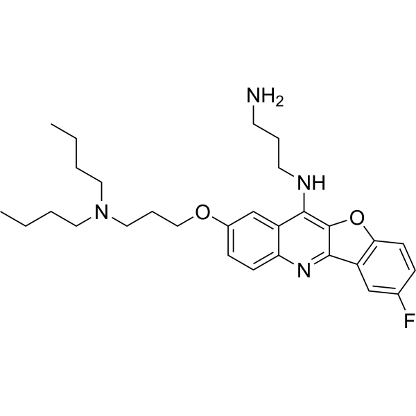
Anticancer agent 207 (compound 10b) is a potent anticancer agent. Anticancer agent 207 bounds to the NRAS rG4 with a KD value of 2.31 µM. Anticancer agent 207 shows cytotoxicity and decreases the expression of NRAS protein. Anticancer agent 207 shows antitumor activity.
For research use only. We do not sell to patients.
- Formula: C29H39FN4O2
- Molecular Weight:494.64
-
Storage:
Please store the product under the recommended conditions in the Certificate of Analysis.

To place orders, for customer services and technical support, please contact: MedChemExpress USA
Tel: 609-228-6898 E-mail: [email protected] [email protected]


-
 Biological Activity
-
Chemical Information
- Protocols
- Purity & Documentation
- References
-
 Help & FAQs




Biological Activity
Description
IC50 & Target
[1]|
NRAS rG4 2.31 μM (Kd) |
Cellular Effect
|
Cell Line

|
Type | Value | Description | References |
|---|---|---|---|---|
| A-375 | IC50 |
4.5 μM
Compound: 10b
|
Cytotoxicity against human A-375 cells incubated for 48 hrs by MTT assay
Cytotoxicity against human A-375 cells incubated for 48 hrs by MTT assay
|
[PMID: 38688064] |
| HepG2 | IC50 |
1.5 μM
Compound: 10b
|
Cytotoxicity against human HepG2 cells incubated for 48 hrs by MTT assay
Cytotoxicity against human HepG2 cells incubated for 48 hrs by MTT assay
|
[PMID: 38688064] |
| HL-60 | IC50 |
2.7 μM
Compound: 10b
|
Cytotoxicity against human HL-60 cells incubated for 48 hrs by MTT assay
Cytotoxicity against human HL-60 cells incubated for 48 hrs by MTT assay
|
[PMID: 38688064] |
| MCF7 | IC50 |
4.1 μM
Compound: 10b
|
Cytotoxicity against human MCF7 cells incubated for 48 hrs by MTT assay
Cytotoxicity against human MCF7 cells incubated for 48 hrs by MTT assay
|
[PMID: 38688064] |
| SK-MEL-2 | IC50 |
2 μM
Compound: 10b
|
Cytotoxicity against human SK-MEL-2 cells incubated for 48 hrs by MTT assay
Cytotoxicity against human SK-MEL-2 cells incubated for 48 hrs by MTT assay
|
[PMID: 38688064] |
In Vitro
Anticancer agent 207 (48 h) shows cytotoxicity for NRAS-mutant melanoma SK-MEL-2 with an IC50 value of 2.0 µM[1].
Anticancer agent 207 (0, 0.5, 1.0 µM; 72 h) decreases the expression of NRAS protein[1].
Anticancer agent 207 (0, 0.13, 0.25 µM; 10 days) inhibits the colony formation of SK-MEL-2 cells[1].
MedChemExpress (MCE) has not independently confirmed the accuracy of these methods. They are for reference only.
-
Cell Line:SK-MEL-2 cells
-
Concentration:0, 0.5, 1.0 µM
-
Incubation Time:72 h
-
Result:Decreased the expression of NRAS protein in a dose-dependent manner.
-
Cell Line:MCF-7, HepG2, HL60, A375 cells
-
Concentration:0-100 µM
-
Incubation Time:48 h
-
Result:Showed cytotoxicity with IC50s of 4.1, 1.5, 2.7, 4.5 µM for MCF-7, HepG2, HL60, A375 cells, respectively.
In Vivo
MedChemExpress (MCE) has not independently confirmed the accuracy of these methods. They are for reference only.
-
Animal Model:Six-week-old male BALB/C nude mice (SK-MEL-2 xenograft mouse model)[1]
-
Dosage:1 mg/kg
-
Administration:I.p.; every day for 21 days
-
Result:Suppressed tumor growth in volume and weight in the xenograft mouse model time-dependently.


Chemical Information
-
Molecular Weight 494.64
-
Formula C29H39FN4O2
-
SMILES
CCCCN(CCCOC1=CC=C2N=C3C4=C(OC3=C(C2=C1)NCCCN)C=CC(F)=C4)CCCC
-
Shipping
Room temperature in continental US; may vary elsewhere.
-
Storage
Please store the product under the recommended conditions in the Certificate of Analysis.


Purity & Documentation
References

Calculators
Concentration (start) × Volume (start) = Concentration (final) × Volume (final)


Anticancer agent 207 Related Classifications
HY-163515 Related Classifications
 Powered by Bioz