



Anticancer agent 69
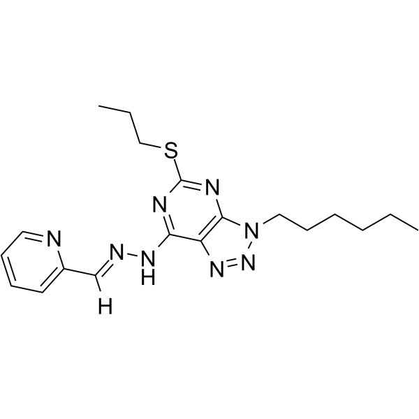
Anticancer agent 69 (Compound 34), a potent and selective anticancer agent , potently and selectively inhibits human prostate cancer cell line PC3 (IC50=26 nM). Anticancer agent 69 increases ROS level, down-regulates EGFR and induces apoptosis.
For research use only. We do not sell to patients.
- Formula: C19H26N8S
- Molecular Weight:398.53
-
Storage:
Please store the product under the recommended conditions in the Certificate of Analysis.

To place orders, for customer services and technical support, please contact: MedChemExpress USA
Tel: 609-228-6898 E-mail: [email protected] [email protected]


-
 Biological Activity
-
Chemical Information
- Protocols
- Purity & Documentation
- References
-
 Help & FAQs


All EGFR Isoforms
More

Biological Activity
Description
In Vitro
Anticancer agent 69 (Compound 34, 0-20 μM approximately, 72 h) has selective anticancer activity in PC3 cells with an IC50 value of 26 nM against other cancer cell lines and normal cell lines[1].
Anticancer agent 69 (1-7 days) inhibits the cell viabilities of PC3 cells and PC9 cells in a time- and dose-dependent manne[1].
Anticancer agent 69 (0-100 nM, 7 days) forms fewer and smaller colonies compared those with DMSO-treated cells[1].
Anticancer agent 69 (0-800 nM, 34/72 h) increases ROS production, and further stimulates Prx I-III expression, phosphorylation level of MAPK ‘s downstream proteins, expression of apoptosis-related proteins[1].
Anticancer agent 69 (0-500 nM, 72 h) decreases the level of EGFR and phosphorylation level of its downstream protein (ERK, AKT), and induces PC3 cells and PC9 cells apoptosis in a dose-dependent manner[1].
MedChemExpress (MCE) has not independently confirmed the accuracy of these methods. They are for reference only.
-
Cell Line:PC3, PC9
-
Concentration:0-800 nM
-
Incubation Time:72 h
-
Result:Stimulated Prx I-III expression, phosphorylation level of MAPK ‘s downstream proteins (P38, JNK), expression of apoptosis-related proteins (Bax, cleaved-Caspased 3). Inhibited the level of EGFR and its downstream protein (p-ERK, p-AKT). Increased pro-apoptotic proteins (Bax and P53) expression and reduces anti-apoptotic protein (Bcl-2) expression.
-
Cell Line:PC3, PC9
-
Concentration:0-100 nM
-
Incubation Time:7 days
-
Result:Formed fewer and smaller colonies compared those with DMSO-treated cells.
-
Cell Line:Cancer cell lines (PC3, MGC-803, PC9, EC9706, SMMC-7721), normal cell lines (Het-1A, L02 and GES-1)
-
Concentration:0-20 μM approximately
-
Incubation Time:72 h
-
Result:Inhibited proliferation of multiple cancer cell lines with IC50 values of 26 nM (PC3), 557 nM (MGC-803), 148 nΜ (PC9), 3.99 μΜ (EC9706), 844 nΜ (SMMC-7721), respectively. Displayed good selectivity against normal cell lines with IC50 values of > 20 μΜ (L02), > 5 μΜ (Het-1A), 1.57 μΜ (GES-1), respectively.


Chemical Information
-
Molecular Weight 398.53
-
Formula C19H26N8S
-
SMILES
[H]/C(C1=CC=CC=N1)=N\NC2=NC(SCCC)=NC3=C2N=NN3CCCCCC
-
Shipping
Room temperature in continental US; may vary elsewhere.
-
Storage
Please store the product under the recommended conditions in the Certificate of Analysis.


Purity & Documentation
References

Calculators
Concentration (start) × Volume (start) = Concentration (final) × Volume (final)


Anticancer agent 69 Related Classifications
HY-146170 Related Classifications
 Powered by Bioz