



P62-RNF168 agonist-1

Based on 1 Customer Validation
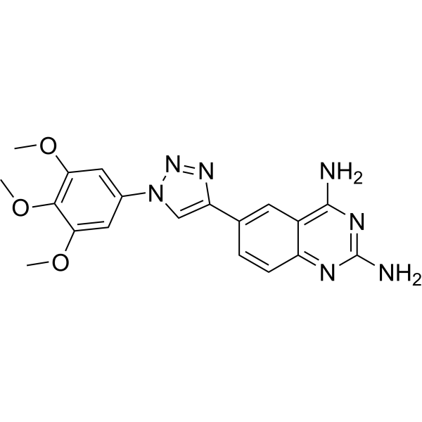
P62-RNF168 agonist-1 (compound 5a) is a low cytotoxicity P62-RNF168 agonist that enhances the interaction between P62 and RNF168. P62-RNF168 agonist-1 induces a reduction in H2A ubiquitination mediated by RNF168 and impairs homologous recombination-mediated DNA repair. P62-RNF168 agonist-1 also inhibits the growth of xenograft tumors in mice in a dose-dependent manner.
Nur für Forschungszwecke. Wir verkaufen nicht an Patienten.
- Reinheit : 98.03%
- CAS. Nr.: 2765180-17-2
- Formel: C19H19N7O3
- Molecular Weight:393.40
-
Speicherung:
4°C, protect from light
* In solvent : -80°C, 6 months; -20°C, 1 month (protect from light)

To place orders, for customer services and technical support, please contact: MedChemExpress USA
Tel: 609-228-6898 E-mail: [email protected] [email protected]


-
 Biologische Aktivität
-
Chemical Information
-
Lösungsmittel & Löslichkeit
- Protokoll
- Reinheit & Dokumentation
- Verweise
-
 Help & FAQs


-
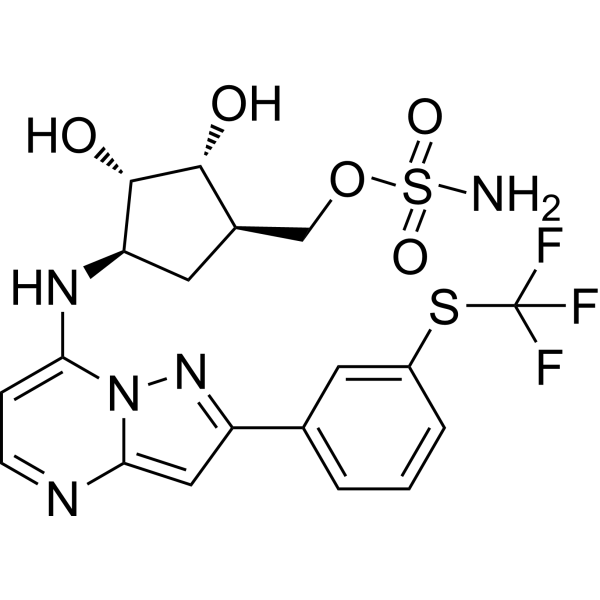
HY-100487
TAK-243UAE/E1 Inhibitor -
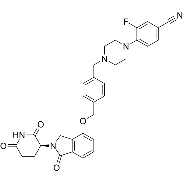
HY-129395
MezigdomideCereblon E3 Ubiquitin Ligase Modulating Agent -
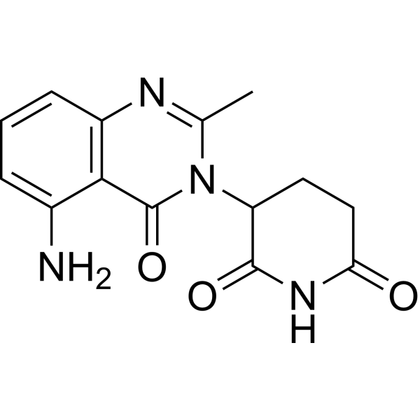
HY-100507
AvadomideCereblon Modulator -
HY-12025
SerdemetanAnticancer Aagent -
HY-15694
SMIP004Apoptosis Inducer -

HY-100507R
Avadomide (Standard)Cereblon Modulator -

HY-100487R
TAK-243 (Standard)UAE/E1 Inhibitor


Biologische Aktivität
Beschreibung
Cellular Effect
|
Cell Line

|
Type | Value | Description | References |
|---|---|---|---|---|
| A-375 | IC50 |
1.93 μM
Compound: 5a
|
Cytotoxicity against human A-375 cells assessed as reduction in cell viability incubated for 72 hrs by MTS assay
Cytotoxicity against human A-375 cells assessed as reduction in cell viability incubated for 72 hrs by MTS assay
|
[PMID: 36331508] |
| A549 | IC50 |
1.18 μM
Compound: 5a
|
Cytotoxicity against human A549 cells assessed as reduction in cell viability incubated for 72 hrs by MTS assay
Cytotoxicity against human A549 cells assessed as reduction in cell viability incubated for 72 hrs by MTS assay
|
[PMID: 36331508] |
| HCT-116 | IC50 |
0.36 μM
Compound: 5a
|
Cytotoxicity against human HCT-116 cells assessed as reduction in cell viability incubated for 72 hrs by MTS assay
Cytotoxicity against human HCT-116 cells assessed as reduction in cell viability incubated for 72 hrs by MTS assay
|
[PMID: 36331508] |
| HeLa | IC50 |
2.48 μM
Compound: 5a
|
Cytotoxicity against human HeLa cells assessed as reduction in cell viability incubated for 72 hrs by MTS assay
Cytotoxicity against human HeLa cells assessed as reduction in cell viability incubated for 72 hrs by MTS assay
|
[PMID: 36331508] |
| HepG2 | IC50 |
19.61 μM
Compound: 5a
|
Cytotoxicity against human HepG2 cells assessed as reduction in cell viability incubated for 72 hrs by MTS assay
Cytotoxicity against human HepG2 cells assessed as reduction in cell viability incubated for 72 hrs by MTS assay
|
[PMID: 36331508] |
| MCF-10A | IC50 |
6.98 μM
Compound: 5a
|
Cytotoxicity against human MCF-10A cells assessed as reduction in cell viability incubated for 72 hrs by MTS assay
Cytotoxicity against human MCF-10A cells assessed as reduction in cell viability incubated for 72 hrs by MTS assay
|
[PMID: 36331508] |
| MCF7 | IC50 |
2.79 μM
Compound: 5a
|
Cytotoxicity against human MCF7 cells assessed as reduction in cell viability incubated for 72 hrs by MTS assay
Cytotoxicity against human MCF7 cells assessed as reduction in cell viability incubated for 72 hrs by MTS assay
|
[PMID: 36331508] |
| MDA-MB-231 | IC50 |
1.17 μM
Compound: 5a
|
Cytotoxicity against human MDA-MB-231 cells assessed as reduction in cell viability incubated for 72 hrs by MTS assay
Cytotoxicity against human MDA-MB-231 cells assessed as reduction in cell viability incubated for 72 hrs by MTS assay
|
[PMID: 36331508] |
| MGC-803 | IC50 |
1.88 μM
Compound: 5a
|
Cytotoxicity against human MGC-803 cells assessed as reduction in cell viability incubated for 72 hrs by MTS assay
Cytotoxicity against human MGC-803 cells assessed as reduction in cell viability incubated for 72 hrs by MTS assay
|
[PMID: 36331508] |
| U2OS | IC50 |
3.64 μM
Compound: 5a
|
Cytotoxicity against human U2OS cells assessed as reduction in cell viability incubated for 72 hrs by MTS assay
Cytotoxicity against human U2OS cells assessed as reduction in cell viability incubated for 72 hrs by MTS assay
|
[PMID: 36331508] |
In Vitro
P62-RNF168 agonist-1 (20 μM, 72 h) has an IC50 of 0.36 μM for the cancer cell line HCT116 and an IC50 of 1.18 μM for A549. It shows cytotoxicity against cancer cells A357, HeLa, HepG2, MCF-7, MDA-MB-231, MGC-803, and H2OS, but does not induce cell apoptosis and has lower toxicity towards normal cells MCF-10A[1].
P62-RNF168 agonist-1 (1-5 μM; 24 h) affects the HR (homologous recombination)-mediated DSB repair in HCT-116 cells, contributing to its antitumor activity[1].
P62-RNF168 agonist-1 enhances the interaction between P62 and RNF168 in HCT-116 cells, with a Kd of 20.97 μM[1].
P62-RNF168 agonist-1 inhibits the catalytic activity of RNF168 and the E3 ligase activity of RNF168 in HCT-116 cells[1].
MedChemExpress (MCE) has not independently confirmed the accuracy of these methods. They are for reference only. Further protocols information, click here.
-
Cell Line:A549, HCT-116, A375, HeLa, HepG2, MCF-7, MDA-MB-231, MGC-803, U2OS and MCF-10A cells
-
Concentration:0-20 µM
-
Incubation Time:72 h
-
Result:Inhibited A549, HCT-116, A375, HeLa, HepG2, MCF-7, MDA-MB-231, MGC-803, U2OS and MCF-10A cells, with IC50 values of 1.18, 0.36, 1.93, 2.48, 19.61, 2.79, 1.17, 1.88, 3.64 and 6.98, respectively.
-
Cell Line:HCT-116 cells
-
Concentration:1, 2, 5 µM
-
Incubation Time:24 h
-
Result:Induced a G2/M arrest but no significant accumulation of sub-G1 phase.
Compromised both HR(homologous recombination)- and NHEJ (non-homologous end joining)- mediated DSB repair in a dose-dependent manner, but HR was the more severely impacted process.
-
Cell Line:HCT-116
-
Concentration:10 µM
-
Incubation Time:Over night
-
Result:Had the expression of RNF168 and GST-RNF168 ,increased the level of P62 in the endogenous RNF-168 immune complex.
-
Cell Line:HCT-116
-
Concentration:5, 10 µM
-
Incubation Time:24 h
-
Result:Made apoptosis less obvious.
In Vivo
P62-RNF168 agonist-1 (5-10 mg/kg, i.p., single every 3 days for 22 days) promotes tumor apoptosis and inhibits tumor growth in mice[1].
| Dose | T1/2 (h) | Cmax (ng/mL) | AUC0-t (h?ng/mL) | CL ((mL/h)/kg) |
| IP (5 mg/kg) | 16.17 | 26.10 | 215.69 | 19647.83 |
| IP (10 mg/kg) | 31.00 | 39.87 | 408.18 | 16554.30 |
| IP (20 mg/kg) | 22.10 | 52.23 | 1003.58 | 14669.81 |
MedChemExpress (MCE) has not independently confirmed the accuracy of these methods. They are for reference only.
-
Animal Model:Female BALB/c nude mice (xenograft tumor model)[1].
-
Dosage:5, 10 mg/kg
-
Administration:Intraperitoneal injection; single every 3 days for 22 days
-
Result:Suppressed tumor growth by promoting apoptosis in xenografted tumorigenesis.
-
Animal Model:Female BALB/c nude mice[1].
-
Dosage:5, 10, 20 mg/kg
-
Administration:Intraperitoneal injection; single
-
Result:Exhibited plasma concentration peaked 1 h after administration.
Showed a relatively high maximum concentration (Cmax= 52.23 ng/mL) and exposure (AUC0-t= 1003.58 h•ng/mL) at a dose of 20 mg/kg.
-
Animal Model:BALB/c mice, Female nude mice[1]
-
Dosage:25, 50, 75, 100 mg/kg, single dose; 12.5, 25, 50 mg/kg, single dose
-
Administration:Intraperitoneal injection
-
Result:Showed in the 100 mg/kg dosage group of BALB/c mice, out of 8 animals, 3 males and 3 females died on the 7th day, and the remaining animals experienced diarrhea. In the 75 mg/kg dosage group, 1 out of 8 animals (1 male) had diarrhea on the 7th day. There were no deaths or diarrhea observed in the ≤50 mg/kg dosage group.
Showed in the female nude mice, both the 25 and 50 mg/kg dosage groups experienced deaths or diarrhea, while the 12.5 mg/kg dosage group had no deaths or diarrhea.


Chemical Information
-
CAS. Nr. 2765180-17-2
-
Appearance Solid
-
Molecular Weight 393.40
-
Formel C19H19N7O3
-
Color Off-white to light yellow
-
SMILES
NC1=C2C(C=CC(C3=CN(C4=CC(OC)=C(C(OC)=C4)OC)N=N3)=C2)=NC(N)=N1
-
Versand
Room temperature in continental US; may vary elsewhere.
-
Speicherung
4°C, protect from light
* In solvent : -80°C, 6 months; -20°C, 1 month (protect from light)


Lösungsmittel & Löslichkeit
In Vitro:
DMSO : ≥ 60 mg/mL (152.52 mM; Hygroscopic DMSO has a significant impact on the solubility of product, please use newly opened DMSO)
* "≥" means soluble, but saturation unknown.
Please refer to the solubility information to select the appropriate solvent. Once prepared, please aliquot and store the solution to prevent product inactivation from repeated freeze-thaw cycles.
Storage method and period of stock solution: -80°C, 6 months; -20°C, 1 month (protect from light). When stored at -80°C, please use it within 6 months. When stored at -20°C, please use it within 1 month.
Please refer to the solubility information to select the appropriate solvent. Once prepared, please aliquot and store the solution to prevent product inactivation from repeated freeze-thaw cycles.
Storage method and period of stock solution: -80°C, 6 months; -20°C, 1 month (protect from light). When stored at -80°C, please use it within 6 months. When stored at -20°C, please use it within 1 month.
Konzentration (Stammlösung) × Volumen (Stammlösung) = Konzentration (Ziellösung) × Volumen (Ziellösung)
In Vivo:
Select the appropriate dissolution method based on your experimental animal and administration route.
- For the following dissolution methods, please ensure to first prepare a clear stock solution using an In Vitro approach and then sequentially add co-solvents:
- To ensure reliable experimental results, the clarified stock solution can be appropriately stored based on storage conditions. As for the working solution for In Vivo experiments, it is recommended to prepare freshly and use it on the same day.
- The percentages shown for the solvents indicate their volumetric ratio in the final prepared solution. If precipitation or phase separation occurs during preparation, heat and/or sonication can be used to aid dissolution.
Add each solvent one by one: 10% DMSO 40% PEG300 5% Tween-80 45% Saline
Solubility: ≥ 2.4 mg/mL (6.10 mM); Clear solution
This protocol yields a clear solution of ≥ 2.4 mg/mL (saturation unknown).
Taking 1 mL working solution as an example, add 100 μL DMSO stock solution (24.0 mg/mL) to 400 μL PEG300, and mix evenly; then add 50 μL Tween-80 and mix evenly; then add 450 μL Saline to adjust the volume to 1 mL.
Preparation of Saline: Dissolve 0.9 g sodium chloride in ddH₂O and dilute to 100 mL to obtain a clear Saline solution.
Add each solvent one by one: 10% DMSO 90% (20% SBE-β-CD in Saline)
Solubility: ≥ 2.4 mg/mL (6.10 mM); Clear solution
This protocol yields a clear solution of ≥ 2.4 mg/mL (saturation unknown).
Taking 1 mL working solution as an example, add 100 μL DMSO stock solution (24.0 mg/mL) to 900 μL 20% SBE-β-CD in Saline, and mix evenly.
Preparation of 20% SBE-β-CD in Saline (4°C, storage for one week): 2 g SBE-β-CD powder is dissolved in 10 mL Saline, completely dissolve until clear.
In Vivo Dissolution Calculator
Please enter the basic information of animal experiments:
-
-
-
-
Recommended: Prepare an additional quantity of animals to account for potential losses during experiments.
Please enter your animal formula composition:
-
%DMSO +
Recommended: Keep the proportion of DMSO in working solution below 2% if your animal is weak.
-
%+
-
+%Tween-80 + +
-
%Saline +
The co-solvents required include: DMSO, . All of co-solvents are available by MedChemExpress (MCE). , Tween 80. All of co-solvents are available by MedChemExpress (MCE).
Working solution concentration: 0.22 mg/mL
Method for preparing stock solution: mg drug dissolved in μL DMSO. Stock solution concentration: mg/mL. * In solvent : -80°C, 6 months; -20°C, 1 month (protect from light)
1. Take μL DMSO stock solution;
2. Add μL .
μL , mix evenly;
3. Then add μL Tween 80, mix evenly;
4. Then add μL
Please ensure that the stock solution in the first step is dissolved to a clear state, and add co-solvents in sequence. You can use ultrasonic heating (ultrasonic cleaner, recommended frequency 20-40 kHz), vortexing, etc. to assist dissolution.

Protokoll
-
CRISPR-Cas9 HDR knock-in/precise editing
CRISPR-Cas9 HDR knock-in uses a guide RNA to direct Cas9 to a genomic target adjacent to a PAM, where Cas9 creates a double-strand break; if a donor DNA template with homology to the cut region is present, cellular HDR can copy the donor sequence into the genome, producing a precise substitution, tag, reporter, or insertion rather than an indel. The readout is the fraction of alleles or cells carrying the intended donor-derived edit, measured by junction PCR, restriction-fragment analysis, Sanger sequencing, amplicon deep sequencing, flow cytometry for reporter knock-in, or clone genotyping; NHEJ indels and partial or non-HDR insertions are measured in parallel because they compete with or confound precise HDR outcomes.
-
Cell Cytotoxicity Assay
Cytotoxicity assays are usually based on the assessment of cell membrane damage, which can also be indirectly detected by measuring cell viability. Detection methods include MTT assay, CKK-8 assay, LDH assay and ATP assay, etc.
-
Mammalian live/dead viability and cytotoxicity staining
Live/dead viability and cytotoxicity staining assays are based on the simultaneous detection of intracellular esterase activity in metabolically active (viable) cells and membrane integrity loss in non-viable cells. In commonly used dual-staining approaches, membrane-permeant fluorogenic substrates are converted by intracellular esterases into fluorescent products in live cells, while impermeant DNA-binding dyes selectively enter cells with compromised plasma membranes and label nucleic acids in dead or dying cells, enabling discrimination between viable and non-viable populations by fluorescence microscopy or flow cytometry.
-
Autophagy
Autophagy is a process in which eukaryotic cells use lysosomes to degrade their own cytoplasmic proteins and damaged organelles under the regulation of autophagy related gene (Atg). Microtubule-associated proteins light chain 3 (LC3) is recognized as autophagy marker, which transfers from cytoplasmic LC3 (LC3-I) to membrane type (LC3-II). LC3-II/I ratio could be detected by Western Blot and fluorescence microscopy.
-
Patient-Derived Xenograft (PDX)
Patient-derived xenograft (PDX) models are generated by engrafting primary human tumor tissue directly into immunodeficient mice, allowing in vivo propagation of patient tumor biology without initial in vitro adaptation. These models are used to preserve key histopathological and molecular characteristics of the original tumor and enable assessment of tumor growth dynamics and therapeutic response in a living organism. The biological readout is tumor engraftment and subsequent growth in the murine host, which reflects the ability of human tumor cells to survive, vascularize, and expand in an immunocompromised microenvironment.
-
Splenic/Portal-Vein Liver Metastasis Xenograft
Splenic and portal-vein liver metastasis xenograft models deliver tumor cells into the portal circulation so that cells reach the liver first and form hepatic metastatic lesions; splenic injection uses the spleen as an access route to the portal system, while direct portal-vein injection introduces cells into the portal vein without requiring splenectomy. The assay detects liver colonization, intrahepatic tumor growth, tumor distribution, treatment response, survival, and liver-metastasis microenvironment changes; readouts include bioluminescence or fluorescence imaging, gross liver nodule counts, liver weight or tumor burden, histology, and survival.
-
Subcutaneous Cell-Line-Derived Xenograft
Subcutaneous cell-line-derived xenograft (CDX) models are established by implanting cultured human cancer cell lines into immunodeficient mice, where the injected cells form localized tumors that can be monitored in vivo as a measure of tumorigenic potential, growth kinetics, and treatment response. These models are widely used in oncology research because they allow reproducible tumor formation and enable comparative assessment of tumor growth between different cell lines or genetic manipulations in a controlled in vivo microenvironment. Subcutaneous implantation of cancer cells in immunodeficient mice is a standard approach for evaluating tumor growth behavior and therapeutic response across multiple cancer types, including prostate, esophageal, pancreatic, and colon cancer models.
-
Macroautophagy Solutions
Macroautophagy is a conserved lysosome-dependent degradation pathway in which cytoplasmic material is sequestered into double-membrane autophagosomes and delivered to lysosomes for degradation and recycling. The pathway supports cellular homeostasis during nutrient limitation, organelle stress, protein-aggregate accumulation, infection, differentiation, and tissue remodeling by coupling cargo sequestration, autophagosome maturation, lysosomal fusion, and degradation of cargo-derived macromolecules. The core molecular sequence includes initiation by nutrient- and stress-regulated autophagy machinery, autophagosome nucleation, LC3/ATG8-family conjugation to autophagosomal membranes, cargo selection through receptors such as SQSTM1/p62, autophagosome-lysosome fusion, and lysosomal degradation. LC3 was identified as a mammalian homolog of yeast Atg8 that localizes to autophagosomal membranes after processing, and p62/SQSTM1 was shown to connect ubiquitinated cargo with autophagic degradati
-
Orthotopic Cell-Line Xenograft
Orthotopic cell-line xenograft models involve implantation of human cancer cell lines into the anatomically corresponding organ of immunodeficient mice to reproduce tumor growth within a native microenvironment, enabling more clinically relevant tumor behavior compared with subcutaneous models. These models are widely used because orthotopic placement better recapitulates tumor progression, including invasion and metastatic spread, which are often underrepresented in heterotopic implantation systems. Compared with conventional xenografts, orthotopic implantation is described as more technically complex but provides improved simulation of tumor-microenvironment interactions and metastatic behavior, making it particularly valuable for translational oncology research. Surgical orthotopic implantation approaches have been emphasized as enabling faithful reproduction of clinical cancer features, including metastasis and disease progression patterns that align with the tumor’s organ of origi
-
Cell Viability Determination by MTT Colorimetric Assay
The following protocol uses the MTT colorimetric assay as a classic literature-established method for assessing cell viability/metabolic activity in cultured mammalian cells. MTT[3-(4,5-dimethylthiazol-2-yl)-2,5-diphenyltetrazolium bromide] is reduced by metabolically active cells to a colored formazan product; the amount of formazan is quantified spectrophotometrically and provides an indirect measure of metabolically active viable cells. Importantly, MTT reduction reflects cellular oxidoreductase/metabolic activity rather than an absolute direct count of living cells, so changes in cellular metabolism can alter the signal independently of cell number.
-
Intraperitoneal/Peritoneal Dissemination Xenograft
Intraperitoneal (IP) or peritoneal dissemination xenograft models are based on the introduction of human cancer cells into the peritoneal cavity of immunodeficient mice, where they attach to peritoneal surfaces, form multicellular aggregates or spheroids, and progressively generate disseminated tumor nodules that mimic advanced peritoneal metastatic disease. These models are widely used to study ovarian cancer progression, tumor-microenvironment interactions, and intraperitoneal therapeutic responses, often incorporating bioluminescence or fluorescence imaging to longitudinally monitor tumor burden in vivo. The biological principle relies on the capacity of tumor cells such as SKOV3 or related ovarian carcinoma lines to survive in suspension, aggregate within ascites-like fluid, adhere to mesothelial surfaces, and invade peritoneal organs, thereby recapitulating human peritoneal carcinomatosis patterns observed in advanced disease.


Reinheit & Dokumentation
-
Data Sheet (280 KB)
-
SDS (252 KB)
- English - EN (252 KB)
- Français - FR (252 KB)
- Deutsch - DE (252 KB)
- Norwegian - NO (252 KB)
- Español - ES (252 KB)
- Swedish - SV (252 KB)
- Italian - IT (252 KB)
- Korean - KR (252 KB)
- Portuguese - PT (252 KB)
-
Handling Instructions (2659 KB)
Verweise


Complete Stock Solution Preparation Table
Please refer to the solubility information to select the appropriate solvent. Once prepared, please aliquot and store the solution to prevent product inactivation from repeated freeze-thaw cycles.
Storage method and period of stock solution: -80°C, 6 months; -20°C, 1 month (protect from light). When stored at -80°C, please use it within 6 months. When stored at -20°C, please use it within 1 month.
| Optional Solvent | Concentration Solvent Mass | 1 mg | 5 mg | 10 mg | 25 mg |
|---|---|---|---|---|---|
| DMSO | 1 mM | 2.5419 mL | 12.7097 mL | 25.4194 mL | 63.5486 mL |
| 5 mM | 0.5084 mL | 2.5419 mL | 5.0839 mL | 12.7097 mL | |
| 10 mM | 0.2542 mL | 1.2710 mL | 2.5419 mL | 6.3549 mL | |
| 15 mM | 0.1695 mL | 0.8473 mL | 1.6946 mL | 4.2366 mL | |
| 20 mM | 0.1271 mL | 0.6355 mL | 1.2710 mL | 3.1774 mL | |
| 25 mM | 0.1017 mL | 0.5084 mL | 1.0168 mL | 2.5419 mL | |
| 30 mM | 0.0847 mL | 0.4237 mL | 0.8473 mL | 2.1183 mL | |
| 40 mM | 0.0635 mL | 0.3177 mL | 0.6355 mL | 1.5887 mL | |
| 50 mM | 0.0508 mL | 0.2542 mL | 0.5084 mL | 1.2710 mL | |
| 60 mM | 0.0424 mL | 0.2118 mL | 0.4237 mL | 1.0591 mL | |
| 80 mM | 0.0318 mL | 0.1589 mL | 0.3177 mL | 0.7944 mL | |
| 100 mM | 0.0254 mL | 0.1271 mL | 0.2542 mL | 0.6355 mL |


P62-RNF168 agonist-1 Related Classifications
HY-151799 Related Classifications
 Powered by Bioz