



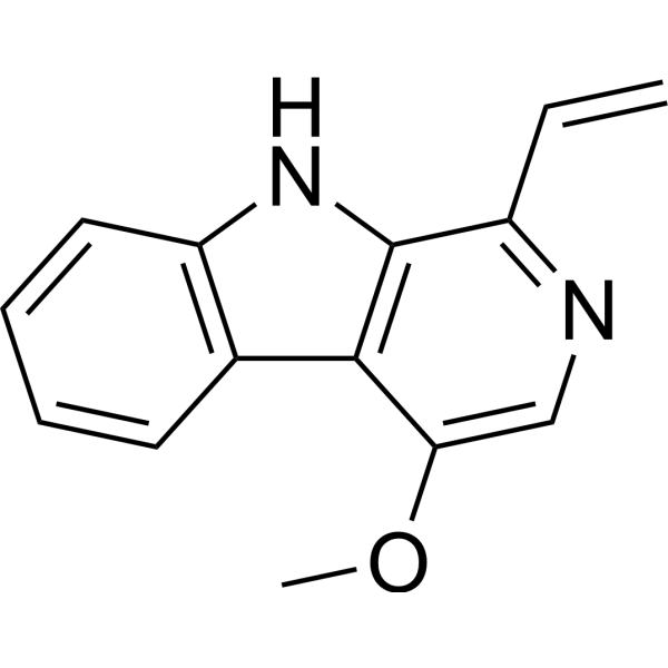
Dehydrocrenatine
Dehydrocrenatidine, a β-carboline alkaloid that can be isolated from Picrasma quassioides. Dehydrocrenatidine induces cell apoptosis by activates ERK and JNK. Dehydrocrenatidine inhibits invasion and migration of cancer cells, it also suppresses neuronal excitability to exert analgesic effects.
Nur für Forschungszwecke. Wir verkaufen nicht an Patienten.
- CAS. Nr.: 26585-13-7
- Formel: C14H12N2O
- Molecular Weight:224.26
-
Speicherung:
Please store the product under the recommended conditions in the Certificate of Analysis.

To place orders, for customer services and technical support, please contact: MedChemExpress USA
Tel: 609-228-6898 E-mail: [email protected] [email protected]


-
 Biologische Aktivität
-
Chemical Information
- Protokoll
- Reinheit & Dokumentation
- Verweise
-
 Help & FAQs




Biologische Aktivität
Beschreibung
Cellular Effect
|
Cell Line

|
Type | Value | Description | References |
|---|---|---|---|---|
| A2780 | IC50 |
4.77 μg/mL
Compound: 50
|
Antiproliferative activity against human A2780 cells
Antiproliferative activity against human A2780 cells
|
[PMID: 34332400] |
| SK-OV-3 | IC50 |
15.72 μg/mL
Compound: 50
|
Antiproliferative activity against human SK-OV-3 cells
Antiproliferative activity against human SK-OV-3 cells
|
[PMID: 34332400] |


Chemical Information
-
CAS. Nr. 26585-13-7
-
Molecular Weight 224.26
-
Formel C14H12N2O
-
SMILES
COC1=CN=C(C=C)C(N2)=C1C3=C2C=CC=C3
-
Structure Classification
-
Initial Source
-
Versand
Room temperature in continental US; may vary elsewhere.
-
Speicherung
Please store the product under the recommended conditions in the Certificate of Analysis.

Protokoll
-
Apoptosis
Apoptosis, also called programmed cell death, is generally characterized by distinct morphological characteristics.
-
TUNEL staining for apoptotic DNA fragmentation
TUNEL staining detects DNA strand breaks by using terminal deoxynucleotidyl transferase to add labeled nucleotides to exposed 3′-OH DNA termini, generating either microscopic staining in fixed cells or tissue sections, or fluorescence/cytometric signal in cell suspensions. TUNEL positivity reflects DNA fragmentation but should not be interpreted alone as definitive apoptosis, because TUNEL can also label necrotic, autolytic, mechanically damaged, or DNA-repair-associated DNA breaks.
-
Annexin V plus membrane-impermeant dye apoptosis staining
Annexin V-based apoptosis assays rely on the detection of phosphatidylserine (PS) externalization from the inner leaflet of the plasma membrane to the outer leaflet, an early biochemical hallmark of apoptosis. Fluorescently labeled Annexin V binds PS in a calcium-dependent manner, enabling identification of early apoptotic cells by flow cytometry or fluorescence microscopy. When combined with a membrane-impermeant DNA-binding dye (e. g. , propidium iodide), this approach allows discrimination between viable (Annexin V−/dye−), early apoptotic (Annexin V+/dye−), and late apoptotic or necrotic (Annexin V+/dye+) cell populations by assessing membrane integrity and PS exposure.
-
Cell migration
Cell migration is a method that plays an important role in wound healing, cell differentiation, embryonic development, etc.
-
Neuronal voltage-sensitive dye imaging
Neuronal voltage-sensitive dye imaging detects membrane-potential-dependent optical changes from dyes associated with neuronal membranes, enabling optical recording of electrical activity from single neurons, dendrites, axons, spines, or neuronal populations in brain slices and cultured neurons. VSD signals are typically reported as fractional fluorescence or absorbance changes over baseline, such as ΔF/F or ΔI/I, and published protocols use high-speed cameras or photodiode arrays because neuronal voltage signals occur on millisecond time scales. Fast VSD imaging can be applied at two common scales: bulk staining of brain slices to measure circuit-level spatiotemporal activity, and single-cell loading or biolistic delivery to record membrane-potential transients from individual neuronal compartments. Optical signals should be interpreted as membrane-potential-related readouts, and validation by simultaneous electrophysiology or pharmacological controls is recommended when the experimen
-
Cell-attached patch-clamp recording
Cell-attached patch-clamp recording measures ionic current through one or more ion channels in a small membrane patch that remains attached to an intact cell; the readout is a time-resolved current trace generated when channels in the sealed patch open and close under controlled pipette voltage or stimulus conditions. Classic applications include single acetylcholine receptor currents in frog skeletal muscle, single sodium-channel currents in cultured rat muscle cells, one-channel NMDA receptor recordings, and mechanically activated PIEZO-channel recordings. The method depends on forming a high-resistance pipette-membrane seal, commonly described as a gigaohm seal, which reduces leak and noise sufficiently to resolve picoampere-scale single-channel currents. In the cell-attached configuration, the patch membrane is not ruptured, so cytosolic composition is not directly dialyzed by the pipette solution.
-
Apoptosis Solutions
Apoptosis is a regulated, generally non-lytic cell-death pathway that removes unwanted, damaged, infected, or abnormal cells through coordinated morphological changes, caspase activation, DNA fragmentation, and membrane remodeling. The intrinsic apoptosis pathway is controlled mainly by mitochondrial outer membrane permeabilization, BCL-2 family proteins, cytochrome c release, apoptosome formation, caspase-9 activation, and downstream executioner caspase-3/7 activation. The extrinsic apoptosis pathway is initiated by death receptors such as Fas, TNFR, and TRAIL receptors, which recruit adaptor proteins and activate caspase-8 before engaging executioner caspases or mitochondrial amplification through BID cleavage. Apoptosis is linked to many phenotypes, including cancer cell killing, tissue homeostasis, immune regulation, neurodegeneration, infection response, and treatment-induced cytotoxicity; unresolved questions include how apoptosis interacts with necroptosis, pyroptosis, ferroptos
-
Cell invasion
Cell invasion is the ability of cells to migrate from one area to another via the extracellular matrix. Cell invasion is the response of normal and cancer cells to chemical and mechanical stimuli. Before migrating to a new region, the extracellular matrix is degraded by proteases within the cell. Cell invasion often occurs during wound repair, vascularization and inflammation, abnormal tissue invasion, and tumor cell metastasis.


Reinheit & Dokumentation
Verweise
[1]. Ho HY, et al. Apoptotic effects of dehydrocrenatidine via JNK and ERK pathway regulation in oral squamous cell carcinoma. Biomed Pharmacother. 2021 May;137:111362. [Content Brief]
[2]. Hsieh MC, et al. Dehydrocrenatidine extracted from Picrasma quassioidesinduces the apoptosis of nasopharyngeal carcinoma cells through the JNK and ERK signaling pathways. Oncol Rep. 2021 Aug;46(2):166. [Content Brief]

Calculators
Konzentration (Stammlösung) × Volumen (Stammlösung) = Konzentration (Ziellösung) × Volumen (Ziellösung)


Dehydrocrenatine Related Classifications
HY-N3711 Related Classifications
 Powered by Bioz