



Dipivefrin hydrochloride

Based on 1 Customer Validation
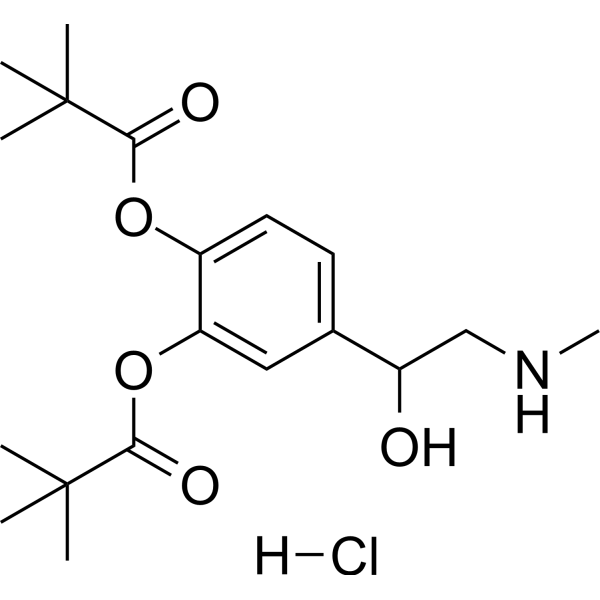
Dipivefrin hydrochloride is an α/β-adrenoceptor agonist and a prodrug of epinephrine. Dipivefrin hydrochloride regulates intraocular pressure by agonizing α/β-adrenoceptors and modulating the aqueous humor outflow pathway. Dipivefrin hydrochloride acts on neuroblastoma cells and downregulates glutamate-induced intracellular calcium levels. Dipivefrin hydrochloride is used in research related to α/β-adrenoceptor-mediated diseases, such as glaucoma.
For research use only. We do not sell to patients.
- Purity : 98.76%
- CAS No.: 64019-93-8
- Formula: C19H30ClNO5
- Molecular Weight:387.90
-
Storage:
-20°C, stored under nitrogen, away from moisture
* In solvent : -80°C, 6 months; -20°C, 1 month (stored under nitrogen, away from moisture)

To place orders, for customer services and technical support, please contact: MedChemExpress USA
Tel: 609-228-6898 E-mail: [email protected] [email protected]


-
 Biological Activity
-
Chemical Information
-
Solvent & Solubility
- Protocols
- Purity & Documentation
- References
-
 Help & FAQs




Biological Activity
Description
In Vitro
Dipivefrin hydrochloride (2.8-280 μM; 100 min) significantly inhibits the proliferation of cultured human corneal stromal cells only at a high concentration of 280 μM[3].
Dipivefrin hydrochloride (0.28-28 mM; 100 seconds) dose-dependently inhibits glutamate-induced elevation of [Ca2+]i and decreases basal [Ca2+]i in cultured N1E-115 neuroblastoma cells[4].
MedChemExpress (MCE) has not independently confirmed the accuracy of these methods. They are for reference only. Further protocols information, click here.
In Vivo
Dipivefrin hydrochloride (10 µg/kg; i.p.; before memory retrieval) administered before memory retrieval enhances the ability of alcohol-associated light cues to serve as conditioned reinforcers, indicating bidirectional modulation of alcohol memory reconsolidation through enhanced central adrenergic signaling[2].
MedChemExpress (MCE) has not independently confirmed the accuracy of these methods. They are for reference only.


Chemical Information
-
CAS No. 64019-93-8
-
Appearance Solid
-
Molecular Weight 387.90
-
Formula C19H30ClNO5
-
Color White to off-white
-
SMILES
CNCC(C1=CC=C(OC(C(C)(C)C)=O)C(OC(C(C)(C)C)=O)=C1)O.Cl
-
Synonyms
Dipivefrine hydrochloride
-
Shipping
Room temperature in continental US; may vary elsewhere.
-
Storage
-20°C, stored under nitrogen, away from moisture
* In solvent : -80°C, 6 months; -20°C, 1 month (stored under nitrogen, away from moisture)


Solvent & Solubility
In Vitro:
DMSO : 250 mg/mL (644.50 mM; Need ultrasonic; Hygroscopic DMSO has a significant impact on the solubility of product, please use newly opened DMSO)
H2O : ≥ 100 mg/mL (257.80 mM)
* "≥" means soluble, but saturation unknown.
Please refer to the solubility information to select the appropriate solvent. Once prepared, please aliquot and store the solution to prevent product inactivation from repeated freeze-thaw cycles.
Storage method and period of stock solution: -80°C, 6 months; -20°C, 1 month (stored under nitrogen, away from moisture). When stored at -80°C, please use it within 6 months. When stored at -20°C, please use it within 1 month.
* Note: If you choose water as the stock solution, please dilute it to the working solution, then filter and sterilize it with a 0.22 μm filter before use.
Please refer to the solubility information to select the appropriate solvent. Once prepared, please aliquot and store the solution to prevent product inactivation from repeated freeze-thaw cycles.
Storage method and period of stock solution: -80°C, 6 months; -20°C, 1 month (stored under nitrogen, away from moisture). When stored at -80°C, please use it within 6 months. When stored at -20°C, please use it within 1 month.
* Note: If you choose water as the stock solution, please dilute it to the working solution, then filter and sterilize it with a 0.22 μm filter before use.
Concentration (start) × Volume (start) = Concentration (final) × Volume (final)
In Vivo:
Select the appropriate dissolution method based on your experimental animal and administration route.
- For the following dissolution methods, please ensure to first prepare a clear stock solution using an In Vitro approach and then sequentially add co-solvents:
- To ensure reliable experimental results, the clarified stock solution can be appropriately stored based on storage conditions. As for the working solution for In Vivo experiments, it is recommended to prepare freshly and use it on the same day.
- The percentages shown for the solvents indicate their volumetric ratio in the final prepared solution. If precipitation or phase separation occurs during preparation, heat and/or sonication can be used to aid dissolution.
Add each solvent one by one: 10% DMSO 40% PEG300 5% Tween-80 45% Saline
Solubility: ≥ 2.08 mg/mL (5.36 mM); Clear solution
This protocol yields a clear solution of ≥ 2.08 mg/mL (saturation unknown).
Taking 1 mL working solution as an example, add 100 μL DMSO stock solution (20.8 mg/mL) to 400 μL PEG300, and mix evenly; then add 50 μL Tween-80 and mix evenly; then add 450 μL Saline to adjust the volume to 1 mL.
Preparation of Saline: Dissolve 0.9 g sodium chloride in ddH₂O and dilute to 100 mL to obtain a clear Saline solution.
Add each solvent one by one: 10% DMSO 90% (20% SBE-β-CD in Saline)
Solubility: ≥ 2.08 mg/mL (5.36 mM); Clear solution
This protocol yields a clear solution of ≥ 2.08 mg/mL (saturation unknown).
Taking 1 mL working solution as an example, add 100 μL DMSO stock solution (20.8 mg/mL) to 900 μL 20% SBE-β-CD in Saline, and mix evenly.
Preparation of 20% SBE-β-CD in Saline (4°C, storage for one week): 2 g SBE-β-CD powder is dissolved in 10 mL Saline, completely dissolve until clear.
In Vivo Dissolution Calculator
Please enter the basic information of animal experiments:
-
-
-
-
Recommended: Prepare an additional quantity of animals to account for potential losses during experiments.
Working solution concentration: 0.22 mg/mL
This product has good water solubility, please refer to the measured solubility data in water/PBS/Saline for details.

Protocols
-
Calcium Spark Assay
Calcium sparks are localized, transient increases in intracellular calcium concentration ([Ca2+]i) that occur in cardiac myocytes and represent elementary events underlying excitation-contraction coupling. These events are generated by the coordinated opening of clusters of ryanodine receptors (RyRs) on the sarcoplasmic reticulum membrane, leading to a brief release of Ca2+ into the cytosol. The detection and analysis of calcium sparks provide insights into the mechanisms of calcium handling and signaling in cardiac cells. Imaging techniques using fluorescent calcium indicators such as Fluo-3 are employed to visualize these subcellular calcium transients with high spatial and temporal resolution. The protocol is based on established methodologies described in primary literature for both experimental measurement and automated analysis of calcium sparks.
-
Ca2+ Staining Technique
Ca2+ staining is an experimental technique that utilizes specific fluorescent probes (such as Fluo-4 AM, Fura-2, etc.) to qualitatively or quantitatively detect dynamic changes in intracellular Ca2+ concentrations; this is achieved by monitoring the changes in fluorescent signals generated when these probes bind to free intracellular calcium ions. The underlying principle relies primarily on the presence of chelating groups within the probe's molecular structure that possess high affinity for calcium ions.


Purity & Documentation
-
Data Sheet (307 KB)
-
SDS (480 KB)
- English - EN (480 KB)
- Français - FR (480 KB)
- Deutsch - DE (480 KB)
- Norwegian - NO (480 KB)
- Español - ES (480 KB)
- Swedish - SV (480 KB)
- Italian - IT (480 KB)
- Korean - KR (480 KB)
- Portuguese - PT (480 KB)
-
Handling Instructions (2659 KB)
References


Complete Stock Solution Preparation Table
Please refer to the solubility information to select the appropriate solvent. Once prepared, please aliquot and store the solution to prevent product inactivation from repeated freeze-thaw cycles.
Storage method and period of stock solution: -80°C, 6 months; -20°C, 1 month (stored under nitrogen, away from moisture). When stored at -80°C, please use it within 6 months. When stored at -20°C, please use it within 1 month.
| Optional Solvent | Concentration Solvent Mass | 1 mg | 5 mg | 10 mg | 25 mg |
|---|---|---|---|---|---|
| H2O / DMSO | 1 mM | 2.5780 mL | 12.8899 mL | 25.7798 mL | 64.4496 mL |
| 5 mM | 0.5156 mL | 2.5780 mL | 5.1560 mL | 12.8899 mL | |
| 10 mM | 0.2578 mL | 1.2890 mL | 2.5780 mL | 6.4450 mL | |
| 15 mM | 0.1719 mL | 0.8593 mL | 1.7187 mL | 4.2966 mL | |
| 20 mM | 0.1289 mL | 0.6445 mL | 1.2890 mL | 3.2225 mL | |
| 25 mM | 0.1031 mL | 0.5156 mL | 1.0312 mL | 2.5780 mL | |
| 30 mM | 0.0859 mL | 0.4297 mL | 0.8593 mL | 2.1483 mL | |
| 40 mM | 0.0644 mL | 0.3222 mL | 0.6445 mL | 1.6112 mL | |
| 50 mM | 0.0516 mL | 0.2578 mL | 0.5156 mL | 1.2890 mL | |
| 60 mM | 0.0430 mL | 0.2148 mL | 0.4297 mL | 1.0742 mL | |
| 80 mM | 0.0322 mL | 0.1611 mL | 0.3222 mL | 0.8056 mL | |
| 100 mM | 0.0258 mL | 0.1289 mL | 0.2578 mL | 0.6445 mL |
* Note: If you choose water as the stock solution, please dilute it to the working solution, then filter and sterilize it with a 0.22 μm filter before use.


Dipivefrin hydrochloride Related Classifications
HY-B1323 Related Classifications
 Powered by Bioz
Keywords
- Dipivefrin
- 64019-93-8
- Dipivefrine
- Endogenous Metabolite
- Adrenergic Receptor
- ocular hypotension
- central adrenergic signaling
- intraocular pressure
- adrenergic prodrug
- N1E-115 neuroblastoma cells
- memory retrieval
- αβ-adrenoceptor agonist
- human corneal keratocytes
- conditioned reinforcer
- glaucoma
- Inhibitor
- inhibitor
- inhibit