



GSK-3β inhibitor 7
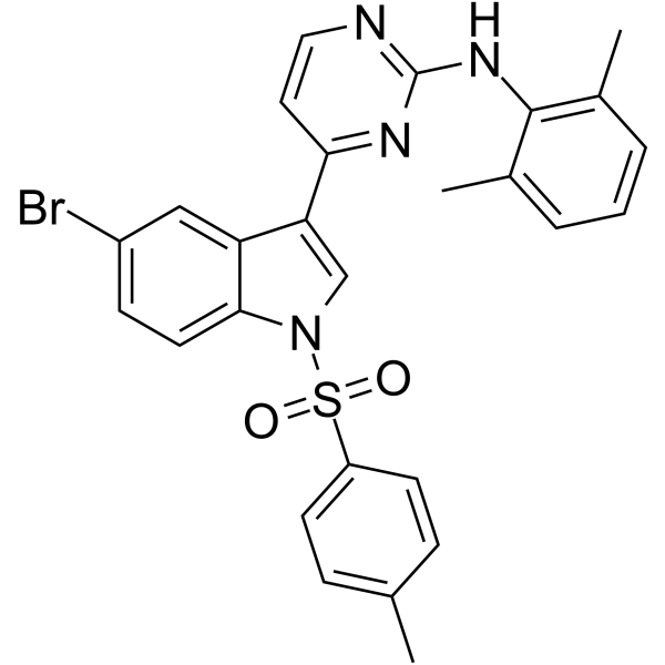
GSK-3β inhibitor 7 is a GSK-3β inhibitor with an IC50 value of 5.25 μM. GSK-3β inhibitor 7 is inserted into the ATP-binding binding pocket of GSK-3β and forms hydrogen-bond. GSK-3β inhibitor 7 shows high hepatocyte glucose uptake (83.5%), and can be used in the research of numerous diseases like diabetes, inflammation, cancer, Alzheimer's disease, and bipolar disorder.
For research use only. We do not sell to patients.
- Formula: C27H23BrN4O2S
- Molecular Weight:547.47
-
Storage:
Please store the product under the recommended conditions in the Certificate of Analysis.

To place orders, for customer services and technical support, please contact: MedChemExpress USA
Tel: 609-228-6898 E-mail: [email protected] [email protected]


-
 Biological Activity
-
Chemical Information
- Protocols
- Purity & Documentation
- References
-
 Help & FAQs




Biological Activity
Description
IC50 & Target
[1]|
GSK-3β 5.25 μM (IC50) |
In Vitro
GSK-3β inhibitor 7 (Compound 6x, 5 μΜ, 3 h) shows high glucose uptake (83.5%) in musle L6 cells[1].
GSK-3β inhibitor 7 (0-30 μΜ, 30 min) inhibits GSK-3β with an IC50 value of 5.25 μM[1].
MedChemExpress (MCE) has not independently confirmed the accuracy of these methods. They are for reference only. Further protocols information, click here.
In Vivo
GSK-3β inhibitor 7 (intragastric administration, 1 g/kg) displays no acute toxicity at a 1 g/kg dose in mice[1].
MedChemExpress (MCE) has not independently confirmed the accuracy of these methods. They are for reference only.
-
Animal Model:Sprague-Dawley rats (pharmacokinetic assay)[1]
-
Dosage:2 mg/kg, 20 mg/kg
-
Administration:Intravenous injection (2 mg/kg), oral administration (20 mg/kg)
-
Result:Pharmacokinetic profile of GSK-3β inhibitor 7 (Compound 6x).
F: oral bioavailability.Compound Route Dose (mg/kg) t1/2 (h) Tmax (h) Cmax (ng/mL) AUC0-t (hr•ng/mL) AUC0-∞ (hr•ng/mL) CL (mL/hr/kg) F (%) GSK-3β inhibitor 7 Oral administration 20 5.40 1.67 507 4265 4501 4536 47.4 GSK-3β inhibitor 7 Intravenous injection 2 8.95 0.08 519 859 948.6 2138.66
-
Animal Model:Male and female mice (acute assay)[1]
-
Dosage:1 g/kg
-
Administration:intragastric administration
-
Result:Increased body weights, caused no death or obvious weight loss.
Showed no marked pathological damage in important organs (brain, heart, liver, spleen, lung, and kidney).


Chemical Information
-
Molecular Weight 547.47
-
Formula C27H23BrN4O2S
-
SMILES
CC1=CC=C(C=C1)S(=O)(N2C=C(C3=CC(Br)=CC=C32)C4=NC(NC5=C(C=CC=C5C)C)=NC=C4)=O
-
Shipping
Room temperature in continental US; may vary elsewhere.
-
Storage
Please store the product under the recommended conditions in the Certificate of Analysis.

Protocols
-
Cell Cytotoxicity Assay
Cytotoxicity assays are usually based on the assessment of cell membrane damage, which can also be indirectly detected by measuring cell viability. Detection methods include MTT assay, CKK-8 assay, LDH assay and ATP assay, etc.
-
Research Protocol for Inflammation-related Diseases
The NLRP3 inflammasome is a cytosolic innate immune signaling platform that integrates priming signals and danger-signal activation to promote caspase-1 activation, maturation of IL-1β and IL-18, and gasdermin D-mediated pyroptotic cell death. The core experimental logic is to determine whether inflammatory disease phenotypes are driven by increased NLRP3 expression, ASC-containing inflammasome assembly, caspase-1 cleavage, GSDMD cleavage, and extracellular release of IL-1β/IL-18 rather than by nonspecific cell injury alone. The pathway is strongly linked to inflammation-related disease phenotypes because monosodium urate crystals activate NALP3/NLRP3 inflammasome signaling in gout-like crystal inflammation, cholesterol crystals activate NLRP3 inflammasomes in atherogenesis models, and DSS-induced intestinal inflammation has been reported to involve NLRP3 inflammasome activity. However, experimental colitis studies also show context-dependent protective effects of NLRP3 inflammasome co
-
Research Protocol for Metabolic Diseases
AMP-activated protein kinase, AMPK, is a conserved cellular energy sensor that responds to reduced cellular energy status and coordinates metabolism by increasing ATP-generating catabolic pathways while suppressing ATP-consuming anabolic processes. In metabolic disease research, the AMPK pathway is experimentally relevant because it regulates hepatic lipid synthesis, fatty acid oxidation, glucose production, skeletal-muscle glucose disposal, mTORC1-linked biosynthesis, autophagy, mitochondrial homeostasis, and whole-body energy balance. The central pathway logic is that energy stress, metformin, exercise-like stimulation, or direct AMPK activators increase AMPKα Thr172 phosphorylation and downstream substrate phosphorylation, including ACC and RAPTOR. Phosphorylation of ACC suppresses lipogenesis and supports fatty acid oxidation, whereas phosphorylation of RAPTOR suppresses mTORC1 signaling and links cellular energy status to growth and protein synthesis control. The pathway is linked
-
Alzheimer’s Disease Modeling
Alzheimer’s Disease (AD) is a neurodegenerative disorder characterized by a progressive decline in cognitive functions and loss of specific types of neurons and synapses. Alzheimer's symptoms can be simulated in mice by injecting drugs (such as Aβ) or genetically modified.


Purity & Documentation
References

Calculators
Concentration (start) × Volume (start) = Concentration (final) × Volume (final)


GSK-3β inhibitor 7 Related Classifications
HY-143261 Related Classifications
 Powered by Bioz