



RET-IN-20
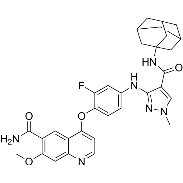
RET-IN-20 is a potent RET inhibitor with an IC50 value of 13.7 nM. RET-IN-20 decreases the expression of p-Ret, p-Shc protein. RET-IN-20 induces apoptosis. RET-IN-20 shows antiproliferative and anti-tumor activity.
연구목적의 판매만을 진행합니다. 환자를 대상으로 한 판매는 하지 않습니다.
- CAS No.: 3033547-06-4
- 화학식: C32H33FN6O4
- 분자량:584.64
-
보관:
Please store the product under the recommended conditions in the Certificate of Analysis.

To place orders, for customer services and technical support, please contact: MedChemExpress USA
Tel: 609-228-6898 E-mail: [email protected] [email protected]


-
 Biological Activity
-
Chemical Information
- Protocol
- 순도&문서
- References
-
 Help & FAQs




Biological Activity
제품 설명
IC50 & Target
IC50: 3.7 nM (RET)[1]
Cellular Effect
|
Cell Line

|
Type | Value | Description | References |
|---|---|---|---|---|
| BaF3 | IC50 |
1001 nM
Compound: 8r
|
Antiproliferative activity against mouse BaF3 cells expressing rearranged during transfection kinase G810C-CCDC6 incubated for 74 to 76 hrs by cell counting kit 8 method
Antiproliferative activity against mouse BaF3 cells expressing rearranged during transfection kinase G810C-CCDC6 incubated for 74 to 76 hrs by cell counting kit 8 method
|
[PMID: 36308779] |
| BaF3 | IC50 |
1360 nM
Compound: 8r
|
Antiproliferative activity against mouse BaF3 cells expressing rearranged during transfection kinase V804M-CCDC6 incubated for 74 to 76 hrs by cell counting kit 8 method
Antiproliferative activity against mouse BaF3 cells expressing rearranged during transfection kinase V804M-CCDC6 incubated for 74 to 76 hrs by cell counting kit 8 method
|
[PMID: 36308779] |
| BaF3 | IC50 |
1639 nM
Compound: 8r
|
Antiproliferative activity against mouse BaF3 cells expressing rearranged during transfection kinase G810R-CCDC6 incubated for 74 to 76 hrs by cell counting kit 8 method
Antiproliferative activity against mouse BaF3 cells expressing rearranged during transfection kinase G810R-CCDC6 incubated for 74 to 76 hrs by cell counting kit 8 method
|
[PMID: 36308779] |
| BaF3 | IC50 |
179 nM
Compound: 8r
|
Antiproliferative activity against mouse BaF3 cells expressing wildtype rearranged during transfection kinase-CCDC6 incubated for 74 to 76 hrs by cell counting kit 8 method
Antiproliferative activity against mouse BaF3 cells expressing wildtype rearranged during transfection kinase-CCDC6 incubated for 74 to 76 hrs by cell counting kit 8 method
|
[PMID: 36308779] |
| BaF3 | IC50 |
>10000 nM
Compound: 8r
|
Antiproliferative activity against mouse BaF3 cells incubated for 74 to 76 hrs by cell counting kit 8 method
Antiproliferative activity against mouse BaF3 cells incubated for 74 to 76 hrs by cell counting kit 8 method
|
[PMID: 36308779] |
In Vitro
RET-IN-20 (compound 8q) (0, 1.2, 3.7, 11.1, 33.3, 100 nM; 4 h) decreases the expression of p-Ret, p-Shc protein in a dose dependent manner[1].
RET-IN-20 (0, 33.3, 100, 300 nM; 48 h) induces apoptosis in a dose dependent manner[1].
MedChemExpress (MCE) has not independently confirmed the accuracy of these methods. They are for reference only.
-
Cell Line:Ba/F3 RETWT, Ba/F3 RETV804M, Ba/F3 RETG810C, Ba/F3 RETG810R, Ba/F3 parental cells
-
Concentration:0-10000 nM
-
Incubation Time:72 h
-
Result:Showed antiproliferative activities with IC50s of 5.6, 24.4, 15.4, 53.2, >10000 nM for Ba/F3 RETWT, Ba/F3 RETV804M, Ba/F3 RETG810C, Ba/F3 RETG810R, Ba/F3 parental, respectively.
-
Cell Line:CCDC6-RET-WT, CCDC6-RET-V804M, CCDC6-RET-G810C, CCDC6-RET-G810R cells
-
Concentration:0, 1.2, 3.7, 11.1, 33.3, 100 nM
-
Incubation Time:4 h
-
Result:Inhibited the expression of p-Ret, p-Shc protein in a dose dependent manner.
-
Cell Line:Ba/F3-CCDC6-RETG810 C/R cells
-
Concentration:0, 33.3, 100, 300 nM
-
Incubation Time:48 h
-
Result:Induced approximately 53.40% and 32.67% of cells undergoing apoptosis at a concentration of 300 nM in Ba/F3-CCDC6-RETG810C and Ba/F3-CCDC6-RETG810R cells, respectively.
In Vivo
MedChemExpress (MCE) has not independently confirmed the accuracy of these methods. They are for reference only.
-
Animal Model:CB17-SCID mice (Ba/F3-CCDC6-RETG810C tumor)[1]
-
Dosage:10, 30 mg/kg
-
Administration:I.p.; once a day for 13 continuous days
-
Result:Suppressed tumor growth in a dose-dependent manner with tumor growth inhibition values (TGI) of 17.1% and 66.9%, respectively.


Chemical Information
-
CAS No. 3033547-06-4
-
분자량 584.64
-
화학식 C32H33FN6O4
-
SMILES
COC1=C(C=C2C(N=CC=C2OC3=CC=C(C=C3F)NC4=NN(C=C4C(NC56CC7CC(C6)CC(C5)C7)=O)C)=C1)C(N)=O
-
선적
Room temperature in continental US; may vary elsewhere.
-
보관
Please store the product under the recommended conditions in the Certificate of Analysis.


순도&문서
References

Calculators
Concentration (start) × Volume (start) = Concentration (final) × Volume (final)


RET-IN-20 Related Classifications
HY-151987 Related Classifications
 Powered by Bioz