


RITA
Based on 3 publication(s) in Google Scholar
RITA is an inhibitor of p53-HDM-2 interaction, binds to p53dN, with a Kd of 1.5 nM, and also induces DNA-DNA cross-links.
For research use only. We do not sell to patients.
- Purity: 99.79%
- CAS No.: 213261-59-7
- Formula: C14H12O3S2
- Molecular Weight:292.37
-
Storage:Powder -20°C, 3 years , 4°C, 2 years ; In solvent -80°C, 2 years , -20°C, 1 year

To place orders, for customer services and technical support, please contact: MedChemExpress USA
Tel: 609-228-6898 E-mail: [email protected] [email protected]


-
 Biological Activity
-
Chemical Information
-
Solvent & Solubility
- Protocol
- Purity & Documentation
- References
-
 Help & FAQs
-


Apoptosis Compound Library
HY-L003
-
Cell Cycle/DNA Damage Compound Library
HY-L004
-
Anti-Cancer Compound Library
HY-L025
-
Clinical Compound Library
HY-L026
-
Autophagy Compound Library
HY-L029
-
Anti-Aging Compound Library
HY-L034
-
Drug Repurposing Compound Library
HY-L035
-
Ferroptosis Compound Library
HY-L051
-
Pyroptosis Compound Library
HY-L059
-
Glutamine Metabolism Compound Library
HY-L064
-
Anti-Lung Cancer Compound Library
HY-L075
-
Anti-Pancreatic Cancer Compound Library
HY-L077
-
Anti-Blood Cancer Compound Library
HY-L079
-
Anti-Cancer Metabolism Compound Library
HY-L083
-
Transcription Factor-Targeted Library
HY-L090
-
Anti-Colorectal Cancer Compound Library
HY-L103
-
Protein-protein Interaction Inhibitor Library
HY-L109
-
Cell Death Library
HY-L162
-
Anti-Hematopathy Compound Library
HY-L171
-
Anti-Ovarian Cancer Compound Library
HY-L173
-
Bioactive Compound Library Max
HY-L181
-
MCE Bioactive Compound Library
HY-L001V
-
Drug Repurposing Compound Library Plus
HY-L035P
-
Clinical Compound Library Plus
HY-L026P
-
Bioactive Compound Library
HY-L001
-
Cell Proliferation Compound Library
HY-L201
-
High-Throughput Bioactive Compound Library
HY-L205


Publications Citing Use of MedChemExpress (MCE) RITA
More Customer Validation & Images

-
WB


Biological Activity
|
Cell Line
|
Type | Value | Description | References |
|---|---|---|---|---|
| HCT-116 | GI50 |
0.11 μM
Compound: RITA
|
Antiproliferative activity against human HCT116 cells
Antiproliferative activity against human HCT116 cells
|
[PMID: 28435526] |
| HCT-116 | GI50 |
0.8 μM
Compound: RITA
|
Growth inhibition of human HCT116 cells after 24 hrs by CCK8 assay
Growth inhibition of human HCT116 cells after 24 hrs by CCK8 assay
|
[PMID: 28435526] |
RITA inhibits p53-HDM-2 interaction, binding to p53dN, with a Kd of 1.5 nM. RITA (10 μM) blocks complex formation between p53 and HDM-2 in HCT116 cells and HDFs and in NHF-ERMyc cells irrespective of c-Myc expression. RITA (0.5 μM) reduces the viability of tumor cells in a wild-type p53-dependent manner. Moreover, RITA (0.1 μM) induces p53-dependent apoptosis. RITA induces p53 but does not via DNA damage-signaling pathway[1]. RITA (NSC 652287) induces DNA-DNA cross-links. RITA induces G2-M cell cycle arrest at 10 nM and causes apoptosis at 100 nM. RITA (100 nM) also elevates p53 and causes dose-dependent effects on p21WAF1 protein levels[2]. RITA inhibits the growth of HeLa and CaSki cells, with IC50s of 1 and 10 μM. In addition, RITA (1 μM) stabilizes p53 by inhibiting p53/E6AP interaction[3].
MedChemExpress (MCE) has not independently confirmed the accuracy of these methods. They are for reference only.
MedChemExpress (MCE) has not independently confirmed the accuracy of these methods. They are for reference only.
| NCT Number | Sponsor | Condition | Start Date |
Phase
|
|---|---|---|---|---|
| NCT01329991 | Plexxikon| | 2011-05 | PHASE1 |


Chemical Information
-
CAS No. 213261-59-7
-
Appearance Solid
-
Molecular Weight 292.37
-
Formula C14H12O3S2
-
Color Brown to reddish brown
-
SMILES
OCC1=CC=C(C2=CC=C(C3=CC=C(CO)S3)O2)S1
-
Synonyms
NSC 652287
-
Shipping
Room temperature in continental US; may vary elsewhere.
-
Storage
Powder -20°C 3 years 4°C 2 years In solvent -80°C 2 years -20°C 1 year


Publications (3)
-
Journal Impact Factor
-
Most Recent
-
J Mol Med (Berl)
2019 Aug;97(8):1183-1193. PMID: 31201471 -
J Cell Mol Med
MircoRNA-1275 promotes proliferation, invasion and migration of glioma cells via SERPINE1. [Abstract]2018 Oct;22(10):4963-4974. PMID: 30024092
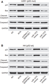
RITA purchased from MedChemExpress. Usage Cited in: J Cell Mol Med. 2018 Oct;22(10):4963-4974. [Abstract]
Western blot of H4 cells: expressions of p21, Bax and cleaved Caspase3 are up-regulated by miR-1275 mimics. RITA treatment and down-regulated by pcDNA3.1-SERPINE1 or miR-1275 inhibitor.
-
Virology
Live cell, image-based high-throughput screen to quantitate p53 stabilization and viability in human papillomavirus positive cancer cells. [Abstract]2021 Aug:560:96-109. PMID: 34051479


Solvent & Solubility
DMSO : ≥ 100 mg/mL (342.03 mM; Hygroscopic DMSO has a significant impact on the solubility of product, please use newly opened DMSO)
* "≥" means soluble, but saturation unknown.
Please refer to the solubility information to select the appropriate solvent. Once prepared, please aliquot and store the solution to prevent product inactivation from repeated freeze-thaw cycles.
Storage method and period of stock solution: -80°C, 2 years; -20°C, 1 year. When stored at -80°C, please use it within 2 years. When stored at -20°C, please use it within 1 year.
Concentration (start) × Volume (start) = Concentration (final) × Volume (final)
Select the appropriate dissolution method based on your experimental animal and administration route.
- For the following dissolution methods, please ensure to first prepare a clear stock solution using an In Vitro approach and then sequentially add co-solvents:
- To ensure reliable experimental results, the clarified stock solution can be appropriately stored based on storage conditions. As for the working solution for In Vivo experiments, it is recommended to prepare freshly and use it on the same day.
- The percentages shown for the solvents indicate their volumetric ratio in the final prepared solution. If precipitation or phase separation occurs during preparation, heat and/or sonication can be used to aid dissolution.
Add each solvent one by one: 10% DMSO 40% PEG300 5% Tween-80 45% Saline
Solubility: ≥ 2.5 mg/mL (8.55 mM); Clear solution
This protocol yields a clear solution of ≥ 2.5 mg/mL (saturation unknown).
Taking 1 mL working solution as an example, add 100 μL DMSO stock solution (25.0 mg/mL) to 400 μL PEG300, and mix evenly; then add 50 μL Tween-80 and mix evenly; then add 450 μL Saline to adjust the volume to 1 mL.
Preparation of Saline: Dissolve 0.9 g sodium chloride in ddH₂O and dilute to 100 mL to obtain a clear Saline solution.
Add each solvent one by one: 10% DMSO 90% (20% SBE-β-CD in Saline)
Solubility: ≥ 2.5 mg/mL (8.55 mM); Clear solution
This protocol yields a clear solution of ≥ 2.5 mg/mL (saturation unknown).
Taking 1 mL working solution as an example, add 100 μL DMSO stock solution (25.0 mg/mL) to 900 μL 20% SBE-β-CD in Saline, and mix evenly.
Preparation of 20% SBE-β-CD in Saline (4°C, storage for one week): 2 g SBE-β-CD powder is dissolved in 10 mL Saline, completely dissolve until clear.
Please enter the basic information of animal experiments:
-
-
-
-
Recommended: Prepare an additional quantity of animals to account for potential losses during experiments.
Please enter your animal formula composition:
-
%DMSO +
Recommended: Keep the proportion of DMSO in working solution below 2% if your animal is weak.
-
%+
-
+%Tween-80 + +
-
%Saline +
The co-solvents required include: DMSO, . All of co-solvents are available by MedChemExpress (MCE). , Tween 80. All of co-solvents are available by MedChemExpress (MCE).
Working solution concentration: 0.22 mg/mL
Method for preparing stock solution: mg drug dissolved in μL DMSO. Stock solution concentration: mg/mL.
1. Take μL DMSO stock solution;
2. Add μL .
μL , mix evenly;
3. Then add μL Tween 80, mix evenly;
4. Then add μL
Please ensure that the stock solution in the first step is dissolved to a clear state, and add co-solvents in sequence. You can use ultrasonic heating (ultrasonic cleaner, recommended frequency 20-40 kHz), vortexing, etc. to assist dissolution.

Protocol
For the cell viability assay, 3,000 cells per well are plated in a 96-well plate and treated with RITA for 48 h, after which cell viability is assessed with the proliferation reagent WST-1. For colony formation assay, cells are seeded in 12-well plates and treated with RITA for 24 h, after which the medium is replaced and the cells are allowed to grow for 10-14 d. The colonies are stained with crystal violet. For growth curves, 3000 cells/mL are plated in 12-well plates, treated with RITA, and counted over 5 d[3].
MedChemExpress (MCE) has not independently confirmed the accuracy of these methods. They are for reference only.
Mice[1]
Female SCID mice, 4-6 weeks old, are implanted with subcutaneous xenografts using 1 × 10⁶ cells in 90% Matrigel. Palpable tumors are established 3-6 d after the cells are injected, at which point RITA treatment is initiated. RITA is administered either 0.1, 1 or 10 mg/kg every day by intravenous or intraperitoneal injection in a total volume of 100 μL phosphate buffered saline. Xenografts are measured every 2 d. Tumor volumes are plotted for control and treated groups by dividing the average tumor volume for each data point by average starting tumor volume[1].
MedChemExpress (MCE) has not independently confirmed the accuracy of these methods. They are for reference only.


Purity & Documentation
-
Data Sheet (276 KB)
-
SDS (393 KB)
- English - EN (393 KB)
- Français - FR (393 KB)
- Deutsch - DE (393 KB)
- Norwegian - NO (393 KB)
- Español - ES (393 KB)
- Swedish - SV (393 KB)
- Italian - IT (393 KB)
- Korean - KR (393 KB)
- Portuguese - PT (393 KB)
-
Handling Instructions (2659 KB)
References
[1]. Issaeva N, et al. Small molecule RITA binds to p53, blocks p53-HDM-2 interaction and activates p53 function in tumors. Nat Med. 2004 Dec;10(12):1321-8. Epub 2004 Nov 21. [Content Brief]
[2]. Nieves-Neira W, et al. DNA protein cross-links produced by NSC 652287, a novel thiophene derivative active against human renal cancer cells. Mol Pharmacol. 1999 Sep;56(3):478-84. [Content Brief]
[3]. Zhao CY, et al. Rescue of p53 function by small-molecule RITA in cervical carcinoma by blocking E6-mediated degradation. Cancer Res. 2010 Apr 15;70(8):3372-81. [Content Brief]


Complete Stock Solution Preparation Table
Please refer to the solubility information to select the appropriate solvent. Once prepared, please aliquot and store the solution to prevent product inactivation from repeated freeze-thaw cycles.
Storage method and period of stock solution: -80°C, 2 years; -20°C, 1 year. When stored at -80°C, please use it within 2 years. When stored at -20°C, please use it within 1 year.
| Optional Solvent | Concentration Solvent Mass | 1 mg | 5 mg | 10 mg | 25 mg |
|---|---|---|---|---|---|
| DMSO | 1 mM | 3.4203 mL | 17.1016 mL | 34.2032 mL | 85.5081 mL |
| 5 mM | 0.6841 mL | 3.4203 mL | 6.8406 mL | 17.1016 mL | |
| 10 mM | 0.3420 mL | 1.7102 mL | 3.4203 mL | 8.5508 mL | |
| 15 mM | 0.2280 mL | 1.1401 mL | 2.2802 mL | 5.7005 mL | |
| 20 mM | 0.1710 mL | 0.8551 mL | 1.7102 mL | 4.2754 mL | |
| 25 mM | 0.1368 mL | 0.6841 mL | 1.3681 mL | 3.4203 mL | |
| 30 mM | 0.1140 mL | 0.5701 mL | 1.1401 mL | 2.8503 mL | |
| 40 mM | 0.0855 mL | 0.4275 mL | 0.8551 mL | 2.1377 mL | |
| 50 mM | 0.0684 mL | 0.3420 mL | 0.6841 mL | 1.7102 mL | |
| 60 mM | 0.0570 mL | 0.2850 mL | 0.5701 mL | 1.4251 mL | |
| 80 mM | 0.0428 mL | 0.2138 mL | 0.4275 mL | 1.0689 mL | |
| 100 mM | 0.0342 mL | 0.1710 mL | 0.3420 mL | 0.8551 mL |


 Powered by Bioz