



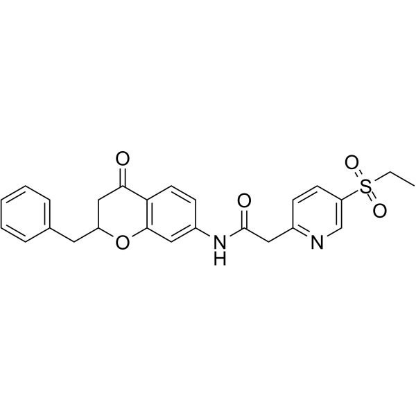
RORγt inverse agonist 29
RORγt inverse agonist 29 is a potent, orally active and selective RORγt inverse agonist (IC50: 21 nM). RORγt inverse agonist 29 can be used in the research of skin inflammation and autoimmune diseases like psoriasis.
Nos produits utilisent uniquement pour la recherche. Nous ne vendons pas aux patients.
- Formule: C25H24N2O5S
- Masse moléculaire:464.53
-
Stockage:
Please store the product under the recommended conditions in the Certificate of Analysis.

To place orders, for customer services and technical support, please contact: MedChemExpress USA
Tel: 609-228-6898 E-mail: [email protected] [email protected]


-
 Activité biologique
-
Chemical Information
- Protocole
- Pureté et documentation
- Références
-
 Help & FAQs




Activité biologique
Description
IC50 & Target
[1]|
RORγt 21 nM (IC50) |
In Vitro
RORγt inverse agonist 29 (compound b12) demonstrates high RORγt transcriptional inhibitory activity (IC50: 28 nM) in human Jurkat cells[1].
RORγt inverse agonist 29 (10 μM) shows good metabolic stabilities in in vitro human liver microsomes, with comparable half-life (T1/2: 4.46 h) and HLM (CLint(liver): 4.8 mL/min/kg)[1].
MedChemExpress (MCE) has not independently confirmed the accuracy of these methods. They are for reference only. Further protocols information, click here.
In Vivo
RORγt inverse agonist 29 (i.v., p.o., 0.3 or 1 mg/kg) displays an acceptable bioavailability and a half-life in rats[1].
MedChemExpress (MCE) has not independently confirmed the accuracy of these methods. They are for reference only.
-
Animal Model:Mouse Imiquimod-induced skin inflammation model[1]
-
Dosage:15 mg/kg, 50 mg/kg, 100 mg/kg
-
Administration:Oral administration, twice a day for 13 days.
-
Result:Inhibited IL-6 and IL-17A protein in the serum, with inhibition rate of 58.06% (IL-6) and 84.07% (IL-17A) at 100 mg/kg.
Reduced the histopathological symptoms on the back skin at dose of 100 mg/kg.
Alleviated symptoms including mononuclear and inflammatory cell infiltration, skin layer thickening, and dermal telangiectasia.
-
Animal Model:Rats (pharmacokinetic assay)[1]
-
Dosage:0.3 mg/kg (i.v.), 1 mg/kg (p.o.)
-
Administration:Intravenous injection, oral administration
-
Result:Pharmacokinetic profile of RORγt inverse agonist 29 (compound b12).
administration route T1/2 (h) Tmax (h) Cmax (ng/mL) AUC0-t (ng•h/mL) AUC0-∞ (ng•h/mL) F (%) i.v. (0.3 mg/kg) 5.8 2.4 88 156 162 p.o. (1 mg/kg) 6.5 3.1 105 340 352 65


Chemical Information
-
Masse moléculaire 464.53
-
Formule C25H24N2O5S
-
SMILES
CCS(=O)(C1=CN=C(C=C1)CC(NC2=CC3=C(C(CC(O3)CC4=CC=CC=C4)=O)C=C2)=O)=O
-
Livraison
Room temperature in continental US; may vary elsewhere.
-
Stockage
Please store the product under the recommended conditions in the Certificate of Analysis.

Protocole
-
Cell Cytotoxicity Assay
Cytotoxicity assays are usually based on the assessment of cell membrane damage, which can also be indirectly detected by measuring cell viability. Detection methods include MTT assay, CKK-8 assay, LDH assay and ATP assay, etc.
-
Imiquimod-Induced Psoriasiform Dermatitis
Imiquimod (IMQ)-induced psoriasiform dermatitis is a widely used murine model in which topical application of IMQ, a Toll-like receptor 7 (TLR7) agonist, triggers innate immune activation in the skin and induces a psoriasis-like inflammatory cascade characterized by epidermal hyperplasia, immune cell infiltration, and cytokine production dominated by the IL-23/IL-17 axis. This inflammatory response is mediated through activation of dendritic cells and downstream induction of IL-23, IL-17A, IL-22, and related pro-inflammatory mediators, recapitulating key features of human plaque psoriasis and enabling mechanistic and therapeutic studies. The model is commonly induced using Aldara (5% IMQ cream) applied topically to murine skin, resulting in rapid onset of erythema, scaling, and thickening that can be quantified as disease severity indices and validated histologically.
-
TPA/Croton Oil Ear Edema and Dermatitis
The TPA (12-O-tetradecanoylphorbol-13-acetate) and croton oil-induced mouse ear edema model is a well-established acute cutaneous inflammation system used to evaluate topical anti-inflammatory activity by measuring edema formation, neutrophil infiltration, vascular permeability, and cytokine-mediated skin responses in vivo. The inflammatory response is triggered by topical application of phorbol esters (TPA) or croton oil constituents, leading to rapid activation of protein kinase C signaling, leukocyte recruitment, and increased vascular permeability, which can be quantified by ear thickness, weight, dye extravasation, and biochemical markers such as myeloperoxidase (MPO) activity and pro-inflammatory mediators in ear tissue homogenates. This model is widely used for screening anti-inflammatory agents, where reductions in edema and inflammatory biomarkers reflect suppression of acute dermal inflammation and immune cell infiltration. Histological evaluation typically confirms epidermal
-
Contact Hypersensitivity Dermatitis
Contact hypersensitivity (CHS) dermatitis is a T cell-mediated delayed-type (Type IV) immune reaction in which low-molecular-weight haptens applied to the skin bind host proteins to form complete antigens, triggering sensitization followed by a secondary inflammatory response upon re-exposure (elicitation phase), which is commonly quantified by ear swelling as a readout of skin inflammation in murine models. This model is widely used to study allergic contact dermatitis because it is antigen-specific, reproducible, and reflects key immunological events including dendritic cell activation, T cell priming in draining lymph nodes, and effector T cell-driven tissue inflammation. DNFB- and oxazolone-induced CHS models are standard systems for evaluating both acute and chronic T cell-dependent skin inflammation and for testing immunomodulatory interventions.
-
Research Protocol for Inflammation-related Diseases
The NLRP3 inflammasome is a cytosolic innate immune signaling platform that integrates priming signals and danger-signal activation to promote caspase-1 activation, maturation of IL-1β and IL-18, and gasdermin D-mediated pyroptotic cell death. The core experimental logic is to determine whether inflammatory disease phenotypes are driven by increased NLRP3 expression, ASC-containing inflammasome assembly, caspase-1 cleavage, GSDMD cleavage, and extracellular release of IL-1β/IL-18 rather than by nonspecific cell injury alone. The pathway is strongly linked to inflammation-related disease phenotypes because monosodium urate crystals activate NALP3/NLRP3 inflammasome signaling in gout-like crystal inflammation, cholesterol crystals activate NLRP3 inflammasomes in atherogenesis models, and DSS-induced intestinal inflammation has been reported to involve NLRP3 inflammasome activity. However, experimental colitis studies also show context-dependent protective effects of NLRP3 inflammasome co


Pureté et documentation
Références

Calculators
Concentration (start) × Volume (start) = Concentration (final) × Volume (final)


RORγt inverse agonist 29 Related Classifications
HY-143271 Related Classifications
 Powered by Bioz