



RUNX1/ETO tetramerization-IN-1

Based on 1 Customer Validation
RUNX1/ETO tetramerization-IN-1 is a small-molecule inhibitor of RUNX1/ETO tetramerization, exhibits anti-leukemic effect. RUNX1/ETO tetramerization-IN-1 specifically targets to NHR2 of RUNX1/ETO (EC50=0.25 μM), restores gene expression down-regulated by RUNX1/ETO. RUNX1/ETO tetramerization-IN-1 inhibits the proliferation of RUNX1/ETO-depending SKNO-1 cells, and reduces the RUNX1/ETO-related tumor growth in a mouse model.
For research use only. We do not sell to patients.
- Purity : 99.36%
- CAS No.: 88755-39-9
- Formula: C18H14O7
- Molecular Weight:342.30
-
Storage:Powder -20°C, 3 years , 4°C, 2 years ; In solvent -80°C, 6 months , -20°C, 1 month

To place orders, for customer services and technical support, please contact: MedChemExpress USA
Tel: 609-228-6898 E-mail: [email protected] [email protected]


-
 Biological Activity
-
Chemical Information
-
Solvent & Solubility
- Protocols
- Purity & Documentation
- References
-
 Help & FAQs




Biological Activity
Description
IC50 & Target
IC50: 630 μM (RUNX1-NHR2 tetramerization)[1]
In Vitro
RUNX1/ETO is composed by the DNA-binding Runt-domain5, the product of the RUNX1 gene, and by four nervy homology regions (NHR1-4), the product of the ETO gene. The NHR2 domain is responsible for the tetramerization of RUNX1/ETO.
RUNX1/ETO tetramerization-IN-1 (compound 7.44) (1 μM and 10 μM; 3, 5, 7 d) selectively reduces the viability of RUNX1/ETO-dependent human leukemic SKNO-1 cells instead of U937 cells[1].
RUNX1/ETO tetramerization-IN-1 (compound 7.44) (25 μM and 50 μM; 5 d) inhibits the growth of and induces myeloid differentiation in RUNX1/ETO-expressing cells (SKNO-1, Kasumi-1, and K562)[2].
RUNX1/ETO tetramerization-IN-1 (100 μM; 7 d) induces growth-arrest and differentiation of RUNX1/ETOtr-expressing CD34+ progenitor cells[2].
RUNX1/ETO tetramerization-IN-1 (compound 7.44) has favorable physicochemical and ADME properties with high aqueous solubility, high stability in buffer and plasma, and a low hepatic intrinsic clearance in vitro, with the aqueous solubility of 60 μg/mL[3].
RUNX1/ETO tetramerization-IN-1 (1 μM and 10 μM) shows a potential to inhibit CYP2B6, 2C9, 2C19, and 3A4[3].
RUNX1/ETO tetramerization-IN-1 (compound 8) (50 μM; 16 h) inhibits c-Jun N-terminal kinase (JNK) and affect the JNK-pathway in cells[4].
MedChemExpress (MCE) has not independently confirmed the accuracy of these methods. They are for reference only. Further protocols information, click here.
-
Cell Line:RUNX1/ETO-dependent human leukemic SKNO-1 and U937 cells
-
Concentration:1 μM and 10 μM
-
Incubation Time:3, 5, 7 days
-
Result:Inhibited the SKNO-1 cell growth specifically.
-
Cell Line:Pharmacokinetic properties of RUNX1/ETO tetramerization-IN-1
-
Concentration:
-
Incubation Time:
-
Result:
Kinetic solubility (99% PBS, 1% DMSO) 177 µM Plasma protein binding (mouse plasma, 60 min) 98.4% Plasma stability (mouse plasma, 0–240 min) No degradation Hepatocyte stability (mouse hepatocytes) 2.5 µL/min/million cells Chemical stability in PBS (0–4 h) No degradation
In Vivo
MedChemExpress (MCE) has not independently confirmed the accuracy of these methods. They are for reference only.
-
Animal Model:NSG immunodeficient mice (NOD.Cg-Prkdcscid Il2rgtm1WjI/SzJ) injected with Kasumi-1 cells[2]
-
Dosage:200-250 μg/Kg
-
Administration:Intraperitoneal injection; 5 times per week, for 130 days
-
Result:Reduced the dissemination of leukemic cells, remained 75% mice alive at day 130 post-treatment.


Chemical Information
-
CAS No. 88755-39-9
-
Appearance Solid
-
Molecular Weight 342.30
-
Formula C18H14O7
-
Color White to off-white
-
SMILES
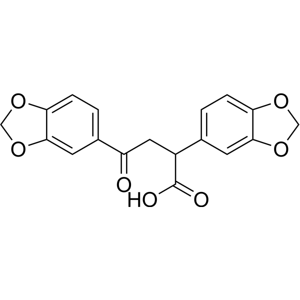
O=C(C(CC(C1=CC=C2OCOC2=C1)=O)C3=CC=C4OCOC4=C3)O
-
Shipping
Room temperature in continental US; may vary elsewhere.
-
Storage
Powder -20°C 3 years 4°C 2 years In solvent -80°C 6 months -20°C 1 month


Solvent & Solubility
In Vitro:
DMSO : 100 mg/mL (292.14 mM; Need ultrasonic; Hygroscopic DMSO has a significant impact on the solubility of product, please use newly opened DMSO)
Please refer to the solubility information to select the appropriate solvent. Once prepared, please aliquot and store the solution to prevent product inactivation from repeated freeze-thaw cycles.
Storage method and period of stock solution: -80°C, 6 months; -20°C, 1 month. When stored at -80°C, please use it within 6 months. When stored at -20°C, please use it within 1 month.
Please refer to the solubility information to select the appropriate solvent. Once prepared, please aliquot and store the solution to prevent product inactivation from repeated freeze-thaw cycles.
Storage method and period of stock solution: -80°C, 6 months; -20°C, 1 month. When stored at -80°C, please use it within 6 months. When stored at -20°C, please use it within 1 month.
Concentration (start) × Volume (start) = Concentration (final) × Volume (final)
In Vivo:
Select the appropriate dissolution method based on your experimental animal and administration route.
- For the following dissolution methods, please ensure to first prepare a clear stock solution using an In Vitro approach and then sequentially add co-solvents:
- To ensure reliable experimental results, the clarified stock solution can be appropriately stored based on storage conditions. As for the working solution for In Vivo experiments, it is recommended to prepare freshly and use it on the same day.
- The percentages shown for the solvents indicate their volumetric ratio in the final prepared solution. If precipitation or phase separation occurs during preparation, heat and/or sonication can be used to aid dissolution.
Add each solvent one by one: 10% DMSO 40% PEG300 5% Tween-80 45% Saline
Solubility: 2.5 mg/mL (7.30 mM); Clear solution; Need ultrasonic
This protocol yields a clear solution of 2.5 mg/mL.
Taking 1 mL working solution as an example, add 100 μL DMSO stock solution (25.0 mg/mL) to 400 μL PEG300, and mix evenly; then add 50 μL Tween-80 and mix evenly; then add 450 μL Saline to adjust the volume to 1 mL.
Preparation of Saline: Dissolve 0.9 g sodium chloride in ddH₂O and dilute to 100 mL to obtain a clear Saline solution.
Add each solvent one by one: 10% DMSO 90% (20% SBE-β-CD in Saline)
Solubility: 2.5 mg/mL (7.30 mM); Clear solution; Need ultrasonic
This protocol yields a clear solution of 2.5 mg/mL.
Taking 1 mL working solution as an example, add 100 μL DMSO stock solution (25.0 mg/mL) to 900 μL 20% SBE-β-CD in Saline, and mix evenly.
Preparation of 20% SBE-β-CD in Saline (4°C, storage for one week): 2 g SBE-β-CD powder is dissolved in 10 mL Saline, completely dissolve until clear.
In Vivo Dissolution Calculator
Please enter the basic information of animal experiments:
-
-
-
-
Recommended: Prepare an additional quantity of animals to account for potential losses during experiments.
Please enter your animal formula composition:
-
%DMSO +
Recommended: Keep the proportion of DMSO in working solution below 2% if your animal is weak.
-
%+
-
+%Tween-80 + +
-
%Saline +
The co-solvents required include: DMSO, . All of co-solvents are available by MedChemExpress (MCE). , Tween 80. All of co-solvents are available by MedChemExpress (MCE).
Working solution concentration: 0.22 mg/mL
Method for preparing stock solution: mg drug dissolved in μL DMSO. Stock solution concentration: mg/mL.
1. Take μL DMSO stock solution;
2. Add μL .
μL , mix evenly;
3. Then add μL Tween 80, mix evenly;
4. Then add μL
Please ensure that the stock solution in the first step is dissolved to a clear state, and add co-solvents in sequence. You can use ultrasonic heating (ultrasonic cleaner, recommended frequency 20-40 kHz), vortexing, etc. to assist dissolution.

Protocols
-
RT-PCR
Reverse transcription technology uses RNA as a template to synthesize DNA. RT-PCR is simple, specific and sensitive, and can be used to detect gene expression levels and expression differences in cells; detect RNA virus content; clone cDNA sequences of specific genes.
-
RNA extraction experimental
By lysing cells, releasing RNA, and removing impurities such as proteins and DNA, high-purity RNA products are finally obtained. The commonly used traditional method is the guanidine isothiocyanate/phenol/chloroform method (Trizol), which is suitable for a variety of animal materials including animal tissues, microorganisms, cultured cells, etc., and most plant materials.
-
Patient-Derived Xenograft (PDX)
Patient-derived xenograft (PDX) models are generated by engrafting primary human tumor tissue directly into immunodeficient mice, allowing in vivo propagation of patient tumor biology without initial in vitro adaptation. These models are used to preserve key histopathological and molecular characteristics of the original tumor and enable assessment of tumor growth dynamics and therapeutic response in a living organism. The biological readout is tumor engraftment and subsequent growth in the murine host, which reflects the ability of human tumor cells to survive, vascularize, and expand in an immunocompromised microenvironment.
-
Patient-Derived Orthotopic Xenograft (PDOX)
Patient-derived orthotopic xenograft (PDOX) modeling implants fresh patient tumor tissue or patient-derived tumor cells into the anatomically corresponding organ or tissue site of immunodeficient mice, usually by surgical orthotopic implantation, to preserve patient tumor histology, local microenvironmental context, invasion, metastatic behavior, and treatment-response features better than subcutaneous implantation. PDOX readouts include tumor engraftment, orthotopic tumor growth, local invasion, metastasis, recurrence after resection, histologic similarity to the donor tumor, biomarker retention, molecular concordance, survival, and response or resistance to therapy. PDOX models are used for preclinical drug testing and individualized therapy evaluation, but engraftment success varies by tumor type and specimen quality.
-
Subcutaneous Cell-Line-Derived Xenograft
Subcutaneous cell-line-derived xenograft (CDX) models are established by implanting cultured human cancer cell lines into immunodeficient mice, where the injected cells form localized tumors that can be monitored in vivo as a measure of tumorigenic potential, growth kinetics, and treatment response. These models are widely used in oncology research because they allow reproducible tumor formation and enable comparative assessment of tumor growth between different cell lines or genetic manipulations in a controlled in vivo microenvironment. Subcutaneous implantation of cancer cells in immunodeficient mice is a standard approach for evaluating tumor growth behavior and therapeutic response across multiple cancer types, including prostate, esophageal, pancreatic, and colon cancer models.
-
Real Time qPCR (Q-PCR)
Real-time quantitative PCR (qPCR) quantifies an amplifiable nucleic-acid target by monitoring fluorescence during PCR cycling rather than measuring product only after amplification. The increase in fluorescence tracks accumulation of PCR product, and the quantification cycle (Cq; historically also Ct/CP) is related to the initial amount of target: samples containing more starting target generally reach the defined fluorescence threshold in fewer cycles.
-
Orthotopic Cell-Line Xenograft
Orthotopic cell-line xenograft models involve implantation of human cancer cell lines into the anatomically corresponding organ of immunodeficient mice to reproduce tumor growth within a native microenvironment, enabling more clinically relevant tumor behavior compared with subcutaneous models. These models are widely used because orthotopic placement better recapitulates tumor progression, including invasion and metastatic spread, which are often underrepresented in heterotopic implantation systems. Compared with conventional xenografts, orthotopic implantation is described as more technically complex but provides improved simulation of tumor-microenvironment interactions and metastatic behavior, making it particularly valuable for translational oncology research. Surgical orthotopic implantation approaches have been emphasized as enabling faithful reproduction of clinical cancer features, including metastasis and disease progression patterns that align with the tumor’s organ of origi
-
Cell Viability Determination by MTT Colorimetric Assay
The following protocol uses the MTT colorimetric assay as a classic literature-established method for assessing cell viability/metabolic activity in cultured mammalian cells. MTT[3-(4,5-dimethylthiazol-2-yl)-2,5-diphenyltetrazolium bromide] is reduced by metabolically active cells to a colored formazan product; the amount of formazan is quantified spectrophotometrically and provides an indirect measure of metabolically active viable cells. Importantly, MTT reduction reflects cellular oxidoreductase/metabolic activity rather than an absolute direct count of living cells, so changes in cellular metabolism can alter the signal independently of cell number.
-
How to Choose the Right Model Animal
Choosing the right model animal is a validity-driven decision in which the species, strain, sex, age, genetic background, disease-induction method, outcome measures, and welfare burden must match the scientific question rather than laboratory tradition or convenience. A model should be selected by judging face validity, construct validity, and predictive validity: whether it resembles the human phenotype, whether it reproduces relevant mechanisms, and whether results are likely to predict human biology or treatment response. Animal studies often fail to translate because of species differences, weak disease resemblance, poor experimental design, inadequate reporting, publication bias, and underuse of randomization, blinding, and sample-size justification. Unresolved questions include how to rank competing models objectively, how much human-disease complexity must be reproduced for a given objective, and when non-animal systems such as organoids, ex vivo tissue, or computational models


Purity & Documentation
-
Data Sheet (280 KB)
-
SDS (251 KB)
- English - EN (251 KB)
- Français - FR (251 KB)
- Deutsch - DE (251 KB)
- Norwegian - NO (251 KB)
- Español - ES (251 KB)
- Swedish - SV (251 KB)
- Italian - IT (251 KB)
- Korean - KR (251 KB)
- Portuguese - PT (251 KB)
-
Handling Instructions (2659 KB)
References
[1]. Metz A, et al. From determinants of RUNX1/ETO tetramerization to small-molecule protein-protein interaction inhibitors targeting acute myeloid leukemia. J Chem Inf Model. 2013 Sep 23;53(9):2197-202. [Content Brief]
[2]. Schanda J, et al. Suppression of RUNX1/ETO oncogenic activity by a small molecule inhibitor of tetramerization. Haematologica. 2017 May;102(5):e170-e174. [Content Brief]
[3]. Gopalswamy M, et al. Biophysical and pharmacokinetic characterization of a small-molecule inhibitor of RUNX1/ETO tetramerization with anti-leukemic effects. Sci Rep. 2022 Aug 19;12(1):14158. [Content Brief]
[4]. Kaoud TS, et al. From in Silico Discovery to intra-Cellular Activity: Targeting JNK-Protein Interactions with Small Molecules. ACS Med Chem Lett. 2012 Aug 6;3(9):721-725. [Content Brief]


Complete Stock Solution Preparation Table
Please refer to the solubility information to select the appropriate solvent. Once prepared, please aliquot and store the solution to prevent product inactivation from repeated freeze-thaw cycles.
Storage method and period of stock solution: -80°C, 6 months; -20°C, 1 month. When stored at -80°C, please use it within 6 months. When stored at -20°C, please use it within 1 month.
| Optional Solvent | Concentration Solvent Mass | 1 mg | 5 mg | 10 mg | 25 mg |
|---|---|---|---|---|---|
| DMSO | 1 mM | 2.9214 mL | 14.6071 mL | 29.2141 mL | 73.0354 mL |
| 5 mM | 0.5843 mL | 2.9214 mL | 5.8428 mL | 14.6071 mL | |
| 10 mM | 0.2921 mL | 1.4607 mL | 2.9214 mL | 7.3035 mL | |
| 15 mM | 0.1948 mL | 0.9738 mL | 1.9476 mL | 4.8690 mL | |
| 20 mM | 0.1461 mL | 0.7304 mL | 1.4607 mL | 3.6518 mL | |
| 25 mM | 0.1169 mL | 0.5843 mL | 1.1686 mL | 2.9214 mL | |
| 30 mM | 0.0974 mL | 0.4869 mL | 0.9738 mL | 2.4345 mL | |
| 40 mM | 0.0730 mL | 0.3652 mL | 0.7304 mL | 1.8259 mL | |
| 50 mM | 0.0584 mL | 0.2921 mL | 0.5843 mL | 1.4607 mL | |
| 60 mM | 0.0487 mL | 0.2435 mL | 0.4869 mL | 1.2173 mL | |
| 80 mM | 0.0365 mL | 0.1826 mL | 0.3652 mL | 0.9129 mL | |
| 100 mM | 0.0292 mL | 0.1461 mL | 0.2921 mL | 0.7304 mL |


RUNX1/ETO tetramerization-IN-1 Related Classifications
HY-151411 Related Classifications
 Powered by Bioz