22 Results for "

SDS-PAGE

" in MedChemExpress (MCE) Product Catalog:
Products (22)

22 Results for "SDS-PAGE" in MCE Product Catalog:

4
4 Cited Publications
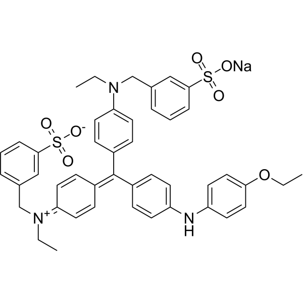
Cat. No.: HY-D0014
CAS No.: 6104-58-1
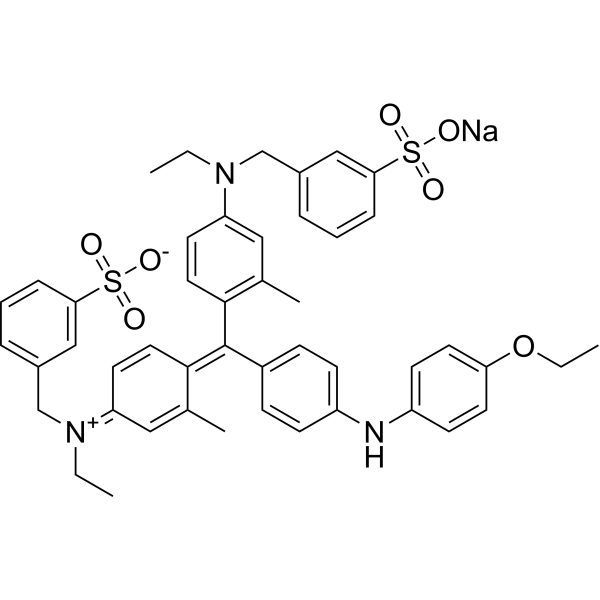
Brilliant Blue G-250 is a dye commonly used for the visualization of proteins separated by SDS-PAGE, offering a simple staining procedure and high quantitation. In the Bradford protein assay, protein concentrations are determined by the absorbance at 595 nm due to the binding of Brilliant Blue G-250 to proteins. Brilliant Blue G-250 is a safe highly selective P2×7R antagonist with promising consequent inactivation of NLRP3 inflammasome .
loading...
    loading...
4
4 Cited Publications
Cat. No.: HY-K1011A

MCE 3-Color Prestained Protein Marker (10-180 kDa) consists of 10 prestained recombinant proteins with an apparent molecular weight range of 10–180 kDa. This marker enables real-time monitoring of protein migration during SDS-PAGE and facilitates evaluation of protein transfer efficiency. The 250 μL is defined as the base specification. All larger sizes correspond to incremental volumes of this base.

loading...
    loading...
4
4 Cited Publications
Cat. No.: HY-K1011

MCE 3-Color Prestained Protein Marker (10-190 kDa) is a three-color protein standard with 10 prestained proteins ranging from 10 kDa to 190 kDa. The 250 μL is defined as the base specification. All larger sizes correspond to incremental volumes of this base.

loading...
    loading...
3
3 Cited Publications
Cat. No.: HY-K1100

SDS-PAGE Sample Loading Buffer (4×) is appropriate for loading protein samples on SDS-PAGE. The 1 mL is defined as the base specification. All larger sizes correspond to incremental volumes of this base.

loading...
    loading...
2
2 Cited Publications
Cat. No.: HY-K1030

MCE SDS dust-free Powder can be directly prepared into 10% SDS solution, and is widely used for protein denaturation.

loading...
    loading...
1
1 Cited Publications
Cat. No.: HY-D0232
CAS No.: 6104-59-2
Synonyms: Brilliant Blue R
Brilliant blue R250 (Brilliant Blue R), an anionic dye, is the most popular stain to detect proteins resolved in SDS-PAGE gels .
loading...
    loading...
1
1 Cited Publications
Cat. No.: HY-K1103

MCE Coomassie Blue Fast Staining Solution (No Heating, 10×) uses Coomassie Brilliant Blue G250 as the dye. It can be used for rapid, sensitive staining of protein gels in SDS-PAGE or Native-PAGE without contamination, or for detecting residual proteins on PAGE gels after Western Blot transfer. The 25 mL is defined as the base specification. All larger sizes correspond to incremental volumes of this base.

loading...
    loading...
Cat. No.: HY-NP002
CAS No.: 9048-46-8
BSA Standard Solution (5 mg/mL) is a bovine serum albumin solution commonly used as a standard for total serum protein detection. BSA Standard Solution (5 mg/mL) can be used for drawing standard curves and calibrating experiments in total protein assays. BSA Standard Solution (5 mg/mL) is also suitable for various biological experiments, including PCR detection inhibition, antibody desalting, chromatography control, SDS-PAGE electrophoresis, and calibration of UV spectrophotometers .
loading...
    loading...
Cat. No.: HY-D0006
CAS No.: 1662-01-7
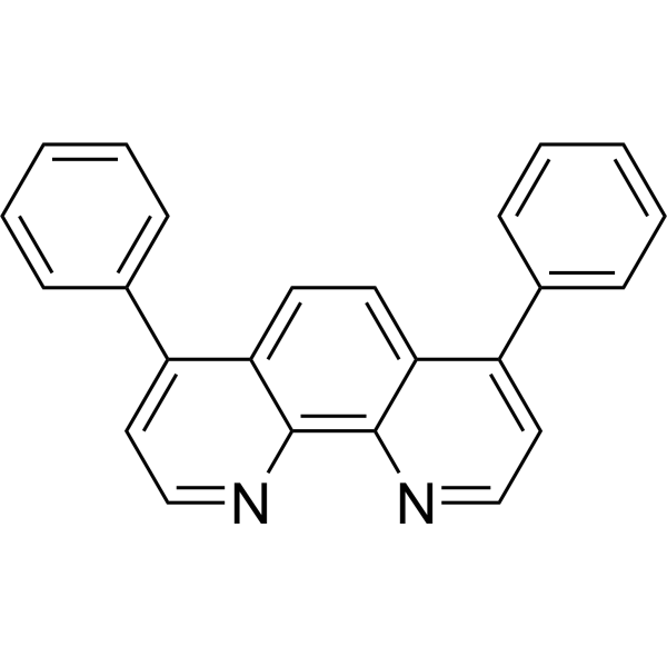
Bathophenanthroline is a nitrogen-containing organic ligand that can form stable complexes with transition metal ions such as Ru 2+. In proteomics, Bathophenanthroline is often used for in-gel protein staining. The negatively charged sulfonic acid groups of Bathophenanthroline interact electrostatically with the positively charged amino acid residues of proteins, thereby achieving specific binding to proteins and enhancing the fluorescent signal of detection. Bathophenanthroline can be used for protein separation and detection in SDS-PAGE gels in the field of proteomics, as well as other fluorescence studies[1]. Ex/Em=532 nm/610 nm[1].
loading...
    loading...
Cat. No.: HY-D1590
CAS No.: 929679-22-1
Target:  

Fluorescent Dye

Research Areas:  

Others

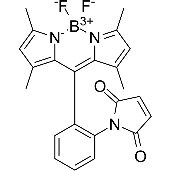
ODIPY Green 8-P2M is a novel thiol-reactive fluorescence probe based on the BODIPY fluorophore, the fluorescence is strongly quenched by d-PeT and then can be restored after reaction with thiol, resulting in an extremely high signal-to-noise ratio. ODIPY Green 8-P2M can be useful for detecting extremely low concentrations of protein in the gel after SDS-PAGE .
loading...
    loading...
Cat. No.: HY-P4279
CAS No.: 1370698-94-4
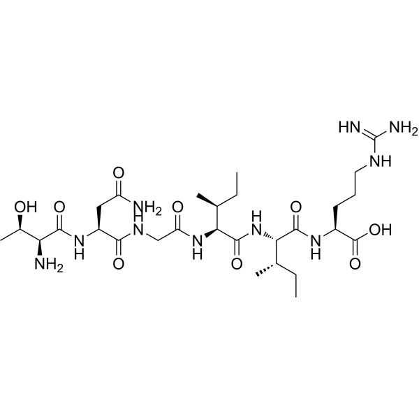
Ovalbumin (154-159) is a protein fragment consisting of residues 154-159 of chicken ovalbumin. Ovalbumin (154-159) is also a poorly conserved sequence (Thr-Asn-Gly-Ile-Ile-Arg) in the α-subunit of the Torpedo acetylcholine receptor, located within the α150-164 epitope region, and exhibits significant angiotensin-converting enzyme (ACE) inhibitory activity .
loading...
    loading...
Cat. No.: HY-179669I
1.5M Tris-HCl Buffer, pH 8.8 is a plant protein extraction buffer. 1.5M Tris-HCl Buffer, pH 8.8 can isolate cytoplasmic proteins from Marchantia, but the protein yield is low and the SDS-PAGE band profile is poor. 1.5M Tris-HCl Buffer, pH 8.8 can be used for proteomic studies on the liverwort Dumortiera hirsuta .
loading...
    loading...
Cat. No.: HY-NP0196
Synonyms: Mouse IgG
Mouse Immunoglobulin G (Mouse IgG) is the most abundant glycoprotein in mouse serum, which bridges innate and adaptive immune responses. A conserved asparagine-297 residue on each heavy chain contains an N-linked glycosylation site. Mouse Immunoglobulin G shares many glycosylation features with human IgG, but contains N-Glycolylneuraminic acid (HY-128965) instead of N-acetylneuraminic acid (HY-I0400). Mouse Immunoglobulin G is widely used as a negative isotype control, immunoassay coating protein and research standard in the life sciences .
loading...
    loading...
Cat. No.: HY-Y0700
CAS No.: 3737-95-9
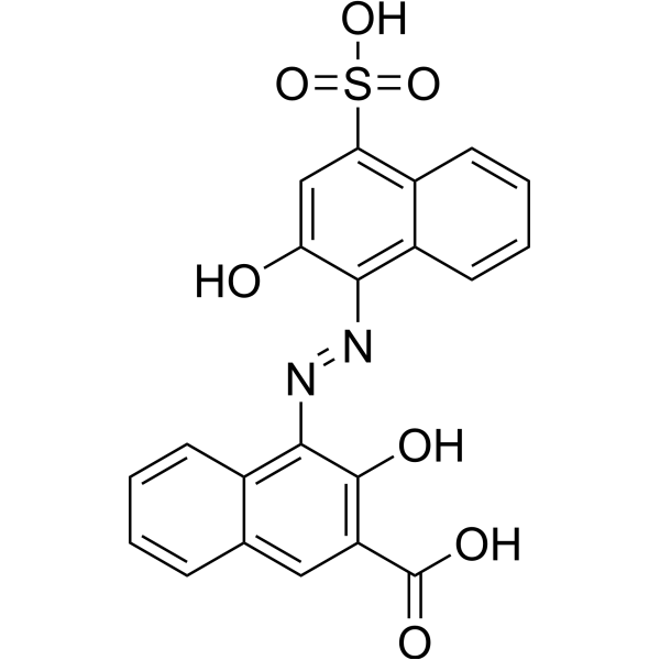
Calconcarboxylic acid, an azo dye, acts as a silver-ion sensitizer to stain protein in SDS-PAGE gels. Calconcarboxylic acid increases silver binding on protein bands or spots by the formation of a silver-dye complex and also increases the reducing power o
loading...
    loading...
Cat. No.: HY-179669H
Research Areas:  

Others

1M Tris-HCl Buffer, pH 8.8 is a buffer solution commonly used in various biochemical and molecular biological applications to maintain a stable pH environment. The pH value of Tris-HCl changes with temperature; generally, the pH decreases by 0.03 units for every degree Celsius increase in temperature. The pH values indicated on the product are the pH values of the Tris-HCl buffer solution at 25°C .
loading...
    loading...
Cat. No.: HY-P990329

Research Areas:  

Inflammation/Immunology

Anti-Albumin Antibody is a monoclonal IgG1 antibody targeting human serum albumin (HSA). Anti-Albumin Antibody binds to immobilized Human HSA-hFc, with an EC50 of 0.04661 μg/mL in ELISA assays using 2 μg/mL HSA-hFc. Anti-Albumin Antibody is applicable for human serum albumin detection and research related to antigen-antibody interactions. The corresponding isotype control is: Human IgG1 lambda2, Isotype Control (HY-P990096) .
loading...
    loading...
Cat. No.: HY-K1108

MCE SDS-PAGE Protein Loading Buffer (non-Reducing, 5×) is appropriate for loading protein samples on SDS-PAGE. The 1 mL is defined as the base specification. All larger sizes correspond to incremental volumes of this base.

loading...
    loading...
Cat. No.: HY-K1020

MCE Tris-Glycine-SDS Powder (1 L of 1×) consists of Tris, Glycine and SDS, and is widely used as an electrophoresis buffer for SDS-PAGE and Western Blot. The 10 pouches are defined as the base specification. All larger sizes correspond to incremental volumes of this base.

loading...
    loading...
Cat. No.: HY-D3348
Target:  

Fluorescent Dye

Research Areas:  

Others

Rhodamine picolyl azide is a fluorescent dye that enables visualization of protein labeling via click chemistry. The detection mechanism of Rhodamine picolyl azide is based on bioorthogonal click chemistry: Rhodamine picolyl azide reacts with the alkyne group on affinity-based protein profiling probes (ABPs) attached to labeled target proteins; this conjugation allows subsequent fluorescence imaging of proteins separated by SDS-PAGE .
loading...
    loading...
Cat. No.: HY-K1101

MCE 3-Color Prestained Protein Marker (2.7-40 kDa) is a three-color protein standard with 9 prestained proteins ranging from 2.7 kDa to 40 kDa. The 250 μL is defined as the base specification. All larger sizes correspond to incremental volumes of this base.

loading...
    loading...

