



Brilliant blue G-250
Based on 4 publication(s) in Google Scholar
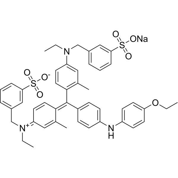
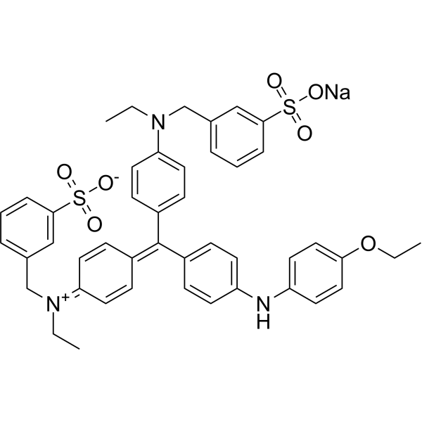
Brilliant Blue G-250 is a dye commonly used for the visualization of proteins separated by SDS-PAGE, offering a simple staining procedure and high quantitation. In the Bradford protein assay, protein concentrations are determined by the absorbance at 595 nm due to the binding of Brilliant Blue G-250 to proteins. Brilliant Blue G-250 is a safe highly selective P2×7R antagonist with promising consequent inactivation of NLRP3 inflammasome.
For research use only. We do not sell to patients.
- Purity : 92.05%
- CAS No.: 6104-58-1
- Formula: C47H48N3NaO7S2
- Molecular Weight:854.02
-
Storage:
4°C, sealed storage, away from moisture and light
* In solvent : -80°C, 6 months; -20°C, 1 month (sealed storage, away from moisture and light)

To place orders, for customer services and technical support, please contact: MedChemExpress USA
Tel: 609-228-6898 E-mail: [email protected] [email protected]


-
 Biological Activity
-
Chemical Information
-
Solvent & Solubility
- Purity & Documentation
- References
-
 Help & FAQs


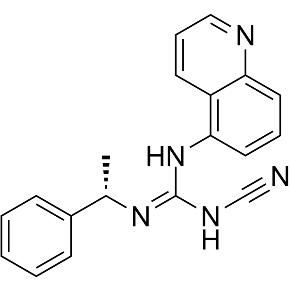
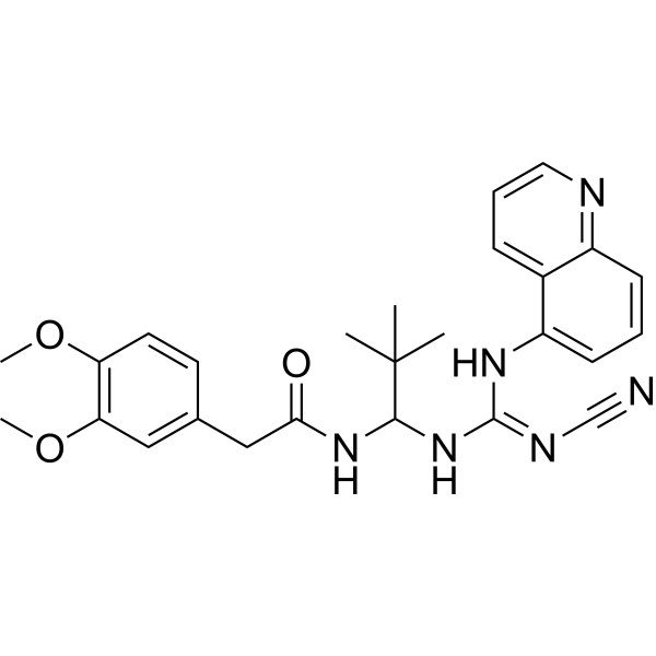
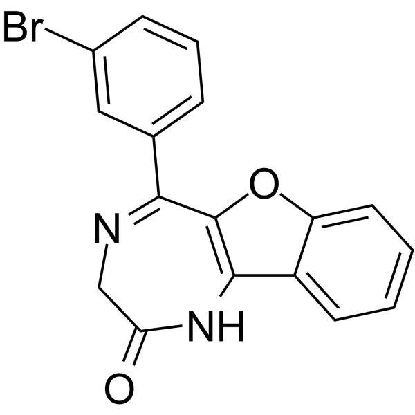
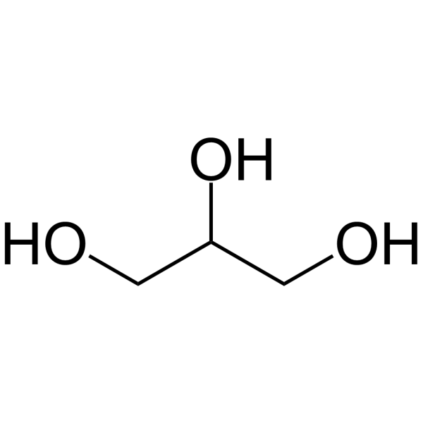
Publications Citing Use of MedChemExpress (MCE) Brilliant blue G-250
More

Biological Activity
Description
Cellular Effect
|
Cell Line

|
Type | Value | Description | References |
|---|---|---|---|---|
| HEK293 | IC50 |
1.37 μM
Compound: BBG
|
Antagonist activity at human P2X7R expressed in HEK293 cells assessed as inhibition of ATP-induced ethidium iodide uptake preincubated for 5 mins followed by ATP induction and measured after 25 mins by fluorescence assay
Antagonist activity at human P2X7R expressed in HEK293 cells assessed as inhibition of ATP-induced ethidium iodide uptake preincubated for 5 mins followed by ATP induction and measured after 25 mins by fluorescence assay
|
[PMID: 30528164] |
| Peritoneal macrophage | CC50 |
87.32 μM
Compound: BBG
|
Cytotoxicity against Swiss Webster mouse peritoneal macrophages by LDH release assay
Cytotoxicity against Swiss Webster mouse peritoneal macrophages by LDH release assay
|
[PMID: 29133043] |
| Peritoneal macrophage | IC50 |
19.77 μM
Compound: BBG
|
Antiinflammatory activity in LPS/IFNgamma-differentiated Swiss Webster mouse peritoneal macrophages assessed as inhibition of ATP-induced IL-1beta release preincubated for 15 mins followed by ATP/LPS challenge measured after 30 mins by sandwich ELISA
Antiinflammatory activity in LPS/IFNgamma-differentiated Swiss Webster mouse peritoneal macrophages assessed as inhibition of ATP-induced IL-1beta release preincubated for 15 mins followed by ATP/LPS challenge measured after 30 mins by sandwich ELISA
|
[PMID: 29133043] |
| THP-1 | IC50 |
19.27 μM
Compound: BBG
|
Antiinflammatory activity in LPS/IFNgamma-differentiated human THP1 cells assessed as inhibition of ATP-induced IL-1beta release after 30 mins by ELISA
Antiinflammatory activity in LPS/IFNgamma-differentiated human THP1 cells assessed as inhibition of ATP-induced IL-1beta release after 30 mins by ELISA
|
[PMID: 29133043] |
In Vitro
Staining Proteins in Polyacrylamide Gel Electrophoresis with Brilliant Blue G-250[1] 1. Solution Preparation Coomassie staining solution: (1) Dissolve 100 g aluminum sulfate (14-18 hydrate) in 2000 mL Milli-Q purified water. Add 200 mL ethanol (96%) and mix well. (2) Add 0.4 g Brilliant Blue G-250 and stir until dissolved. (3) Slowly add 47 mL phosphoric acid (85%) while stirring. (4) Finally, adjust the volume to 2000 mL with Milli-Q purified water. Note: Do not filter the solution. It should appear colloidal with suspended particles. Destaining solution: Prepare 2000 mL Milli-Q purified water, adding 200 mL ethanol (96%) and 47 mL phosphoric acid (85%). 2. Staining Procedure (1) After protein electrophoresis, carefully remove the gel from the glass plates. Wash the gel with Milli-Q purified water 3 times for 10 minutes each to remove SDS. Shake the Coomassie staining solution before use to disperse the colloidal particles. Immerse the gel in the staining solution and agitate on a shaker for 2-12 hours. (2) Protein spots will begin to appear after 10 minutes, and 80% of staining will be completed within 2 hours. For optimal results, overnight staining is recommended. (3) After staining, remove the Coomassie solution and rinse the gel twice with Milli-Q purified water. Place the gel in the destaining solution and agitate for 10-60 minutes to remove excess dye. 3. Rinsing and Storage Rinse the gel twice with Milli-Q purified water to restore its original thickness. The gel can be stored in a refrigerator with an acidic solution to prevent mold contamination. 4. Notes (1) Ensure the gel is thoroughly washed before staining, as residual SDS will interfere with dye-protein binding. (2) The staining solution can be reused as long as particles remain. (3) Store the staining solution in a dark bottle to extend its shelf life.
MedChemExpress (MCE) has not independently confirmed the accuracy of these methods. They are for reference only.


Chemical Information
-
CAS No. 6104-58-1
-
Appearance Solid
-
Molecular Weight 854.02
-
Formula C47H48N3NaO7S2
-
Color Dark purple to black
-
SMILES
O=S(C1=CC(CN(C2=CC(C)=C(/C(C3=CC=C(NC4=CC=C(OCC)C=C4)C=C3)=C5C(C)=C/C(C=C/5)=[N+](CC)/CC6=CC=CC(S(=O)([O-])=O)=C6)C=C2)CC)=CC=C1)(O[Na])=O
-
Shipping
Room temperature in continental US; may vary elsewhere.
-
Storage
4°C, sealed storage, away from moisture and light
* In solvent : -80°C, 6 months; -20°C, 1 month (sealed storage, away from moisture and light)


Publications (4)
-
Journal Impact Factor
-
Most Recent
-
Adv Sci (Weinh)
SRSF5-Mediated Alternative Splicing of M Gene is Essential for Influenza A Virus Replication: A Host-Directed Target Against Influenza Virus. [Abstract]2022 Dec;9(34):e2203088. PMID: 36257906 -
J Neuroinflammation
P2X7 receptor antagonists modulate experimental autoimmune neuritis via regulation of NLRP3 inflammasome activation and Th17 and Th1 cell differentiation. [Abstract]2024 Mar 25;21(1):73. PMID: 38528529 -
Molecules
Enhancing Functional Properties and Protein Structure of Almond Protein Isolate Using High-Power Ultrasound Treatment. [Abstract]2024 Jul 30;29(15):3590. PMID: 39124994 -


Solvent & Solubility
In Vitro:
DMSO : 12.5 mg/mL (14.64 mM; ultrasonic and warming and heat to 60°C; Hygroscopic DMSO has a significant impact on the solubility of product, please use newly opened DMSO)
H2O : 10 mg/mL (11.71 mM; Need ultrasonic)
Please refer to the solubility information to select the appropriate solvent. Once prepared, please aliquot and store the solution to prevent product inactivation from repeated freeze-thaw cycles.
Storage method and period of stock solution: -80°C, 6 months; -20°C, 1 month (sealed storage, away from moisture and light). When stored at -80°C, please use it within 6 months. When stored at -20°C, please use it within 1 month.
* Note: If you choose water as the stock solution, please dilute it to the working solution, then filter and sterilize it with a 0.22 μm filter before use.
Please refer to the solubility information to select the appropriate solvent. Once prepared, please aliquot and store the solution to prevent product inactivation from repeated freeze-thaw cycles.
Storage method and period of stock solution: -80°C, 6 months; -20°C, 1 month (sealed storage, away from moisture and light). When stored at -80°C, please use it within 6 months. When stored at -20°C, please use it within 1 month.
* Note: If you choose water as the stock solution, please dilute it to the working solution, then filter and sterilize it with a 0.22 μm filter before use.
Concentration (start) × Volume (start) = Concentration (final) × Volume (final)
In Vivo:
Select the appropriate dissolution method based on your experimental animal and administration route.
- For the following dissolution methods, please ensure to first prepare a clear stock solution using an In Vitro approach and then sequentially add co-solvents:
- To ensure reliable experimental results, the clarified stock solution can be appropriately stored based on storage conditions. As for the working solution for In Vivo experiments, it is recommended to prepare freshly and use it on the same day.
- The percentages shown for the solvents indicate their volumetric ratio in the final prepared solution. If precipitation or phase separation occurs during preparation, heat and/or sonication can be used to aid dissolution.
Add each solvent one by one: 10% DMSO 40% PEG300 5% Tween-80 45% Saline
Solubility: ≥ 0.56 mg/mL (0.66 mM); Clear solution
This protocol yields a clear solution of ≥ 0.56 mg/mL (saturation unknown).
Taking 1 mL working solution as an example, add 100 μL DMSO stock solution (5.6 mg/mL) to 400 μL PEG300, and mix evenly; then add 50 μL Tween-80 and mix evenly; then add 450 μL Saline to adjust the volume to 1 mL.
Preparation of Saline: Dissolve 0.9 g sodium chloride in ddH₂O and dilute to 100 mL to obtain a clear Saline solution.
Add each solvent one by one: 10% DMSO 90% (20% SBE-β-CD in Saline)
Solubility: ≥ 0.56 mg/mL (0.66 mM); Clear solution
This protocol yields a clear solution of ≥ 0.56 mg/mL (saturation unknown).
Taking 1 mL working solution as an example, add 100 μL DMSO stock solution (5.6 mg/mL) to 900 μL 20% SBE-β-CD in Saline, and mix evenly.
Preparation of 20% SBE-β-CD in Saline (4°C, storage for one week): 2 g SBE-β-CD powder is dissolved in 10 mL Saline, completely dissolve until clear.
In Vivo Dissolution Calculator
Please enter the basic information of animal experiments:
-
-
-
-
Recommended: Prepare an additional quantity of animals to account for potential losses during experiments.
Please enter your animal formula composition:
-
%DMSO +
Recommended: Keep the proportion of DMSO in working solution below 2% if your animal is weak.
-
%+
-
+%Tween-80 + +
-
%Saline +
The co-solvents required include: DMSO, . All of co-solvents are available by MedChemExpress (MCE). , Tween 80. All of co-solvents are available by MedChemExpress (MCE).
Working solution concentration: 0.22 mg/mL
Method for preparing stock solution: mg drug dissolved in μL DMSO. Stock solution concentration: mg/mL. * In solvent : -80°C, 6 months; -20°C, 1 month (sealed storage, away from moisture and light)
1. Take μL DMSO stock solution;
2. Add μL .
μL , mix evenly;
3. Then add μL Tween 80, mix evenly;
4. Then add μL
Please ensure that the stock solution in the first step is dissolved to a clear state, and add co-solvents in sequence. You can use ultrasonic heating (ultrasonic cleaner, recommended frequency 20-40 kHz), vortexing, etc. to assist dissolution.


Purity & Documentation
-
Data Sheet (281 KB)
-
SDS (393 KB)
- English - EN (393 KB)
- Français - FR (393 KB)
- Deutsch - DE (393 KB)
- Norwegian - NO (393 KB)
- Español - ES (393 KB)
- Swedish - SV (393 KB)
- Italian - IT (393 KB)
- Korean - KR (393 KB)
- Portuguese - PT (393 KB)
-
Handling Instructions (2659 KB)
References
[1]. Dyballa N, Metzger S. Fast and sensitive colloidal coomassie G-250 staining for proteins in polyacrylamide gels. J Vis Exp. 2009;(30):1431. Published 2009 Aug 3. [Content Brief]
[2]. Han XX, et al. Highly sensitive protein concentration assay over a wide range via surface-enhanced Raman scattering of Coomassie brilliant blue. Anal Chem. 2010;82(11):4325-4328. [Content Brief]
[3]. Zohny MH, et al. Coomassie brilliant blue G-250 dye attenuates bleomycin-induced lung fibrosis by regulating the NF-κB and NLRP3 crosstalk: A novel approach for filling an unmet medical need [published online ahead of print, 2022 Feb 21]. Biomed Pharmacother. 2022;148:112723. [Content Brief]


Complete Stock Solution Preparation Table
Please refer to the solubility information to select the appropriate solvent. Once prepared, please aliquot and store the solution to prevent product inactivation from repeated freeze-thaw cycles.
Storage method and period of stock solution: -80°C, 6 months; -20°C, 1 month (sealed storage, away from moisture and light). When stored at -80°C, please use it within 6 months. When stored at -20°C, please use it within 1 month.
| Optional Solvent | Concentration Solvent Mass | 1 mg | 5 mg | 10 mg | 25 mg |
|---|---|---|---|---|---|
| H2O / DMSO | 1 mM | 1.1709 mL | 5.8547 mL | 11.7093 mL | 29.2733 mL |
| 5 mM | 0.2342 mL | 1.1709 mL | 2.3419 mL | 5.8547 mL | |
| 10 mM | 0.1171 mL | 0.5855 mL | 1.1709 mL | 2.9273 mL |
* Note: If you choose water as the stock solution, please dilute it to the working solution, then filter and sterilize it with a 0.22 μm filter before use.


Brilliant blue G-250 Related Classifications
HY-D0014 Related Classifications
 Powered by Bioz