11 Results for "

charge-transfer state

" in MedChemExpress (MCE) Product Catalog:
Products (11)

11 Results for "charge-transfer state" in MCE Product Catalog:

1
1 Cited Publications
Cat. No.: HY-W127832
CAS No.: 123148-15-2
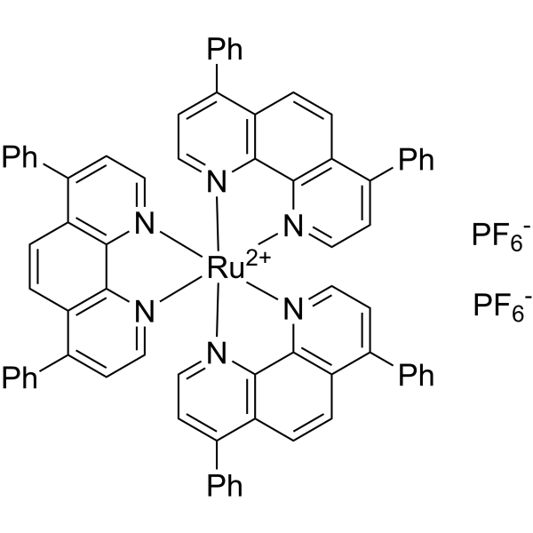
Ru(dpp)3(PF6)2 is a ruthenium (II) polypyridine complex and oxygen-sensing luminophore with excellent photostability and bright orange emission properties based on metal-to-ligand charge transfer (MLCT). Ru(dpp)3(PF6)2 undergoes oxygen-induced collisional luminescence quenching following the Stern-Volmer relationship, and can also engage in Förster-type resonance energy transfer with closed-form BTF6. Featuring excellent photophysical and electrochemical properties, Ru(dpp)3(PF6)2 is applied in studies on quenching-type oxygen sensors based on fluorinated ORMOSIL/xerogels and solid-state light-emitting devices .
loading...
    loading...
Cat. No.: HY-I0259
CAS No.: 118-46-7
8-Amino-2-naphthol is a photoactive charge transfer compounds, which can be used as fluorescent probe. 8-Amino-2-naphthol undergoes excited-state proton transfer (ESPT) to form a zwitterion under acidic conditions, where the photoacidity of its hydroxyl group is regulated by the protonation state of the amino group, enabling pH to act as an on/off switch for photoacidity. 8-Amino-2-naphthol is also utilized as chiral organocatalyst .
loading...
    loading...
Cat. No.: HY-W034043
CAS No.: 1335047-34-1
[Ir(dF(Me)ppy)2(dtbbpy)]PF6 is a blue-green luminescent material. [Ir(dF(Me)ppy)2(dtbbpy)]PF6 exhibits irregular emission, with its emission peak maxima located at 498, 512, and 519 nM, respectively. [Ir(dF(Me)ppy)2(dtbbpy)]PF6 can serve as an electrochemiluminescence-based biological probe .
loading...
    loading...
Cat. No.: HY-149203
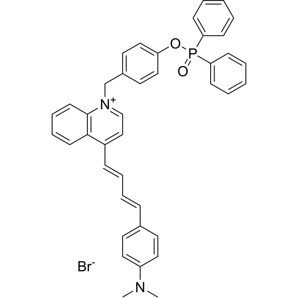
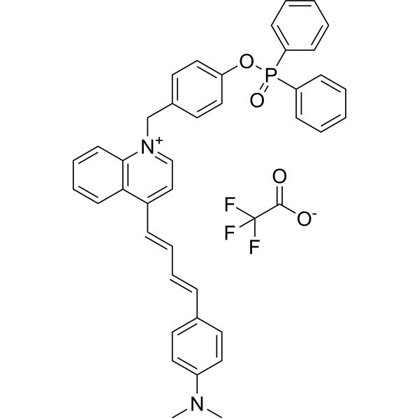
MQA-P is a multifunctional near-infrared (NIR) fluorescent probe for simultaneously detecting ONOO -, viscosity, and polarity within mitochondria. MQA-P exhibits a remarkable turn-on response to ONOO -em=645 nm) and is highly sensitive to viscosity/polarity in the NIR channel with λem>704 nm. MQA-P exhibits excited-state intramolecular charge transfer (ESICT) feature that is highly polarity-sensitive by engineering N,N-dimethylamino as the electron donor and a quinoline cationic unit as the electron acceptor. MQA-P is used for ferroptosis or cancer diagnosis in vitro and in vivo via dual-channel images .
loading...
    loading...
Cat. No.: HY-149203A
Purity:  97.36%
Target:  

Fluorescent Dye

Research Areas:  

Others

MQA-P is a multifunctional near-infrared (NIR) fluorescent probe that simultaneously detects ONOO -, viscosity, and polarity within mitochondria. MQA-P exhibits significant response to ONOO -, λem=645 nm; and NIR channel at λem>704 nm Medium is highly sensitive to viscosity/polarity. MQA-P possesses excited-state intramolecular charge transfer (ESICT) properties that are highly sensitive to polarity by designing the N,N-dimethylamino group as the electron donor and the quinoline cation unit as the electron acceptor. MQA-P is used for ferroptosis or cancer diagnosis in vitro and in vivo via dual-channel images .
loading...
    loading...
Cat. No.: HY-D3092
CAS No.: 2757682-01-0
Target:  

Fluorescent Dye

Research Areas:  

Others

NRERf is an endoplasmic reticulum (ER)-targeted solvatochromic fluorescent probe based on Nile Red (HY-D0718). It possesses ER-targeting capability and can sense changes in local polarity and lipid order of biological membranes. NRERf undergoes excited-state charge transfer, with its emission color changing according to local polarity: the tightly packed, low-polarity liquid-ordered phase (Lo) induces a blue-shifted emission, while the high-polarity liquid-disordered phase (Ld) induces a red-shifted emission. The emission wavelength of NRERf varies with the environment. NRERf can be used to detect changes in lipid order and polarity induced in the ER by cholesterol extraction/enrichment, oxidative stress, and hyperosmotic shock .
loading...
    loading...
Cat. No.: HY-D3391
RMR-Tre is a fluorescent probe targeting the mycobacterial acyltransferase Ag85. Under the catalysis of Ag85, RMR-Tre undergoes 6-position mycoloylation and anchors to the mycobacterial membrane, while achieving fluorescence activation by inhibiting the intramolecular twisted charge transfer state transition. RMR-Tre can distinguish live mycobacteria from dead ones through metabolism-driven labeling, enabling rapid, wash-free, low-background detection of viable bacteria. RMR-Tre reports the drug resistance of Mycobacterium tuberculosis via the trehalose catalytic shift activity readout associated with TreS. In addition, RMR-Tre can be combined with flow cytometry or high-content imaging techniques to visualize and quantitatively analyze the metabolic heterogeneity of Mycobacterium tuberculosis related to persistence and drug resistance. RMR-Tre is widely used in tuberculosis-related research .
loading...
    loading...
Cat. No.: HY-D3090
CAS No.: 2757682-04-3
Target:  

Fluorescent Dye

Research Areas:  

Others

NRLD is a lipid droplet-targeting solvatochromic fluorescent probe developed based on Nile Red (HY-D0718), which can target lipid droplets and sense the non-polar lipid microenvironment inside lipid droplets. NRLD exhibits superior lipid droplet-targeting selectivity compared to the parent Nile Red dye in HeLa cell imaging. NRLD achieves detection relying on the solvatochromic effect generated by excited-state charge transfer, and its emission wavelength shifts according to the local polarity changes of the microenvironment it locates in: the more compact and ordered the lipid packing and the lower the environmental polarity, the more blue-shifted the emission; the looser the lipid packing or the stronger the hydration and the higher the environmental polarity, the more red-shifted the emission. NRLD shows blue shift in the non-polar oil core of lipid droplets and red shift in high-polarity environments such as phosphate buffer .
loading...
    loading...
Cat. No.: HY-D3108
CAS No.: 2569032-08-0
BV-1 is a fluorescent probe for detecting mitochondrial viscosity in living cells. The detection mechanism of BV-1 relies on a twisted intramolecular charge transfer (TICT) mechanism associated with environmental viscosity: in low-viscosity media, the unsaturated domain at the meso-position of the BODIPY fluorophore undergoes TICT, achieving efficient excited-state deactivation through molecular rotation, thereby quenching fluorescence; under high-viscosity conditions, such molecular rotation is restricted, TICT is inhibited, and the conjugated system of the probe extends, which leads to significant fluorescence enhancement accompanied by a red shift in emission wavelength. BV-1 exhibits low cytotoxicity. Its detection wavelengths include excitation wavelengths of 480 nm or 488 nm, with an emission wavelength of 545 nm in low-viscosity environments and 570 nm in high-viscosity environments; in addition, its absorption peak locates at 528 nm in ethanol and red-shifts to 531 nm in glycerol .
loading...
    loading...
Cat. No.: HY-D3127
CAS No.: 455251-97-5
Target:  

Fluorescent Dye

Research Areas:  

Others

DCA-Mln is a Fluorescent probe for ferric iron (Fe³⁺) detection. The probe works via an internal charge transfer (ICT) process from its diethylamino group to its dicyano group, which produces bright pink fluorescence in its unbound state; when Fe³⁺ is present, two molecules of DCA-Mln chelate with one Fe³⁺ ion, with one cyano group of each DCA-Mln participating in the complexation, and this binding triggers a photoinduced electron transfer (PET) process that provides a nonradiative deactivation pathway, resulting in fluorescence quenching; additionally, the probe exhibits a visible color change from purple to peach pink upon Fe³⁺ binding, enabling dual-channel detection. The probe has an excitation wavelength of 570 nm and an emission wavelength of 670 nm, and it shows a rapid response, with fluorescence stabilizing within 15 seconds of Fe³⁺ addition. DCA-Mln is cell-membrane-permeable, nontoxic at the imaging concentration of 10 μM, and can be used for Fe³⁺ detection in actual water samples, Fe³⁺ imaging in living cells, and as an anti-counterfeiting ink[1].
loading...
    loading...
Cat. No.: HY-D3117
CAS No.: 2146114-18-1
Target:  

Fluorescent Dye

Research Areas:  

Others

MBCB is a two-photon Fluorescent probe for dual-detection of mitochondrial SO₂ derivatives and viscosity. For SO₂ derivatives detection, the probe utilizes a Michael addition mechanism: nucleophilic addition of SO₂ derivatives to the C=C bond between the carbazole skeleton and 3-methylbenzothiazolium moiety destroys the strong intramolecular charge transfer (ICT) system between these groups, while enhancing the weak ICT system between the benzothiazole group and carbazole framework; this causes the red emission at 600 nm to decrease and the blue emission at 434 nm to increase, creating a ratiometric response based on the I₄₃₄ₙₘ/I₆₀₀ₙₘ intensity ratio. For viscosity detection, in low-viscosity environments, steric hindrance creates a twisted ICT (TICT) system with weak fluorescence, while in high-viscosity environments, intramolecular rotation is blocked, the TICT state is disrupted, and the strong ICT system is recovered, leading to a strong red emission at 567 nm with negligible change to the short-wavelength emission at 415 nm, creating a ratiometric response based on the I₅₆₇ₙₘ/I₄₁₅ₙₘ intensity ratio that has a logarithmic linear relationship with viscosity. The probe has excitation/emission wavelengths of Ex/Em = 351/434, 600 nm for SO₂ derivatives detection and Ex/Em = 351/567 nm for viscosity detection, with two-photon excitation at 740 nm for bioimaging; it also exhibits good mitochondrial targeting ability with a Pearson's colocalization coefficient of 0.93 when paired with Mito-Tracker Green. The probe shows high sensitivity and selectivity for SO₂ derivatives, has low cell cytotoxicity, and can be applied to detect exogenous/endogenous HSO₃⁻ in living cells and in vivo, as well as visualize mitochondrial viscosity changes induced by nystatin[1].
loading...
    loading...
  • 1