14 Results for "

imaging technique

" in MedChemExpress (MCE) Product Catalog:
Products (14)

14 Results for "imaging technique" in MCE Product Catalog:

Cat. No.: HY-P10932A
Research Areas:  

Neurological Disease

pGk13a TFA is an azide (azide group can be combined with fluorophore)-containing amphiphilic membrane labeling probe. pGk13a TFA enables high-resolution imaging of cell membranes in the ultrastructural membrane expansion microscopy (umExM) technique, facilitating the observation of membrane-associated structures and proteins. pGk13a TFA can be used for neuronal structural studies .
loading...
    loading...
Cat. No.: HY-19997
CAS No.: 92051-23-5
Synonyms: TATM
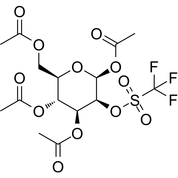
Mannose triflate is a glucose analog. Mannose triflate is a precursor for 18F-FDG synthesis for PET applications. Mannose triflate binds to 18F via SN2 nucleophilic substitution reaction. Mannose triflate can be used for an imaging technique in detection of cancer .
loading...
    loading...
Cat. No.: HY-NP163A
Synonyms: WGA-AF488
Wheat germ agglutinin-AF488 (WGA-AF488) is a cell membrane-specific staining agent prepared by conjugating wheat germ agglutinin with the Alexa Fluor 488 (HY-D1304) fluorescent dye, and it binds to cell surface glycoproteins with high affinity. Wheat germ agglutinin-AF488 is applied in fluorescence microscopy and confocal imaging techniques, and it can clearly label the membrane structures of various cells including breast cancer cells, enabling high-resolution visual observation. Wheat germ agglutinin-AF488 is used in studies of breast cancer and triple-negative breast cancer to observe cell morphology and membrane dynamic changes .
loading...
    loading...
Cat. No.: HY-P10932
Research Areas:  

Neurological Disease

pGk13a is an amphipathic membrane-labeling probe containing an azide group, which can bind to fluorophores. pGk13a enables high-resolution imaging of cell membranes in the ultrastructure expansion microscopy (umExM) technique, facilitating the observation of membrane-associated structures and proteins. pGk13a is applicable to neuronal structure research .
loading...
    loading...
Cat. No.: HY-P5114A
Target:  

Calcium Channel

Research Areas:  

Others

Maurocalcine TFA is an agonist of ryanodine receptor (RyR) channel types 1, 2 and 3 with cellular permeability. Maurocalcine TFA induces [ 3H]ryanodine binding on RyR1 with an EC50 value of 2558 nM. Maurocalcine TFA exhibits a apparent affinity of 14 nM for RyR2. Maurocalcine TFA can be applied to in vivo cell tracking or other cell imaging techniques .
loading...
    loading...
Cat. No.: HY-169868
CAS No.: 2374782-69-9
Research Areas:  

Cancer

NOTA-FAPI-52 is a tracer with potent tumor uptake. NOTA-FAPI-52 can be radiolabeled to enable PET imaging or other bioimaging techniques. NOTA-FAPI-52 can be used to study diseases in which fibroblast activation protein (FAP) is overexpressed .
loading...
    loading...
Cat. No.: HY-160276
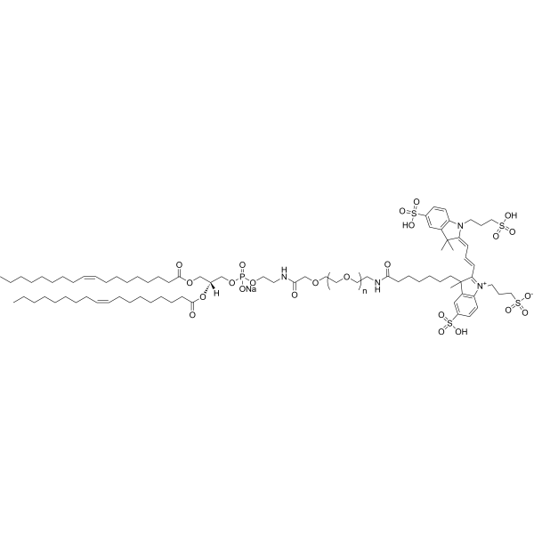
DOPE-PEG5000-Fluor 555 is consist of a DOPE phospholipid which is an unsaturated phospholipid and a Fluor 555 dye which is a bright orange cyanine dye that can be used in fluorescence microscopy, FRET and other in vivo imaging techniques.
loading...
    loading...
Cat. No.: HY-W440920
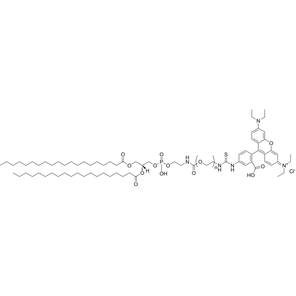
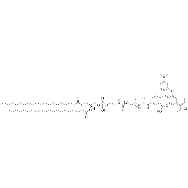
DSPE-PEG3400-Rhodamine is a phospholipid polyPEG with red fluorescent. The polymer can form lipid bilayer and be used to prepare nanoparticles or liposomes for targeted drug delivery. Rhodamine has maximum absorption at 570 nm and emission around 595 nm and can be easily traced using an imaging technique.
loading...
    loading...
Cat. No.: HY-W440921
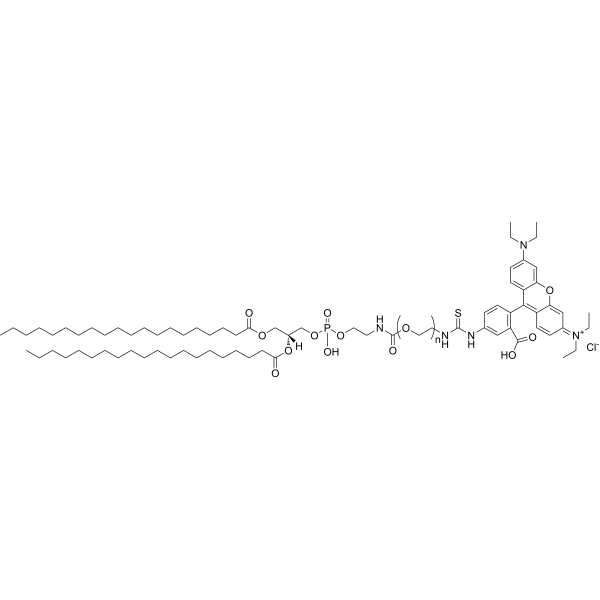
DSPE-PEG5000-Rhodamine is a phospholipid polyPEG with red fluorescent. The polymer can form lipid bilayer and be used to prepare nanoparticles or liposomes for targeted drug delivery. Rhodamine has maximum absorption at 570 nm and emission around 595 nm and can be easily traced using an imaging technique.
loading...
    loading...
Cat. No.: HY-W440919
DSPE-PEG2000-Rhodamine is a dye functionalized phospholipid. The amphiphilic polymer can form lipid bilayer in water and be used to encapsulate therapeutic agents, such as liposomal anticancer drug or mRNA vaccine. Rhodamine has maximum absorption at 570 nm and emission around 595 nm and can be easily traced using an imaging technique.
loading...
    loading...
Cat. No.: HY-P5114
CAS No.: 269745-22-4
Target:  

Calcium Channel

Research Areas:  

Others

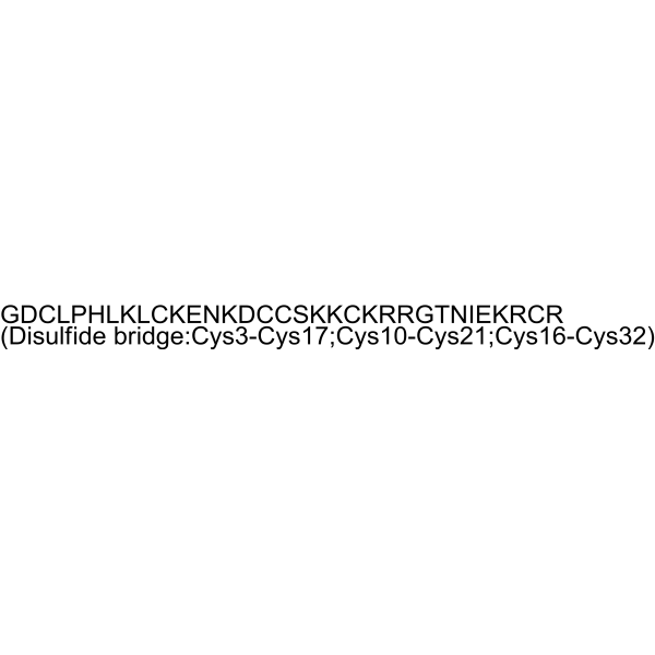
Maurocalcine is an agonist of ryanodine receptor (RyR) channel types 1, 2 and 3 with cellular permeability. Maurocalcine induces [ 3H]ryanodine binding on RyR1 with an EC50 value of 2558 nM. Maurocalcine exhibits a apparent affinity of 14 nM for RyR2. Maurocalcine can be applied to in vivo cell tracking or other cell imaging techniques .
loading...
    loading...
Cat. No.: HY-161476
Target:  

Fluorescent Dye

Research Areas:  

Cancer

Ir-ImNO is a mitochondria-targeted two-photon probe with excellent cell membrane permeability, capable of detecting both endogenous and exogenous cell membrane permeability. Ir-ImNO can monitor different immune response states of macrophages through various imaging techniques, including one-photon and two-photon phosphorescence imaging as well as phosphorescence lifetime imaging. Ir-ImNO can be utilized in the research to assist in the clinical monitoring of immunotherapy .
loading...
    loading...
Cat. No.: HY-114118F4
Synonyms: Semaglutide-Lys(FITC)
Research Areas:  

Metabolic Disease

Semaglutide-FITC (Semaglutide-Lys(FITC)) is a FITC-labeled Semaglutide (a GLP-1R agonist) (HY-114118). Semaglutide-FITC can be used to directly track the distribution, cellular uptake, and transmembrane transport process of vesicles through techniques such as fluorescence imaging and flow cytometry. Semaglutide-FITC can be employed to study the movement and penetration ability of mixed vesicles in porcine intestinal mucus in vitro .
loading...
    loading...
Cat. No.: HY-D3391
RMR-Tre is a fluorescent probe targeting the mycobacterial acyltransferase Ag85. Under the catalysis of Ag85, RMR-Tre undergoes 6-position mycoloylation and anchors to the mycobacterial membrane, while achieving fluorescence activation by inhibiting the intramolecular twisted charge transfer state transition. RMR-Tre can distinguish live mycobacteria from dead ones through metabolism-driven labeling, enabling rapid, wash-free, low-background detection of viable bacteria. RMR-Tre reports the drug resistance of Mycobacterium tuberculosis via the trehalose catalytic shift activity readout associated with TreS. In addition, RMR-Tre can be combined with flow cytometry or high-content imaging techniques to visualize and quantitatively analyze the metabolic heterogeneity of Mycobacterium tuberculosis related to persistence and drug resistance. RMR-Tre is widely used in tuberculosis-related research .
loading...
    loading...
  • 1

