



Tesetaxel
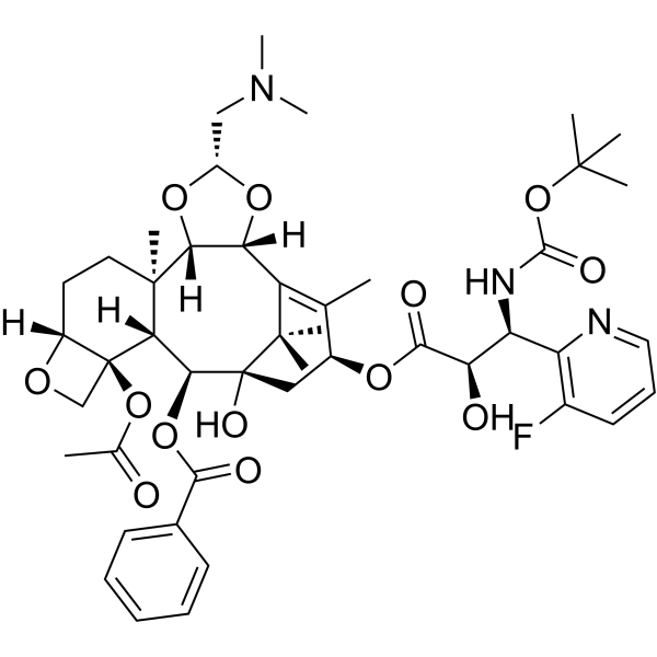
Tesetaxel (DJ-927) is an orally active and brain-penetrant taxane tubulin inhibitor. Tesetaxel inhibits tubulin depolymerization with an IC50 of 0.44 μM. Tesetaxel inhibits cancer cells proliferation and shows potent antitumor activity against P-glycoprotein-positive cancer cells. Tesetaxel can be used for the research of cancer, such as solid tumors, liver metastasis, and advanced breast cancer.
For research use only. We do not sell to patients.
- CAS No.: 333754-36-2
- Formula: C46H60FN3O13
- Molecular Weight:881.98
-
Storage:
Please store the product under the recommended conditions in the Certificate of Analysis.

To place orders, for customer services and technical support, please contact: MedChemExpress USA
Tel: 609-228-6898 E-mail: [email protected] [email protected]


-
 Biological Activity
-
Chemical Information
- Protocols
- Purity & Documentation
- References
-
 Help & FAQs




Biological Activity
Description
Cellular Effect
|
Cell Line

|
Type | Value | Description | References |
|---|---|---|---|---|
| PC-6 | GI50 |
0.38 ng/mL
Compound: 165
|
Growth inhibition of human PC-6 cells
Growth inhibition of human PC-6 cells
|
[PMID: 34213340] |
In Vitro
Tesetaxel inhibits porcine brain tubulin depolymerization with an IC50 of 0.44 μM and does not inhibit tubulin polymerization (IC50 >2.0 μM)[1].
Tesetaxel shows mean GI50 value of 0.550 ng/mL against the 23 different human tumor cells[1].
Tesetaxel exhibits greater cytotoxicity than Paclitaxel (HY-B0015) and Docetaxel (HY-B0011) against P-gp-overexpressing resistant PC-6 and HCT116 cell lines[1].
Tesetaxel (10 days) inhibits colony formation of M5076 cells with an IC50 of 5.5 ng/mL, and its activity is unaffected by the P-gp modulator Verapamil (HY-14275)[1].
MedChemExpress (MCE) has not independently confirmed the accuracy of these methods. They are for reference only.
-
Cell Line:murine ovarian histiocytoma M5076 cells (moderate P-gp expression)
-
Concentration:5.5 ng/mL
-
Incubation Time:10 days
-
Result:Inhibited colony formation with an IC50 of 5.5 ng/mL, which was 10- to 100-fold lower than Paclitaxel and Docetaxel.
Showed unaltered activity with simultaneous addition of Verapamil.
In Vivo
Tesetaxel (12 mg/kg; p.o.; single dose) achieves greater than 90% tumor growth inhibition against P-glycoprotein-positive human breast cancer DU4475 xenografts in male BALB/c-nu/nu mice[1].
Tesetaxel (9.8 mg/kg; p.o. or i.v.; once every 8 days; 2 doses) prolongs the life o in male C57BL/6 mice with P-glycoprotein-positive M5076 liver metastatic tumors[1].
MedChemExpress (MCE) has not independently confirmed the accuracy of these methods. They are for reference only.
-
Animal Model:BALB/c-nu/nu with DLD-1 xenografts (male)[1]
-
Dosage:9.80 mg/kg
-
Administration:p.o.; once every 8 days; 2 doses
-
Result:Achieved an inhibition rate (IR) greater than 90% against DLD-1 xenografts.
-
Animal Model:C57BL/6 with DLD-1 M5076 xenografts (male)[1]
-
Dosage:9.8 mg/kg
-
Administration:p.o. or i.v.; once every 8 days; 2 doses
-
Result:Produced an increase in life span (ILS) of 135% and 155%.
Clinical Trial
| NCT Number | Sponsor | Condition | Start Date |
Phase
|
|---|---|---|---|---|
| NCT01329991 | 2011-05 | PHASE1 |


Chemical Information
-
CAS No. 333754-36-2
-
Molecular Weight 881.98
-
Formula C46H60FN3O13
-
SMILES
CC(O[C@]12[C@]3([H])[C@]([C@@]4([H])[C@@](O[C@H](O4)CN(C)C)([H])C5=C(C)[C@@H](OC([C@H](O)[C@H](C6=C(C=CC=N6)F)NC(OC(C)(C)C)=O)=O)C[C@@](O)([C@H]3OC(C7=CC=CC=C7)=O)C5(C)C)(CC[C@@]1([H])OC2)C)=O
-
Synonyms
DJ-927
-
Shipping
Room temperature in continental US; may vary elsewhere.
-
Storage
Please store the product under the recommended conditions in the Certificate of Analysis.


Purity & Documentation
References

Calculators
Concentration (start) × Volume (start) = Concentration (final) × Volume (final)


Tesetaxel Related Classifications
HY-16491 Related Classifications
 Powered by Bioz