



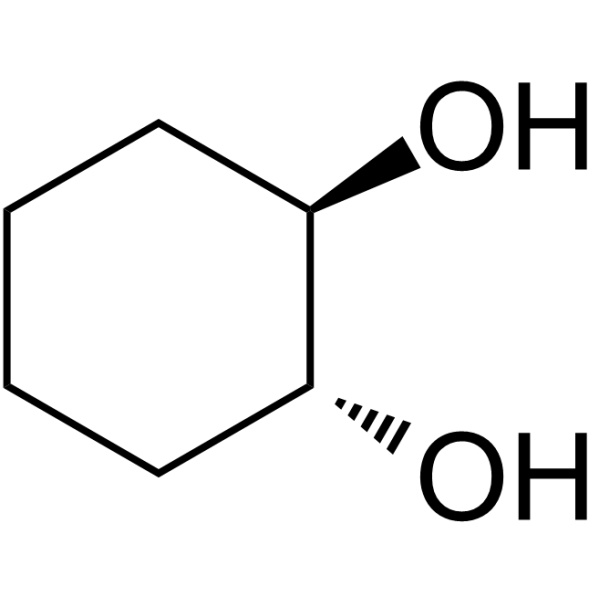
trans-Cyclohexane-1,2-diol

Based on 1 Customer Validation
trans-Cyclohexane-1,2-diol (TCHD) is a transient dilator of the nuclear pore complex (NPC). By interacting with the hydrophobic core (FG nucleoporin) of the NPC, trans-Cyclohexane-1,2-diol can disrupt the NPC structure and reversibly increase the permeability of the nuclear pore, allowing macromolecules larger than 40 kDa (such as plasmid DNA) to enter the cell nucleus by passive diffusion, thereby enhancing the nuclear import efficiency of non-viral vectors. trans-Cyclohexane-1,2-diol can improve the efficiency of in vitro electrotransfection or lipid-mediated gene transfection, especially significantly increasing gene expression in differentiated airway epithelial cells.
For research use only. We do not sell to patients.
- Purity : 99.74%
- CAS No.: 1460-57-7
- Formula: C6H12O2
- Molecular Weight:116.16
-
Storage:
Store at room temperature 3 years.
In solvent -80°C, 2 years , -20°C, 1 year

To place orders, for customer services and technical support, please contact: MedChemExpress USA
Tel: 609-228-6898 E-mail: [email protected] [email protected]


-
 Biological Activity
-
Chemical Information
-
Solvent & Solubility
- Protocols
- Purity & Documentation
- References
-
 Help & FAQs


-

HY-W010505
Hexane-1,6-diolSubstrate -

HY-W010514R
trans-Cyclohexane-1,2-diol (Standard)Nuclear Pore Complex Dilator -
HY-D0843
N-EthylmaleimideCysteine Residues Modifier -
HY-W013712
GI-530159TREK1/2 Channels Opener -
HY-D1005
Poloxamer 407 (F127)LPL Receptor Inhibitor -
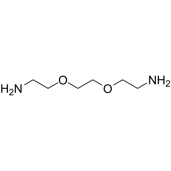
HY-128833
Bis-NH2-PEG2PROTAC Linkers -
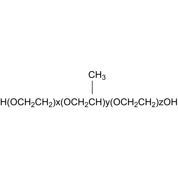
HY-D1005A
Poloxamer 188Linear Copolymer -
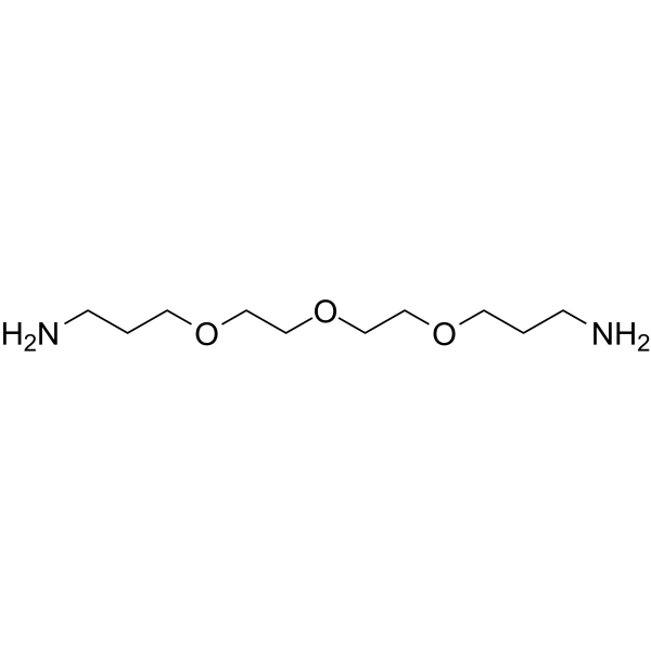
HY-128844
Bis-NH2-C1-PEG3PROTAC Linkers -
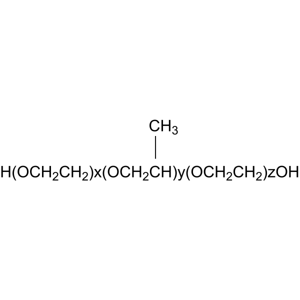
HY-D1005H
Poloxamer 188 (F68)Nonionic Linear Copolymer -
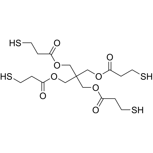
HY-W250309
Pentaerythritol tetra3-mercaptopropionateBiochemical Assay Reagent -

HY-W017061
1,6-BismaleimidohexaneBiochemical Assay Reagent -
HY-12444
Y15FAK Inhibitor
All Endogenous Metabolite Isoforms
More

Biological Activity
Description
In Vitro
Lipid-mediated transfection experiment:
trans-Cyclohexane-1,2-diol (0.5%-2%; 1 h) dose-dependently increases Lipofectamine- and GL67A-mediated luciferase gene expression in 293T cells. Concentrations of ≥1.5% results in 80% cell protein loss, indicating significant cytotoxicity[1].
Electroporation experiment: trans-Cyclohexane-1,2-diol (1%; 10-30 min) significantly increases GFP/Tomato gene expression in both B16F10 and CHO cells (1.5-2.5-fold increase in fluorescence intensity), and cell viability is not inhibited[2].
Trans-Cyclohexane-1,2-diol (TCHD) In Vitro Electroporation Protocol[2]
Materials:
(1) B16F10 mouse melanoma cells and CHO (Chinese hamster ovary) cells were cultured to 70% confluence, trypsinized and resuspended in electroporation buffer (PB: 10 mM K2HPO4/KH2PO4, 1 mM MgCl2, 250 mM sucrose, pH 7.4, cell concentration adjusted to 5×10⁶ cells/mL.
(2) Plasmid DNA: pCMV-eGFP-C1 or pCMV-CpGfree-tdTomato, concentration 20 μg/mL (dissolved in electroporation buffer).
(3) Reagents: TCHD solution: 1% w/v (mass/volume) , dissolved in electroporation buffer, and sterilized by filtration. Fetal bovine serum (FBS), crystal violet staining solution, PBS buffer.
(4) Equipment: electroporator (such as Electrocell S20), parallel stainless steel electrodes (spacing 0.4 cm), fluorescence microscope, flow cytometer.
Operation steps
Cell preparation: Collect cells in logarithmic growth phase, wash twice with PB buffer after centrifugation, resuspend cells to 5×10⁶ cells/mL, add plasmid DNA to a final concentration of 20 μg/mL, and mix well.
Electrotransfection parameter setting:
Low field long pulse (LF-LP): 6 square wave pulses, 600 V/cm, 5 ms pulse duration, frequency 1 Hz.
High field short pulse (HF-SP): 4 square wave pulses, 1200 V/cm, 100 μs pulse duration, frequency 1 Hz. 100 μL of cell-DNA suspension was added to the electrode gap and the electric pulses were applied at room temperature.
TCHD treatment:
Immediately after the pulse: Immediately after the pulse, 20 μL of 1% TCHD solution (final concentration approximately 0.18% w/v) was added and gently mixed. After incubation for 10 minutes after the pulse, another 20 μL of 1% TCHD solution was added. Both treatments were incubated at 37°C for 10, 20, or 30 minutes, and the control group was added with an equal volume of electroporation buffer.
Cell culture:
After the incubation period, the cells were transferred to complete medium (DMEM or EMEM) containing 10% FBS, seeded in 12-well plates, and cultured at 37°C, 5% CO2 for 24 hours.
Detection and analysis:
(1) After 24 hours, cell viability was detected by crystal violet staining. The culture medium was discarded, the cells were washed with PBS, and 0.1% crystal violet solution was added for staining for 20 minutes. After dissolving in 10% acetic acid, the absorbance at 595 nm was measured.
(2) Flow cytometry is used to detect the percentage of GFP/Tomato positive cells and the mean fluorescence intensity (MFI). The cells were trypsinized, washed with PBS, and resuspended in FACS buffer (containing 2.5% FBS) and analyzed by flow cytometry.
(3) After fixing the cells, the distribution of fluorescent protein expression was observed using a fluorescence microscope (GFP excitation light 480/40 nm, emission light 527/40 nm; Tomato excitation light 560/40 nm, emission light 630/75 nm).
Key parameters and optimized TCHD concentration:
The optimal final concentration is 0.18% w/v (i.e., 1% stock solution is added at a volume ratio of 1:6). Higher concentrations (such as 2%) may cause cytotoxicity.
MedChemExpress (MCE) has not independently confirmed the accuracy of these methods. They are for reference only.


Chemical Information
-
CAS No. 1460-57-7
-
Appearance Solid
-
Molecular Weight 116.16
-
Formula C6H12O2
-
Color White to off-white
-
SMILES
O[C@H]1[C@H](O)CCCC1
-
Structure Classification
-
Initial Source
-
Shipping
Room temperature in continental US; may vary elsewhere.
-
Storage
Store at room temperature 3 years
In solvent -80°C 2 years -20°C 1 year


Solvent & Solubility
In Vitro:
H2O : 100 mg/mL (860.88 mM; Need ultrasonic)
DMSO : 50 mg/mL (430.44 mM; Need ultrasonic; Hygroscopic DMSO has a significant impact on the solubility of product, please use newly opened DMSO)
Please refer to the solubility information to select the appropriate solvent. Once prepared, please aliquot and store the solution to prevent product inactivation from repeated freeze-thaw cycles.
Storage method and period of stock solution: -80°C, 2 years; -20°C, 1 year. When stored at -80°C, please use it within 2 years. When stored at -20°C, please use it within 1 year.
* Note: If you choose water as the stock solution, please dilute it to the working solution, then filter and sterilize it with a 0.22 μm filter before use.
Please refer to the solubility information to select the appropriate solvent. Once prepared, please aliquot and store the solution to prevent product inactivation from repeated freeze-thaw cycles.
Storage method and period of stock solution: -80°C, 2 years; -20°C, 1 year. When stored at -80°C, please use it within 2 years. When stored at -20°C, please use it within 1 year.
* Note: If you choose water as the stock solution, please dilute it to the working solution, then filter and sterilize it with a 0.22 μm filter before use.
Concentration (start) × Volume (start) = Concentration (final) × Volume (final)
In Vivo:
Select the appropriate dissolution method based on your experimental animal and administration route.
- For the following dissolution methods, please ensure to first prepare a clear stock solution using an In Vitro approach and then sequentially add co-solvents:
- To ensure reliable experimental results, the clarified stock solution can be appropriately stored based on storage conditions. As for the working solution for In Vivo experiments, it is recommended to prepare freshly and use it on the same day.
- The percentages shown for the solvents indicate their volumetric ratio in the final prepared solution. If precipitation or phase separation occurs during preparation, heat and/or sonication can be used to aid dissolution.
Add each solvent one by one: 10% DMSO 40% PEG300 5% Tween-80 45% Saline
Solubility: ≥ 2.5 mg/mL (21.52 mM); Clear solution
This protocol yields a clear solution of ≥ 2.5 mg/mL (saturation unknown).
Taking 1 mL working solution as an example, add 100 μL DMSO stock solution (25.0 mg/mL) to 400 μL PEG300, and mix evenly; then add 50 μL Tween-80 and mix evenly; then add 450 μL Saline to adjust the volume to 1 mL.
Preparation of Saline: Dissolve 0.9 g sodium chloride in ddH₂O and dilute to 100 mL to obtain a clear Saline solution.
Add each solvent one by one: 10% DMSO 90% (20% SBE-β-CD in Saline)
Solubility: ≥ 2.5 mg/mL (21.52 mM); Clear solution
This protocol yields a clear solution of ≥ 2.5 mg/mL (saturation unknown).
Taking 1 mL working solution as an example, add 100 μL DMSO stock solution (25.0 mg/mL) to 900 μL 20% SBE-β-CD in Saline, and mix evenly.
Preparation of 20% SBE-β-CD in Saline (4°C, storage for one week): 2 g SBE-β-CD powder is dissolved in 10 mL Saline, completely dissolve until clear.
For the following dissolution methods, please prepare the working solution directly:
It is recommended to prepare fresh solutions and use them promptly within a short period of time.
The percentages shown for the solvents indicate their volumetric ratio in the final prepared solution. If precipitation or phase separation occurs during preparation, heat and/or sonication can be used to aid dissolution.
Add each solvent one by one: PBS
Solubility: 110 mg/mL (946.97 mM); Clear solution; Need ultrasonic
In Vivo Dissolution Calculator
Please enter the basic information of animal experiments:
-
-
-
-
Recommended: Prepare an additional quantity of animals to account for potential losses during experiments.
Working solution concentration: 0.22 mg/mL
This product has good water solubility, please refer to the measured solubility data in water/PBS/Saline for details.


Purity & Documentation
-
Data Sheet (280 KB)
-
SDS (394 KB)
- English - EN (394 KB)
- Français - FR (394 KB)
- Deutsch - DE (394 KB)
- Norwegian - NO (394 KB)
- Español - ES (394 KB)
- Swedish - SV (394 KB)
- Italian - IT (394 KB)
- Korean - KR (394 KB)
- Portuguese - PT (394 KB)
-
Handling Instructions (2659 KB)
References
[1]. Griesenbach U, et al. Assessment of the nuclear pore dilating agent trans-cyclohexane-1,2-diol in differentiated airway epithelium. J Gene Med. 2012 Jul;14(7):491-500. [Content Brief]
[2]. Pasquet L, et al. Post-pulse addition of trans-cyclohexane-1,2-diol improves electrotransfer mediated gene expression in mammalian cells. Biochem Biophys Rep. 2016 Jul 17;7:287-294. [Content Brief]


Complete Stock Solution Preparation Table
Please refer to the solubility information to select the appropriate solvent. Once prepared, please aliquot and store the solution to prevent product inactivation from repeated freeze-thaw cycles.
Storage method and period of stock solution: -80°C, 2 years; -20°C, 1 year. When stored at -80°C, please use it within 2 years. When stored at -20°C, please use it within 1 year.
| Optional Solvent | Concentration Solvent Mass | 1 mg | 5 mg | 10 mg | 25 mg |
|---|---|---|---|---|---|
| DMSO / H2O | 1 mM | 8.6088 mL | 43.0441 mL | 86.0882 mL | 215.2204 mL |
| 5 mM | 1.7218 mL | 8.6088 mL | 17.2176 mL | 43.0441 mL | |
| 10 mM | 0.8609 mL | 4.3044 mL | 8.6088 mL | 21.5220 mL | |
| 15 mM | 0.5739 mL | 2.8696 mL | 5.7392 mL | 14.3480 mL | |
| 20 mM | 0.4304 mL | 2.1522 mL | 4.3044 mL | 10.7610 mL | |
| 25 mM | 0.3444 mL | 1.7218 mL | 3.4435 mL | 8.6088 mL | |
| 30 mM | 0.2870 mL | 1.4348 mL | 2.8696 mL | 7.1740 mL | |
| 40 mM | 0.2152 mL | 1.0761 mL | 2.1522 mL | 5.3805 mL | |
| 50 mM | 0.1722 mL | 0.8609 mL | 1.7218 mL | 4.3044 mL | |
| 60 mM | 0.1435 mL | 0.7174 mL | 1.4348 mL | 3.5870 mL | |
| 80 mM | 0.1076 mL | 0.5381 mL | 1.0761 mL | 2.6903 mL | |
| 100 mM | 0.0861 mL | 0.4304 mL | 0.8609 mL | 2.1522 mL |
* Note: If you choose water as the stock solution, please dilute it to the working solution, then filter and sterilize it with a 0.22 μm filter before use.


trans-Cyclohexane-1,2-diol Related Classifications
HY-W010514 Related Classifications
 Powered by Bioz