



trans-Doxercalciferol

Based on 1 Customer Validation
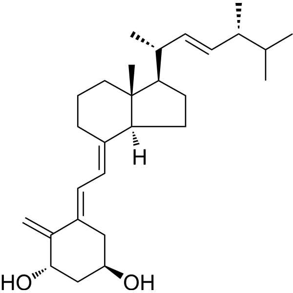
trans-Doxercalciferol is an isomer of Doxercalciferol (HY-32348). Doxercalciferol is a vitamin D receptor agonist. Doxercalciferol reduces proteinuria, podocyte injury, mesangial expansion, extracellular matrix accumulation, macrophage infiltration, oxidative stress, proinflammatory cytokines, profibrotic growth factors, renal neutral lipid accumulation, albuminuria and glomerulosclerosis. Doxercalciferol alleviates high-salt diet-induced myocardial hypertrophy. trans-Doxercalciferol is used for the research of diet-induced obesity-related nephropathy, diabetic nephropathy, myocardial hypertrophy, cardiac dysfunction and secondary hyperparathyroidism.
For research use only. We do not sell to patients.
- Purity : 98.20%
- CAS No.: 74007-20-8
- Formula: C28H44O2
- Molecular Weight:412.65
-
Storage:Powder -20°C, 3 years , 4°C, 2 years ; In solvent -80°C, 2 years , -20°C, 1 year

To place orders, for customer services and technical support, please contact: MedChemExpress USA
Tel: 609-228-6898 E-mail: [email protected] [email protected]


-
 Biological Activity
-
Chemical Information
-
Solvent & Solubility
- Protocols
- Purity & Documentation
- References
-
 Help & FAQs


-
HY-10001
CalcipotriolVitamin D3 Analog -
HY-50919
ParicalcitolVitamin D Receptor Agonist -
HY-32339
MaxacalcitolVDR Agonist -
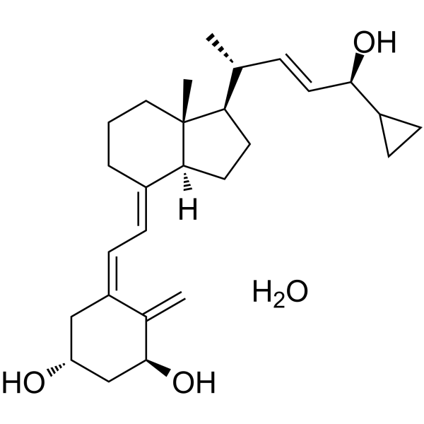
HY-10001A
Calcipotriol monohydrateVitamin D3 Analog -
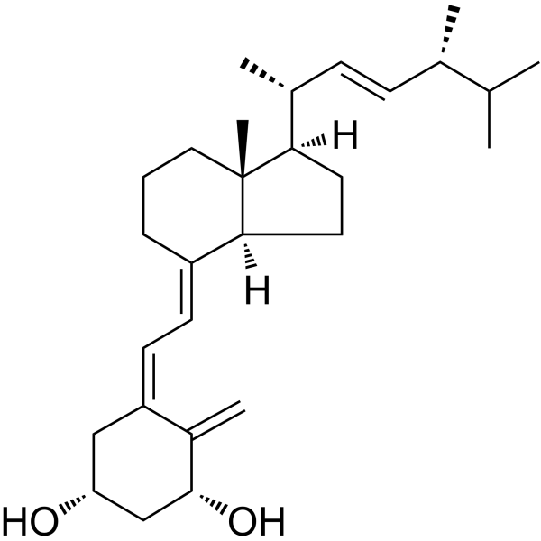
HY-76937
Impurity of DoxercalciferolImpurity of Doxercalciferol -
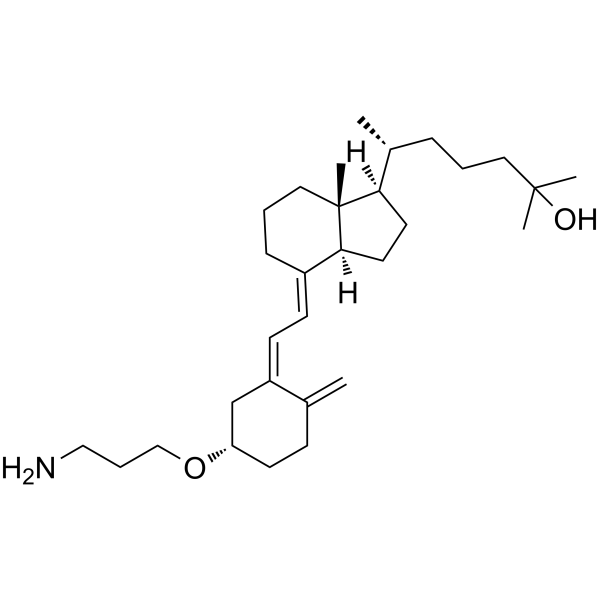
HY-15254
3-O-(2-Aminoethyl)-25-hydroxyvitamin D3Vitamin D3 Derivative -
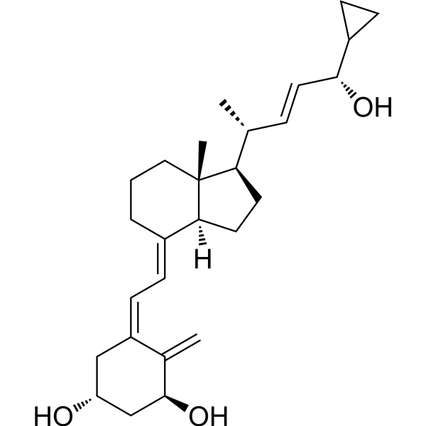
HY-10001R
Calcipotriol (Standard)Vitamin D3 Analog -
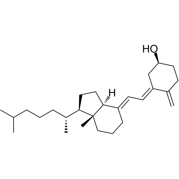
HY-15398A
5,6-trans-Vitamin D3Photoproduct of Vitamin D3 -
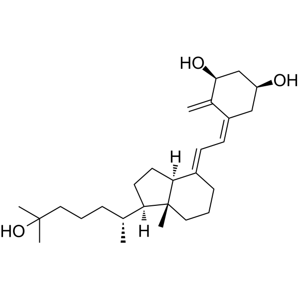
HY-10002A
(1S)-Calcitriol1α,25(OH)2D3 Metabolite -
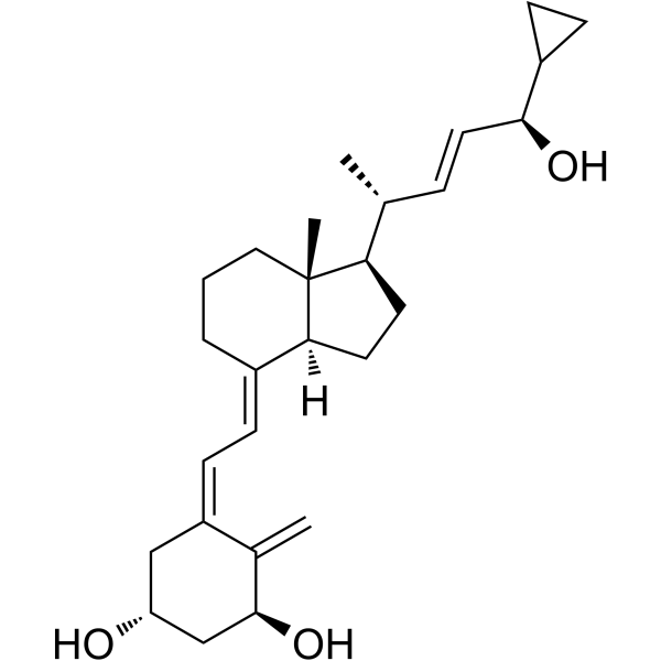
HY-15266
24R-CalcipotriolVitamin D Analog -
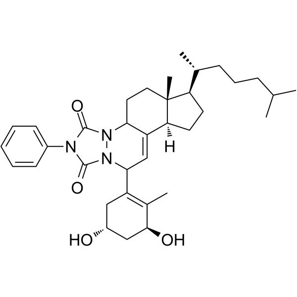
HY-13294
Impurity C of AlfacalcidolAlfacalcidol Impurity -
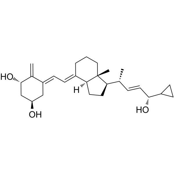
HY-75035
Calcipotriol Impurity CCalcipotriol Impurity


Biological Activity
Description
In Vivo
MedChemExpress (MCE) has not independently confirmed the accuracy of these methods. They are for reference only.
-
Animal Model:Dahl salt sensitive (DSS) (male, 6 weeks of age, cardiac hypertrophy induced via 6-week high-salt (6% NaCl) diet)[3]
-
Dosage:150 ng; 250 ng
-
Administration:i.p.; 3 times weekly; 6 weeks
-
Result:Reduced heart weight/body weight ratio by 11%.
Reduced lung weight/body weight ratio by 29%.
Reduced diastolic left ventricle posterior wall thickness by 13%.
Improved fractional shortening by 17%.
Attenuated increased left ventricular mass.
Significantly reduced serum brain natriuretic peptide (BNP) levels.
Reduced left ventricular tissue atrial natriuretic factor (ANF) mRNA levels.
Reduced cardiomyocyte cross-sectional diameter by 22%.
Blocked high-salt diet-induced increases in cardiac protein kinase C-α (PKCα) protein level, membrane translocation of PKCα, and phosphorylated PKCα expression.
Did not cause significant changes in serum calcium levels (150 ng and 250 ng doses).
Had no significant effect on organ weights or serum calcium levels in normal diet-fed rats (150 ng dose).
- Fibrosis/Collagen Morphometry
- Phagocytosis Functional Assay
- Collagen: Sirius Red Staining
- 3T3-L1 preadipocyte-to-adipocyte differentiation
- ROS/oxidative-stress fluorescent staining
- ECM-Embedded Organoid (Matrigel/Dome) Culture
- Matrigel/ECM Transwell Invasion Assay
- Research Protocol for Metabolic Diseases
- Matrigel Transwell/Boyden Chamber Invasion Assay
- Lipid Droplets: Oil Red O/Sudan Dye Lipid Staining


Chemical Information
-
CAS No. 74007-20-8
-
Appearance Solid
-
Molecular Weight 412.65
-
Formula C28H44O2
-
Color White to off-white
-
SMILES
C=C([C@H](C[C@H](O)C/1)O)C1=C\C=C2[C@@]3([H])[C@@](CCC\2)(C)[C@@H]([C@H](C)/C=C/[C@H](C)C(C)C)CC3
-
Shipping
Room temperature in continental US; may vary elsewhere.
-
Storage
Powder -20°C 3 years 4°C 2 years In solvent -80°C 2 years -20°C 1 year


Solvent & Solubility
In Vitro:
DMSO : 50 mg/mL (121.17 mM; Need ultrasonic; Hygroscopic DMSO has a significant impact on the solubility of product, please use newly opened DMSO)
Please refer to the solubility information to select the appropriate solvent. Once prepared, please aliquot and store the solution to prevent product inactivation from repeated freeze-thaw cycles.
Storage method and period of stock solution: -80°C, 2 years; -20°C, 1 year. When stored at -80°C, please use it within 2 years. When stored at -20°C, please use it within 1 year.
Please refer to the solubility information to select the appropriate solvent. Once prepared, please aliquot and store the solution to prevent product inactivation from repeated freeze-thaw cycles.
Storage method and period of stock solution: -80°C, 2 years; -20°C, 1 year. When stored at -80°C, please use it within 2 years. When stored at -20°C, please use it within 1 year.
Concentration (start) × Volume (start) = Concentration (final) × Volume (final)


Purity & Documentation
-
Data Sheet (276 KB)
-
SDS (479 KB)
- English - EN (479 KB)
- Français - FR (479 KB)
- Deutsch - DE (479 KB)
- Norwegian - NO (479 KB)
- Español - ES (479 KB)
- Swedish - SV (479 KB)
- Italian - IT (479 KB)
- Korean - KR (479 KB)
- Portuguese - PT (479 KB)
-
Handling Instructions (2659 KB)
References
[1]. Wang XX, et al. Vitamin D receptor agonist doxercalciferol modulates dietary fat-induced renal disease and renal lipid metabolism. Am J Physiol Renal Physiol. 2011;300(3):F801-F810. [Content Brief]
[2]. Zhang Y, et al. Long-term therapeutic effect of vitamin D analog doxercalciferol on diabetic nephropathy: strong synergism with AT1 receptor antagonist. Am J Physiol Renal Physiol. 2009 Sep;297(3):F791-801. [Content Brief]
[3]. Choi JH, et al. Doxercalciferol, a pro-hormone of vitamin D, prevents the development of cardiac hypertrophy in rats. J Card Fail. 2011;17(12):1051-1058. [Content Brief]
[4]. Kubodera N, et al. A new look at the most successful prodrugs for active vitamin D (D hormone): alfacalcidol and doxercalciferol. Molecules. 2009;14(10):3869-3880. Published 2009 Sep 29. [Content Brief]
[5]. Wesseling-Perry K, et al. Calcitriol and doxercalciferol are equivalent in controlling bone turnover, suppressing parathyroid hormone, and increasing fibroblast growth factor-23 in secondary hyperparathyroidism. Kidney Int. 2011;79(1):112-119. [Content Brief]


Complete Stock Solution Preparation Table
Please refer to the solubility information to select the appropriate solvent. Once prepared, please aliquot and store the solution to prevent product inactivation from repeated freeze-thaw cycles.
Storage method and period of stock solution: -80°C, 2 years; -20°C, 1 year. When stored at -80°C, please use it within 2 years. When stored at -20°C, please use it within 1 year.
| Optional Solvent | Concentration Solvent Mass | 1 mg | 5 mg | 10 mg | 25 mg |
|---|---|---|---|---|---|
| DMSO | 1 mM | 2.4234 mL | 12.1168 mL | 24.2336 mL | 60.5840 mL |
| 5 mM | 0.4847 mL | 2.4234 mL | 4.8467 mL | 12.1168 mL | |
| 10 mM | 0.2423 mL | 1.2117 mL | 2.4234 mL | 6.0584 mL | |
| 15 mM | 0.1616 mL | 0.8078 mL | 1.6156 mL | 4.0389 mL | |
| 20 mM | 0.1212 mL | 0.6058 mL | 1.2117 mL | 3.0292 mL | |
| 25 mM | 0.0969 mL | 0.4847 mL | 0.9693 mL | 2.4234 mL | |
| 30 mM | 0.0808 mL | 0.4039 mL | 0.8078 mL | 2.0195 mL | |
| 40 mM | 0.0606 mL | 0.3029 mL | 0.6058 mL | 1.5146 mL | |
| 50 mM | 0.0485 mL | 0.2423 mL | 0.4847 mL | 1.2117 mL | |
| 60 mM | 0.0404 mL | 0.2019 mL | 0.4039 mL | 1.0097 mL | |
| 80 mM | 0.0303 mL | 0.1515 mL | 0.3029 mL | 0.7573 mL | |
| 100 mM | 0.0242 mL | 0.1212 mL | 0.2423 mL | 0.6058 mL |


trans-Doxercalciferol Related Classifications
HY-75992 Related Classifications
 Powered by Bioz
Keywords
- trans-Doxercalciferol
- 74007-20-8
- Drug Isomer
- renin
- protein kinase C-α
- farnesoid X receptor
- mineralocorticoid receptor
- vitamin D receptor
- angiotensin II type 1 receptor
- angiotensinogen
- sterol regulatory element binding protein 2
- sterol regulatory element binding protein 1
- parathyroid hormone
- Inhibitor
- inhibitor
- inhibit