



TrxR-IN-2
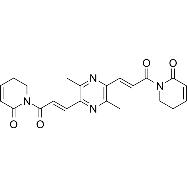
TrxR-IN-2 is a thioredoxin reductase (TrxR) inhibitor. TrxR-IN-2 increases reactive oxidative species (ROS) levels and decreases mitochondrial transmembrane potential levels. TrxR-IN-2 triggers DNA damage via H2AX regulation, and induces autophagy via LC3, beclin-1, and p62 regulation. TrxR-IN-2 can be used for the research of drug-resistant hepatocellular carcinoma[1].
For research use only. We do not sell to patients.
- CAS No.: 2866261-50-7
- Formula: C22H22N4O4
- Molecular Weight:406.43
-
Storage:
Please store the product under the recommended conditions in the Certificate of Analysis.

To place orders, for customer services and technical support, please contact: MedChemExpress USA
Tel: 609-228-6898 E-mail: [email protected] [email protected]


-
 Biological Activity
-
Chemical Information
- Protocols
- Purity & Documentation
- References
-
 Help & FAQs




Biological Activity
Description
Cellular Effect
|
Cell Line

|
Type | Value | Description | References |
|---|---|---|---|---|
| Bel-7402 | IC50 |
0.8 μM
Compound: 11
|
Antiproliferative activity against human Bel-7402 cells incubated for 24 hrs by MTT assay
Antiproliferative activity against human Bel-7402 cells incubated for 24 hrs by MTT assay
|
[PMID: 34806369] |
| Bel7402/5-FU | IC50 |
0.9 μM
Compound: 11
|
Antiproliferative activity against human BEL-7402/5-FU cells incubated for 24 hrs by MTT assay
Antiproliferative activity against human BEL-7402/5-FU cells incubated for 24 hrs by MTT assay
|
[PMID: 34806369] |
| HepG2 | IC50 |
1.1 μM
Compound: 11
|
Antiproliferative activity against human HepG2 cells incubated for 24 hrs by MTT assay
Antiproliferative activity against human HepG2 cells incubated for 24 hrs by MTT assay
|
[PMID: 34806369] |
| HGC-27 | IC50 |
1.4 μM
Compound: 11
|
Antiproliferative activity against human HGC-27 cells incubated for 24 hrs by MTT assay
Antiproliferative activity against human HGC-27 cells incubated for 24 hrs by MTT assay
|
[PMID: 34806369] |
In Vitro
TrxR-IN-2 (Compound 11) (72 h) potently inhibits the proliferation of Bel7402, HepG2, HGC27, and Bel-7402/5-FU cells with IC50 values ranging from 0.8 μM to 1.4 μM[1].
TrxR-IN-2 (0.2 μM) potently inhibits TrxR activity in Bel-7402/5-FU cells with an IC50 of 0.2 μM[1].
TrxR-IN-2 (0.3-3.0 μM; 24 h) induces dose-dependent ROS accumulation in Bel-7402/5-FU cells[1].
TrxR-IN-2 (1-10 μM) significantly reduces mitochondrial transmembrane potential in Bel-7402/5-FU cells[1].
TrxR-IN-2 (1.0-3.0 μM; 72 h) triggers DNA damage in Bel-7402/5-FU cells by up-regulating the expression of H2AX.S139ph, p-p53, and p53[1].
TrxR-IN-2 (1.0-3.0 μM; 24 h) induces dose-dependent autophagosome formation in Bel-7402/5-FU cells[1].
TrxR-IN-2 (0.3-3.0 μM; 72 h) triggers autophagy in Bel-7402/5-FU cells by dose-dependently increasing the LC3-II/LC3-I ratio and beclin-1 expression, and decreasing p62 expression[1].
MedChemExpress (MCE) has not independently confirmed the accuracy of these methods. They are for reference only.
-
Cell Line:Bel-7402/5-FU drug-resistant human liver carcinoma cells
-
Concentration:1.0 μM; 3.0 μM
-
Incubation Time:72 h
-
Result:Increased the expression of DNA damage biomarker H2AX.S139ph, phosphorylated p53 (p-p53), and total p53 in a dose-dependent manner.
-
Cell Line:Bel-7402/5-FU drug-resistant human liver carcinoma cells
-
Concentration:1.0 μM; 3.0 μM
-
Incubation Time:24 h
-
Result:Induced autophagosome formation in a dose-dependent manner; at 3.0 μM, triggered autophagy more pronouncedly than piperlongumine at the same concentration.
-
Cell Line:Bel-7402/5-FU drug-resistant human liver carcinoma cells
-
Concentration:0.3 μM; 1.0 μM; 3.0 μM
-
Incubation Time:72 h
-
Result:Up-regulated the conversion of LC3-I to LC3-II (increasing the LC3-II/LC3-I ratio in a dose-dependent manner) and the expression of beclin-1, while down-regulating the expression of p62.
Was more potent than piperlongumine in modulating these protein levels.
In Vivo
TrxR-IN-2 (2-5 mg/kg; i.p.; 21 days) exhibits potent in vivo antitumor activity in Bel-7402/5-FU xenograft nude mice[1].
MedChemExpress (MCE) has not independently confirmed the accuracy of these methods. They are for reference only.
-
Animal Model:ICR mice (female)[1]
-
Dosage:60.0 mg/kg; 90.0 mg/kg; 135.0 mg/kg; 202.5 mg/kg; 303.8 mg/kg
-
Administration:i.p.; single dose
-
Result:Achieved 100% survival of mice 14 days post-administration at 60.0 mg/kg.
Achieved 90% survival of mice 14 days post-administration at 90.0 mg/kg.
Achieved 60% survival of mice 14 days post-administration at 135.0 mg/kg.
Achieved 30% survival of mice 14 days post-administration at 202.5 mg/kg.
Achieved 10% survival of mice 14 days post-administration at 303.8 mg/kg.
Calculated a median lethal dose (LD50) of 178 mg/kg.
-
Animal Model:Nude mice (Bel-7402/5-FU xenograft tumor model)[1]
-
Dosage:2 mg/kg; 5 mg/kg
-
Administration:i.p.; 21 days
-
Result:Significantly suppressed tumor growth compared to the control group at 2 mg/kg, reducing mean tumor weight to ~0.7 g.
Suppressed tumor growth by 76% compared to the control group at 5 mg/kg, reducing mean tumor weight to ~0.45 g.
Showed greater tumor growth inhibition than the 5 mg/kg dose of piperlongumine at 5 mg/kg.


Chemical Information
-
CAS No. 2866261-50-7
-
Molecular Weight 406.43
-
Formula C22H22N4O4
-
SMILES
O=C1C=CCCN1C(/C=C/C2=NC(C)=C(/C=C/C(N3CCC=CC3=O)=O)N=C2C)=O
-
Shipping
Room temperature in continental US; may vary elsewhere.
-
Storage
Please store the product under the recommended conditions in the Certificate of Analysis.


Purity & Documentation
References

Calculators
Concentration (start) × Volume (start) = Concentration (final) × Volume (final)


TrxR-IN-2 Related Classifications
HY-132972 Related Classifications
 Powered by Bioz