



VS 8
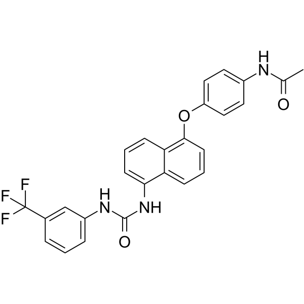
VS 8 (Compound VS 8) is a potent, orally active VEGFR-2 inhibitor with significant anti-angiogenic effects. VS 8 induces cancer cell apoptosis and migration. VS 8 is active against CSCs (Cancer stem cells).
For research use only. We do not sell to patients.
- CAS No.: 2471865-38-8
- Formula: C26H20F3N3O3
- Molecular Weight:479.45
-
Storage:
Please store the product under the recommended conditions in the Certificate of Analysis.

To place orders, for customer services and technical support, please contact: MedChemExpress USA
Tel: 609-228-6898 E-mail: [email protected] [email protected]


-
 Biological Activity
-
Chemical Information
- Protocols
- Purity & Documentation
- References
-
 Help & FAQs


All VEGFR Isoforms
More

Biological Activity
Description
IC50 & Target
|
VEGFR-2 |
Cellular Effect
|
Cell Line

|
Type | Value | Description | References |
|---|---|---|---|---|
| HepG2 | IC50 |
1166 nM
Compound: VS8
|
Cytotoxicity against human HepG2 cells assessed as reduction in cell viability by trypan blue assay assay
Cytotoxicity against human HepG2 cells assessed as reduction in cell viability by trypan blue assay assay
|
[PMID: 33002846] |
| HepG2 | IC50 |
2201 nM
Compound: VS8
|
Antiproliferative activity against human HepG2 cells harboring CD133+ assessed as reduction in cell viability incubated for 24 hrs by MTT assay
Antiproliferative activity against human HepG2 cells harboring CD133+ assessed as reduction in cell viability incubated for 24 hrs by MTT assay
|
[PMID: 33002846] |
| HepG2 | IC50 |
257.8 nM
Compound: VS8
|
Cytotoxicity against human HepG2 cells assessed as reduction in cell viability incubated for 24 hrs by MTT assay
Cytotoxicity against human HepG2 cells assessed as reduction in cell viability incubated for 24 hrs by MTT assay
|
[PMID: 33002846] |
| HepG2 | IC50 |
3290 nM
Compound: VS8
|
Antiproliferative activity against human HepG2 cells harboring CD44+ assessed as reduction in cell viability incubated for 24 hrs by MTT assay
Antiproliferative activity against human HepG2 cells harboring CD44+ assessed as reduction in cell viability incubated for 24 hrs by MTT assay
|
[PMID: 33002846] |
| HUVEC | IC50 |
9643 nM
Compound: VS8
|
Antiproliferative activity against human HUVEC assessed as reduction in cell viability incubated for 24 hrs by MTT assay
Antiproliferative activity against human HUVEC assessed as reduction in cell viability incubated for 24 hrs by MTT assay
|
[PMID: 33002846] |
| MCF7 | IC50 |
953.3 nM
Compound: VS8
|
Cytotoxicity against human MCF7 cells assessed as reduction in cell viability incubated for 24 hrs by MTT assay
Cytotoxicity against human MCF7 cells assessed as reduction in cell viability incubated for 24 hrs by MTT assay
|
[PMID: 33002846] |
| MDA-MB-231 | IC50 |
1413 nM
Compound: VS8
|
Cytotoxicity against human MDA-MB-231 cells assessed as reduction in cell viability incubated for 24 hrs by MTT assay
Cytotoxicity against human MDA-MB-231 cells assessed as reduction in cell viability incubated for 24 hrs by MTT assay
|
[PMID: 33002846] |
| MDA-MB-231 | IC50 |
3577 nM
Compound: VS8
|
Cytotoxicity against human MDA-MB-231 cells assessed as reduction in cell viability by trypan blue assay assay
Cytotoxicity against human MDA-MB-231 cells assessed as reduction in cell viability by trypan blue assay assay
|
[PMID: 33002846] |
In Vitro
VS 8 (Compound VS 8) (0.01-100 µM, 24 h) shows potent anti-proliferative activity against MCF-7, MDA-MB-231, Hep G2, and HUVECs cells[1].
VS 8 induces early apoptosis in MDA-MB-231 (1413 nM, 72 h), Hep G2 (257.80 nM, 24 h), and HUVECs (1954 nM, 24 h) cells[1].
VS 8 is shown to be a pro-oxidant molecule that enhances the ROS level in Hep G2 cells[1].
VS 8 inhibits wound healing and migration of MCF-7 cancer cells[1].
VS 8 downregulates human vascular endothelial growth factor (hVEGF) and hVEGFR-2 expression in HUVECs[1].
VS 8 (257.80 nM, 48 h) arrests cell cycle at ‘G0/G1’ and ‘S’ phase in CD44+ and CD133+ CSCs, respectively[1].
VS 8 inhibits TGF-β-induced epithelial-mesenchymal transition (EMT) in hepatocellular carcinoma by the upregulation of E-cadherin and the suppression of vimentin and SNAIL[1].
MedChemExpress (MCE) has not independently confirmed the accuracy of these methods. They are for reference only.
-
Cell Line:MCF-7, MDA-MB-231, Hep G2, and HUVECs cells
-
Concentration:0.01, 0.1, 1, 10, 50, and 100 µM
-
Incubation Time:24 h
-
Result:Showed significantly potent anti-proliferative activity against all the selected cell lines in a dose-dependent manner, with IC50 values of 953.30, 1413, 257.80, and 1954 nM against MCF-7, MDA-MB-231, Hep G2, and HUVECs cells.
-
Cell Line:MDA-MB-231, Hep G2, and HUVECs cells
-
Concentration:1413, 257.80, and 1954 nM for MDA-MB-231, Hep G2, and HUVECs cells, respectively.
-
Incubation Time:72 h for MDA-MB-231 cells; 24 h for Hep G2 and HUVECs cells
-
Result:Resulted in high population of early apoptotic MDA-MB-231 cells (68.34 ± 0.18%). A significant increase in % apoptotic index (~86.66%) was observed in Hep G2 cells. The percentage of early apoptotic cells were found to be ~37.53% in HUVECs cells.
-
Cell Line:CD44+ and CD133+ CSCs isolated from Hep G2 cells
-
Concentration:257.80 nM
-
Incubation Time:48 h
-
Result:Arrested cell cycle at ‘G0/G1’ and ‘S’ phase in CD44+ and CD133+ CSCs, respectively.
In Vivo
MedChemExpress (MCE) has not independently confirmed the accuracy of these methods. They are for reference only.
-
Animal Model:Male Wistar rats (180-220 gm)[1]
-
Dosage:5 mg/kg
-
Administration:Oral administration (Pharmacokinetic Analysis)
-
Result:Pharmacokinetic parameters for VS 8 in rats after administration of oral dose (5 mg/ kg) [1]
Pharmacokinetic parameters Unit Value Cmax μg/mL 39.7193 ± 0.36 Tmax hrs 6 ± 0 AUC(0-72) mg/mL*hrs 621.3236 ± 1.843 AUC(0-∞) mg/mL*hrs 625.2219 ± 1.864 AUMC(0-∞) (mg/mL*hrs2) 8929.284 ± 72.85 MRT hrs 14.2817 ± 0.102 t1/2 hrs 11.9277 ± 0.324
Data represented as mean ± SD (n = 3); t1/2, Half-Life; Cmax, Maximum Observed Concentration; Tmax, Maximum Observed Time; AUC, Area Under Curve; AUMC Area Under Movement Curve, MRT, Mean Residence Time.


Chemical Information
-
CAS No. 2471865-38-8
-
Molecular Weight 479.45
-
Formula C26H20F3N3O3
-
SMILES
CC(NC1=CC=C(OC2=C3C=CC=C(NC(NC4=CC=CC(C(F)(F)F)=C4)=O)C3=CC=C2)C=C1)=O
-
Shipping
Room temperature in continental US; may vary elsewhere.
-
Storage
Please store the product under the recommended conditions in the Certificate of Analysis.


Purity & Documentation
References

Calculators
Concentration (start) × Volume (start) = Concentration (final) × Volume (final)


VS 8 Related Classifications
HY-143491 Related Classifications
 Powered by Bioz