


XRK3F2 free base
Based on 7 publication(s) in Google Scholar
XRK3F2 free base is a p62 (sequestosome-1) ZZ domain inhibitor that has specificity for the p62-ZZ domain over other p62 signaling domains. XRK3F2 free base blocks TNFα effects and upregulation in bone marrow stromal cells, and induces multiple myeloma cell apoptosis. XRK3F2 free base can be used for the research of multiple myeloma bone disease, acute myeloid leukemia, and multiple myeloma.
For research use only. We do not sell to patients.
- CAS No.: 2375193-42-1
- Formula: C23H23F2NO3
- Molecular Weight:399.43
-
Storage:
Please store the product under the recommended conditions in the Certificate of Analysis.

To place orders, for customer services and technical support, please contact: MedChemExpress USA
Tel: 609-228-6898 E-mail: [email protected] [email protected]


-
 Biological Activity
-
Chemical Information
- Purity & Documentation
- References
-
 Help & FAQs


Publications Citing Use of MedChemExpress (MCE) XRK3F2 free base
More Customer Validation & Images

-
Cell Proliferation/Viability Assay
-
In Vivo Efficacy Study
-
Histological Imaging/Staining
-
WB


Biological Activity
XRK3F2 free base (5 μM; 48 h co-culture + 4 days osteogenic culture) prevents MM-induced Runx2 suppression and Gfi1 upregulation in MC4 pre-osteoblasts during co-culture and subsequent osteogenic differentiation. XRK3F2 also rescues alkaline phosphatase activity in MC4 pre-osteoblasts[1].
XRK3F2 free base (5 μM; 48 h) blocks Gfi1 upregulation, Runx2 repression, and IL6 induction in primary murine BMSC treated with MM1.S conditioned media or TNFα plus IL7. XRK3F2 prevents MM-induced GFI1 and HDAC1 recruitment to the Runx2-P1 promoter and preserves H3K9ac marks in MC4 pre-osteoblasts[1].
XRK3F2 free base (2.5-5 μM; 4 days) rescues Runx2 expression, osteogenic target gene expression, and H3K9ac marks, and reduces GFI1 binding at the Runx2-P1 promoter in MM-exposed MC4 pre-osteoblasts[1].
XRK3F2 free base (5 μM; 5 days) rescues Runx2 mRNA expression in MM-exposed HD-hBMSC during osteogenic differentiation[1].
XRK3F2 free base (0-20 μM; 72 h) inhibits the viability of K562, HL-60, K562/A02, and HL60/ADR leukemia cells with IC50 values ranging from 6.41 to 8.76 μM[2].
XRK3F2 free base (5 μM) induces apoptosis of primary human AML CD34+CD38− cells (LIC-like population) at 5 μM[2].
XRK3F2 free base (0.1-10 μM, 48 h ) inhibits TNFα-induced differentiation of human CD116+ OCL precursors[3].
XRK3F2 free base (10 μM; 48 h, 72 h) directly inhibits growth of human MM cell lines, primary MM cells, and murine 5TGM1-gfp cells by inducing apoptosis, with an IC50 of 4.35 μM for 5TGM1 cells[3].
MedChemExpress (MCE) has not independently confirmed the accuracy of these methods. They are for reference only.
-
Cell Line:murine pre-osteoblast MC4 cells
-
Concentration:3 μM
-
Incubation Time:48 h (mRNA analysis); 7 days (alkaline phosphatase staining)
-
Result:Prevented TNFα-induced Gfi1 mRNA upregulation and Runx2 mRNA repression; rescued alkaline phosphatase staining (a marker of osteogenic differentiation) in TNFα-treated MC4 cells
-
Cell Line:murine pre-osteoblast MC4 cells, 5TGM1 multiple myeloma (MM) cells
-
Concentration:2.5-5 μM
-
Incubation Time:4 days (osteogenic media culture)
-
Result:Significantly elevated Runx2 mRNA and downstream osteogenic target genes (Ocn, Bsp, Osx); reduced GFI1 binding at the Runx2-P1 promoter; increased H3K9ac levels at the Runx2-P1 promoter; rescued alkaline phosphatase staining
-
Cell Line:healthy donor human BMSC (HD-hBMSC), MM1.S cells
-
Concentration:5 μM
-
Incubation Time:5 days (osteogenic media culture after MM removal)
-
Result:Rescued Runx2 mRNA levels in MM-exposed HD-hBMSC; decreased Gfi1 mRNA (difference not significant)
-
Cell Line:K562, HL-60, K562/A02, HL60/ADR
-
Concentration:0-20 μM
-
Incubation Time:72 h
-
Result:Exhibited cytotoxicity toward K562 (IC50 = 6.41 μM), HL-60 (IC50 = 8.23 μM), K562/A02 (IC50 = 6.42 μM), and HL60/ADR (IC50 = 8.76 μM) cells, with comparable sensitivity between parental and drug-resistant lines.
-
Cell Line:human MM cell lines, primary MM cells, murine 5TGM1-gfp cells
-
Concentration:10 μM
-
Incubation Time:48 h (human MM cell lines, primary MM cells); 72 h (murine 5TGM1-gfp cells)
-
Result:Significantly inhibited growth of all 6 human MM cell lines and primary human MM cells; achieved an IC50 of 4.35 μM for 5TGM1 cells and 4.6 μM for MM1.S cells; induced cleavage of caspases 9, 7, and 3 in MM1.S cells after 16 hours.
XRK3F2 free base (27, 40 mg/kg/day; i.p.; 5 consecutive days/week; 2 weeks) induces significant new cortical bone formation restricted to MM-containing bones in an immunocompetent multiple myeloma model[3].
MedChemExpress (MCE) has not independently confirmed the accuracy of these methods. They are for reference only.
-
Animal Model:NOG (sublethally irradiated 120 cGy; AML PDX model)[2]
-
Dosage:40 mg/kg
-
Administration:i.p.; daily; 10 consecutive days
-
Result:Markedly reduced leukemic burden of hCD45+ cells in the bone marrow compared to the vehicle group.
-
Animal Model:C57BL/KaLwRij (male, 6-12 weeks of age, multiple myeloma model via tibial inoculation of 1×10⁵ 5TGM1-gfp cells)[3]
-
Dosage:27 mg/kg/day; 40 mg/kg/day
-
Administration:i.p.; 5 consecutive days/week; 2 weeks
-
Result:Increased cortical bone volume (measured by new bone volume to total bone volume ratio) in all treated mice; Observed greater cortical bone formation in treated mice with high tumor burden (high IgG₂b levels); Detected no new bone formation in non-MM-bearing legs; Confirmed new woven bone formation localized to MM-containing bones via histology, with active osteoblasts and osteoclasts present on new bone surfaces.


Chemical Information
-
CAS No. 2375193-42-1
-
Molecular Weight 399.43
-
Formula C23H23F2NO3
-
SMILES
FC1=CC=C(COC2=CC=C(CNCCO)C=C2OCC3=CC=C(C=C3)F)C=C1
-
Shipping
Room temperature in continental US; may vary elsewhere.
-
Storage
Please store the product under the recommended conditions in the Certificate of Analysis.


Publications (7)
-
Journal Impact Factor
-
Most Recent
-
-
Mol Med
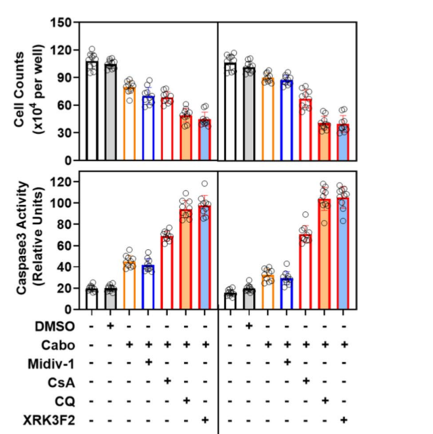
Overexpression of c-Myc triggers p62 aggregation-mediated mitochondrial mitophagy in cabozantinib resistance of hepatocellular carcinoma. [Abstract]2025 May 27;31(1):209. PMID: 40426058
XRK3F2 free base purchased from MedChemExpress. Usage Cited in: Mol Med. 2025 May 27;31(1):209. [Abstract]
Cell viability (live cells counted using hemocytometer after trypan blue exclusion) and intracellular Caspase3 activity (Caspase 3/7 Activity Apoptosis Assay) were assessed after treating cabozantinib-resistant HCC cells with XRK3F2.
-
Eur J Pharmacol
Doxorubicin-mediated retardation of aggresome formation enhances Carfilzomib-induced cell death synergistically by augmenting ER stress and proapoptotic signaling. [Abstract]2025 Dec 5:1008:178303. PMID: 41183585 -
Front Cell Dev Biol
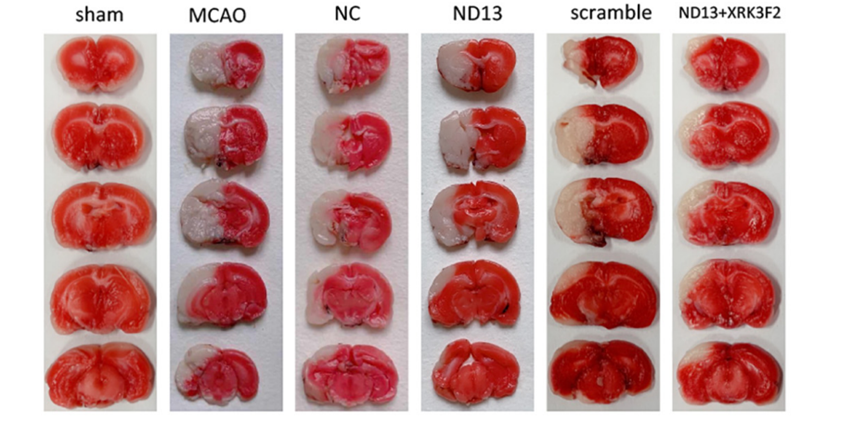
DJ-1 Regulates Microglial Polarization Through P62-Mediated TRAF6/IRF5 Signaling in Cerebral Ischemia-Reperfusion. [Abstract]2020 Dec 17:8:593890. PMID: 33392187
XRK3F2 free base purchased from MedChemExpress. Usage Cited in: Front Cell Dev Biol. 2020 Dec 17:8:593890. [Abstract]
The infarct volume was significantly decreased compared with that of the ND13 group.after treatment with XRK3F2 (2.5 μg/μL).
XRK3F2 free base purchased from MedChemExpress. Usage Cited in: Front Cell Dev Biol. 2020 Dec 17:8:593890. [Abstract]
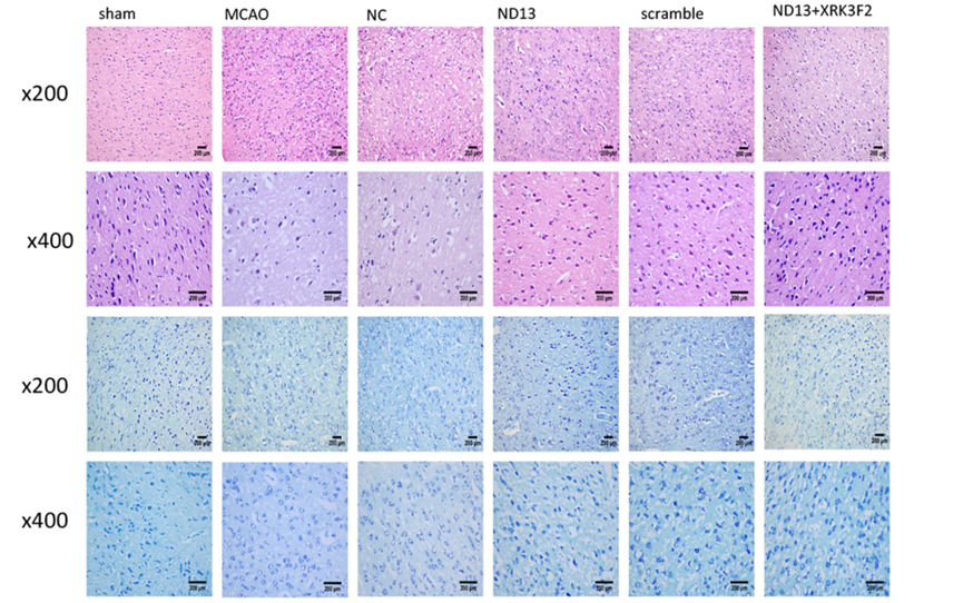
HE staining showed that after treatment with XRK3F2 (2.5 μg/μL), the injuries condition improved.
XRK3F2 free base purchased from MedChemExpress. Usage Cited in: Front Cell Dev Biol. 2020 Dec 17:8:593890. [Abstract]
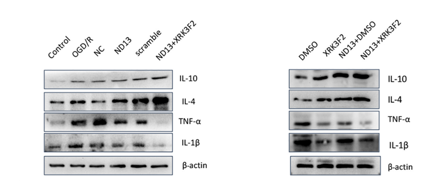
After combined treatment with XRK3F2 (2.5 μg/μL) and ND13, the expression levels of IL-10 and IL-4 were higher and the expression levels of TNF-α and IL-1β were lower than those of the ND13 group.
-
-
-


Purity & Documentation
References
[1]. Adamik J, et al. XRK3F2 Inhibition of p62-ZZ Domain Signaling Rescues Myeloma-Induced GFI1-Driven Epigenetic Repression of the Runx2 Gene in Pre-osteoblasts to Overcome Differentiation Suppression. Front Endocrinol (Lausanne). 2018;9:344. Published 2018 Jun 29. [Content Brief]
[2]. Li Y, et al. A mitophagy inhibitor targeting p62 attenuates the leukemia-initiation potential of acute myeloid leukemia cells. Cancer Lett. 2021;510:24-36. [Content Brief]
[3]. Teramachi J, et al. Blocking the ZZ domain of sequestosome1/p62 suppresses myeloma growth and osteoclast formation in vitro and induces dramatic bone formation in myeloma-bearing bones in vivo. Leukemia. 2016;30(2):390-398. [Content Brief]

Calculators
Concentration (start) × Volume (start) = Concentration (final) × Volume (final)


 Powered by Bioz