



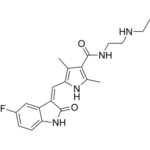
N-Desethyl Sunitinib
Based on 7 publication(s) in Google Scholar
N-Desethyl Sunitinib (SU-12662) is a metabolite of Sunitinib (HY-10255A). N-Desethyl Sunitinib serves as a good transport substrate for human ABCB1, ABCG2 and murine ABCG2.
For research use only. We do not sell to patients.
- Purity : 99.81%
- CAS No.: 356068-97-8
- Formula: C20H23FN4O2
- Molecular Weight:370.42
-
Storage:Powder -20°C, 3 years , 4°C, 2 years ; In solvent -80°C, 6 months , -20°C, 1 month

To place orders, for customer services and technical support, please contact: MedChemExpress USA
Tel: 609-228-6898 E-mail: [email protected] [email protected]


-
 Biological Activity
-
Chemical Information
-
Solvent & Solubility
- Purity & Documentation
- References
-
 Help & FAQs


-
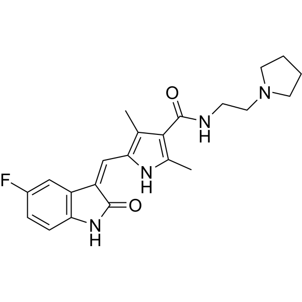
HY-10330
ToceranibRTK Inhibitor -

HY-15141
StaurosporinePKC/PKA Inhibitor -
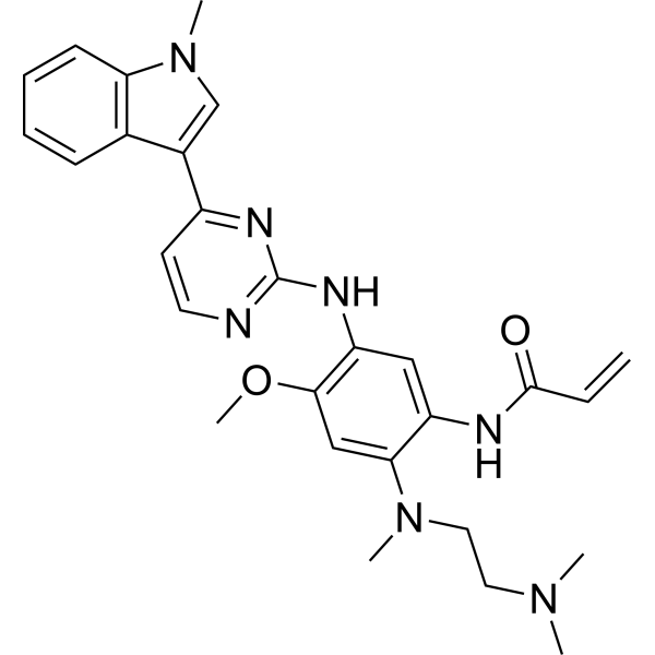
HY-15772
OsimertinibMutant-Selective EGFR Inhibitor -
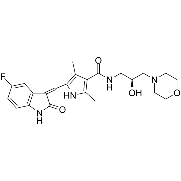
HY-10501
SU14813Multiple RTK Inhibitor -
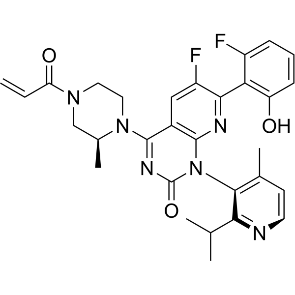
HY-114277
SotorasibKRAS G12C Inhibitor -
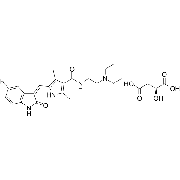
HY-10255
Sunitinib MalateTyrosine Kinase Inhibitor -
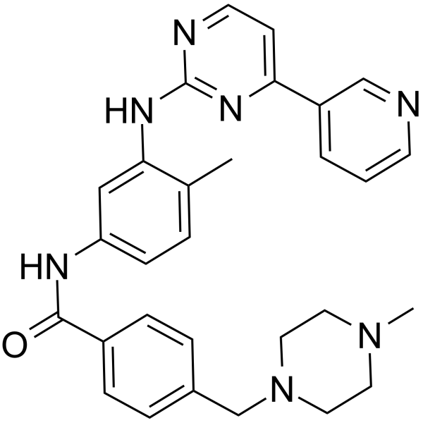
HY-15463
ImatinibTyrosine Kinases Inhibitor -
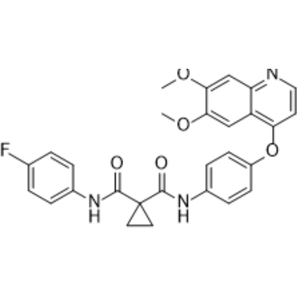
HY-13016
CabozantinibVEGFR2/MET Inhibitor -
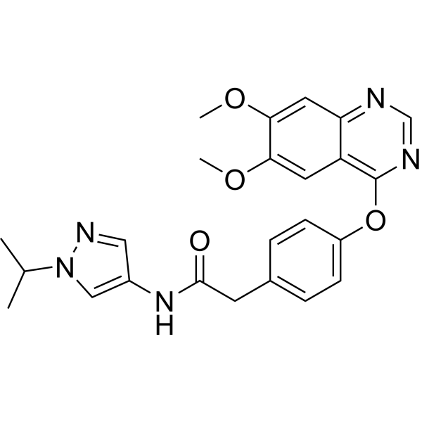
HY-18179
AZD2932c-Kit Inhibitor -
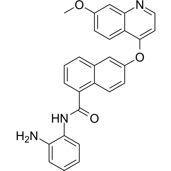
HY-124526
ChiauranibMulti-target Inhibitor -
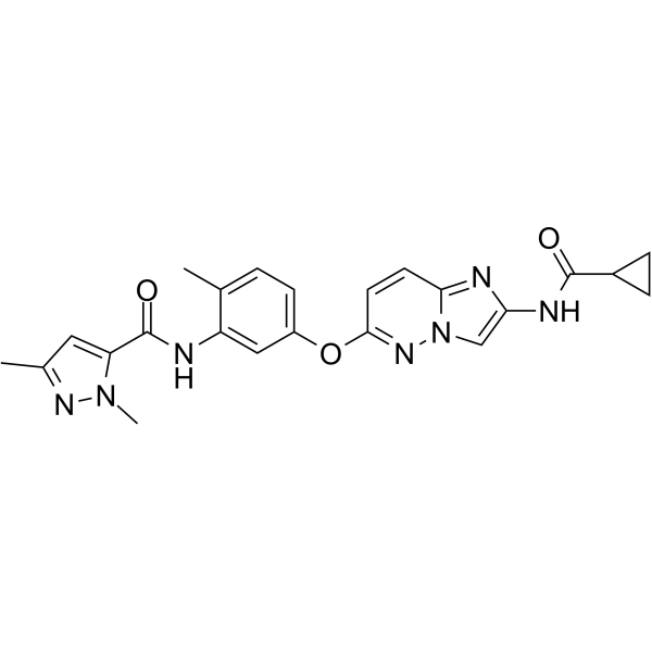
HY-15506
TAK-593VEGFR/PDGFR Inhibitor -
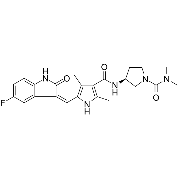
HY-109019
VorolanibVEGFR/PDGFR Inhibitor
Publications Citing Use of MedChemExpress (MCE) N-Desethyl Sunitinib
More- Acta Pharmacol Sin. 2016 Jul;37(7):930-40. [Abstract]
- Pharmaceutics. 2024 Aug 28;16(9):1138. [Abstract]
- J Pharmacol Sci. 2026 Oct;162(2):99-108.
- J Chromatogr B Analyt Technol Biomed Life Sci. 2024 Apr 15:1237:124100. [Abstract]
- Biol Pharm Bull. 2021;44(10):1565-1570. [Abstract]
- Biomed Chromatogr. 2015 May;29(5):679-88. [Abstract]
- SSRN. 23 Sep 2021.


Biological Activity
Description
Cellular Effect
|
Cell Line

|
Type | Value | Description | References |
|---|---|---|---|---|
| K562 | EC50 |
11.88 μM
Compound: 21
|
Cytotoxicity against human K562 cells assessed as reduction in cell viability incubated for 48 hrs by celltox-green assay
Cytotoxicity against human K562 cells assessed as reduction in cell viability incubated for 48 hrs by celltox-green assay
|
[PMID: 32334266] |
| K562 | EC50 |
12.89 μM
Compound: 21
|
Cytotoxicity against human K562 cells assessed as reduction in cell viability incubated for 24 hrs by celltox-green assay
Cytotoxicity against human K562 cells assessed as reduction in cell viability incubated for 24 hrs by celltox-green assay
|
[PMID: 32334266] |
| K562 | EC50 |
13.59 μM
Compound: 21
|
Cytotoxicity against human K562 cells assessed as reduction in cell viability incubated for 72 hrs by MTS assay
Cytotoxicity against human K562 cells assessed as reduction in cell viability incubated for 72 hrs by MTS assay
|
[PMID: 32334266] |
| K562 | EC50 |
15.48 μM
Compound: 21
|
Cytotoxicity against human K562 cells assessed as reduction in cell viability incubated for 24 hrs by cell-titer glo assay
Cytotoxicity against human K562 cells assessed as reduction in cell viability incubated for 24 hrs by cell-titer glo assay
|
[PMID: 32334266] |
In Vitro
N-Desethyl Sunitinib (5 μM; 2-4 h) serves as a good transport substrate for human ABCB1, ABCG2 and murine ABCG2[2].
MedChemExpress (MCE) has not independently confirmed the accuracy of these methods. They are for reference only. .


Chemical Information
-
CAS No. 356068-97-8
-
Appearance Solid
-
Molecular Weight 370.42
-
Formula C20H23FN4O2
-
Color Light yellow to orange
-
SMILES
O=C1NC2=CC=C(F)C=C2/C1=C/C3=C(C)C(C(NCCNCC)=O)=C(C)N3
-
Synonyms
SU-12662
-
Shipping
Room temperature in continental US; may vary elsewhere.
-
Storage
Powder -20°C 3 years 4°C 2 years In solvent -80°C 6 months -20°C 1 month


Publications (7)
-
Journal Impact Factor
-
Most Recent
-
Acta Pharmacol Sin
Preclinical PK/PD model for combined administration of erlotinib and sunitinib in the treatment of A549 human NSCLC xenograft mice. [Abstract]2016 Jul;37(7):930-40. PMID: 27180983 -
Pharmaceutics
Development of Simultaneous Drug Concentration Measurement Method Using an Automated Pretreatment Liquid Chromatography/Tandem Mass Spectrometry System for Therapeutic Drug Monitoring. [Abstract]2024 Aug 28;16(9):1138. PMID: 39339175 -
-
J Chromatogr B Analyt Technol Biomed Life Sci
Simultaneous determination of 11 oral targeted antineoplastic drugs and 2 active metabolites by LC-MS/MS in human plasma and its application to therapeutic drug monitoring in cancer patients. [Abstract]2024 Apr 15:1237:124100. PMID: 38547701 -
Biol Pharm Bull
Development of a Competitive Enzyme-Linked Immunosorbent Assay for the Determination of Sunitinib Unaffected by Light-Induced Isomerization. [Abstract]2021;44(10):1565-1570. PMID: 34602567 -
Biomed Chromatogr
Determination of sunitinib and its active metabolite, N-desethyl sunitinib in mouse plasma and tissues by UPLC-MS/MS: assay development and application to pharmacokinetic and tissue distribution studies. [Abstract]2015 May;29(5):679-88. PMID: 25294592 -


Solvent & Solubility
In Vitro:
DMSO : 6.25 mg/mL (16.87 mM; ultrasonic and warming and heat to 60°C; Hygroscopic DMSO has a significant impact on the solubility of product, please use newly opened DMSO)
Please refer to the solubility information to select the appropriate solvent. Once prepared, please aliquot and store the solution to prevent product inactivation from repeated freeze-thaw cycles.
Storage method and period of stock solution: -80°C, 6 months; -20°C, 1 month. When stored at -80°C, please use it within 6 months. When stored at -20°C, please use it within 1 month.
Please refer to the solubility information to select the appropriate solvent. Once prepared, please aliquot and store the solution to prevent product inactivation from repeated freeze-thaw cycles.
Storage method and period of stock solution: -80°C, 6 months; -20°C, 1 month. When stored at -80°C, please use it within 6 months. When stored at -20°C, please use it within 1 month.
Concentration (start) × Volume (start) = Concentration (final) × Volume (final)
In Vivo:
Select the appropriate dissolution method based on your experimental animal and administration route.
- For the following dissolution methods, please ensure to first prepare a clear stock solution using an In Vitro approach and then sequentially add co-solvents:
- To ensure reliable experimental results, the clarified stock solution can be appropriately stored based on storage conditions. As for the working solution for In Vivo experiments, it is recommended to prepare freshly and use it on the same day.
- The percentages shown for the solvents indicate their volumetric ratio in the final prepared solution. If precipitation or phase separation occurs during preparation, heat and/or sonication can be used to aid dissolution.
Add each solvent one by one: 10% DMSO 40% PEG300 5% Tween-80 45% Saline
Solubility: ≥ 0.62 mg/mL (1.67 mM); Clear solution
This protocol yields a clear solution of ≥ 0.62 mg/mL (saturation unknown).
Taking 1 mL working solution as an example, add 100 μL DMSO stock solution (6.2 mg/mL) to 400 μL PEG300, and mix evenly; then add 50 μL Tween-80 and mix evenly; then add 450 μL Saline to adjust the volume to 1 mL.
Preparation of Saline: Dissolve 0.9 g sodium chloride in ddH₂O and dilute to 100 mL to obtain a clear Saline solution.
Add each solvent one by one: 10% DMSO 90% (20% SBE-β-CD in Saline)
Solubility: 0.62 mg/mL (1.67 mM); Suspended solution; Need ultrasonic
This protocol yields a suspended solution of 0.62 mg/mL. Suspended solution can be used for oral and intraperitoneal injection.
Taking 1 mL working solution as an example, add 100 μL DMSO stock solution (6.2 mg/mL) to 900 μL 20% SBE-β-CD in Saline, and mix evenly.
Preparation of 20% SBE-β-CD in Saline (4°C, storage for one week): 2 g SBE-β-CD powder is dissolved in 10 mL Saline, completely dissolve until clear.
In Vivo Dissolution Calculator
Please enter the basic information of animal experiments:
-
-
-
-
Recommended: Prepare an additional quantity of animals to account for potential losses during experiments.
Please enter your animal formula composition:
-
%DMSO +
Recommended: Keep the proportion of DMSO in working solution below 2% if your animal is weak.
-
%+
-
+%Tween-80 + +
-
%Saline +
The co-solvents required include: DMSO, . All of co-solvents are available by MedChemExpress (MCE). , Tween 80. All of co-solvents are available by MedChemExpress (MCE).
Working solution concentration: 0.22 mg/mL
Method for preparing stock solution: mg drug dissolved in μL DMSO. Stock solution concentration: mg/mL.
1. Take μL DMSO stock solution;
2. Add μL .
μL , mix evenly;
3. Then add μL Tween 80, mix evenly;
4. Then add μL
Please ensure that the stock solution in the first step is dissolved to a clear state, and add co-solvents in sequence. You can use ultrasonic heating (ultrasonic cleaner, recommended frequency 20-40 kHz), vortexing, etc. to assist dissolution.


Purity & Documentation
-
Data Sheet (280 KB)
-
SDS (623 KB)
- English - EN (623 KB)
- Français - FR (623 KB)
- Deutsch - DE (623 KB)
- Norwegian - NO (623 KB)
- Español - ES (623 KB)
- Swedish - SV (623 KB)
- Italian - IT (623 KB)
- Korean - KR (623 KB)
- Portuguese - PT (623 KB)
-
Handling Instructions (2659 KB)
References
[2]. Tang SC, et al. P-glycoprotein (ABCB1) and breast cancer resistance protein (ABCG2) restrict brain accumulation of the active sunitinib metabolite N-desethyl sunitinib. J Pharmacol Exp Ther. 2012;341(1):164-173. [Content Brief]
[3]. Li JY,et al. Preclinical PK/PD model for combined administration of erlotinib and sunitinib in the treatment of A549 human NSCLC xenograft mice. Acta Pharmacol Sin. 2016 Jul;37(7):930-40. [Content Brief]


Complete Stock Solution Preparation Table
Please refer to the solubility information to select the appropriate solvent. Once prepared, please aliquot and store the solution to prevent product inactivation from repeated freeze-thaw cycles.
Storage method and period of stock solution: -80°C, 6 months; -20°C, 1 month. When stored at -80°C, please use it within 6 months. When stored at -20°C, please use it within 1 month.
| Optional Solvent | Concentration Solvent Mass | 1 mg | 5 mg | 10 mg | 25 mg |
|---|---|---|---|---|---|
| DMSO | 1 mM | 2.6996 mL | 13.4982 mL | 26.9964 mL | 67.4910 mL |
| 5 mM | 0.5399 mL | 2.6996 mL | 5.3993 mL | 13.4982 mL | |
| 10 mM | 0.2700 mL | 1.3498 mL | 2.6996 mL | 6.7491 mL | |
| 15 mM | 0.1800 mL | 0.8999 mL | 1.7998 mL | 4.4994 mL |


N-Desethyl Sunitinib Related Classifications
HY-10873 Related Classifications
 Powered by Bioz