



CP-31398
CP-31398 can stabilize the active conformation of p53 and promote p53 activity in cancer cells with either mutant or wild-type p53. In addition, CP-31398 can upregulate p53 target genes, such as p21WAF1/Cip1 and KILLER/DR5. CP-31398 exerts an inhibitory effect on tumor growth.
Nur für Forschungszwecke. Wir verkaufen nicht an Patienten.
- CAS. Nr.: 259199-65-0
- Formel: C22H26N4O
- Molecular Weight:362.47
-
Speicherung:
Please store the product under the recommended conditions in the Certificate of Analysis.


To place orders, for customer services and technical support, please contact: MedChemExpress USA
Tel: 609-228-6898 E-mail: [email protected] [email protected]


-
 Biologische Aktivität
-
Chemical Information
- Protokoll
- Reinheit & Dokumentation
- Verweise
-
 Help & FAQs


-
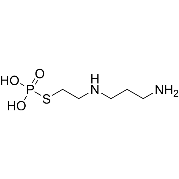
HY-B0639
AmifostineCytoprotective Agent -
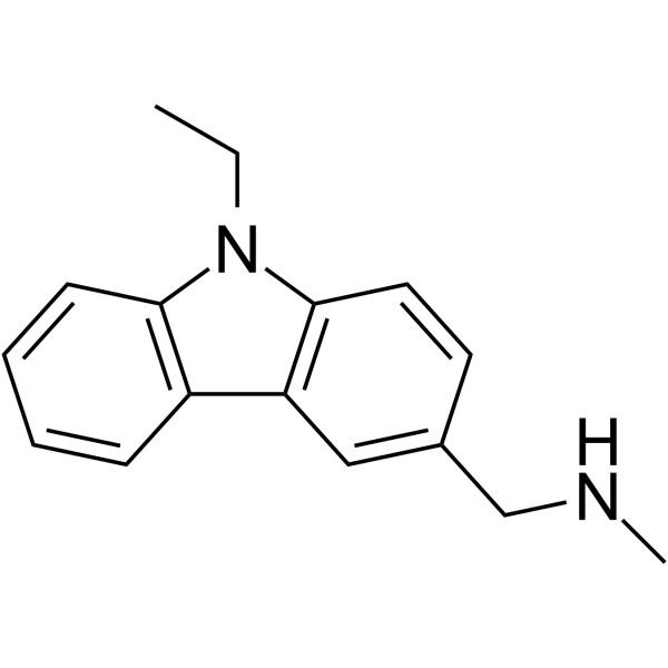
HY-108637
PhiKan 083MDM-2/p53 p53 Activator -
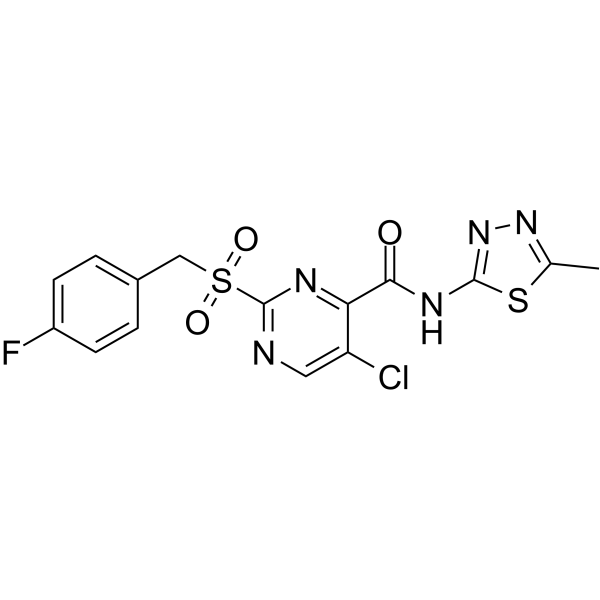
HY-128784
PK11007Anti-p53 Agent -
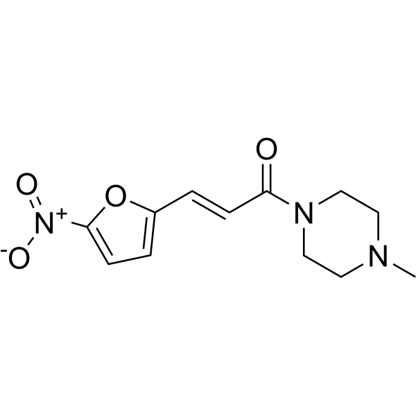
HY-19726
NSC59984MDM-2/p53 p53 Activator -
HY-18343A
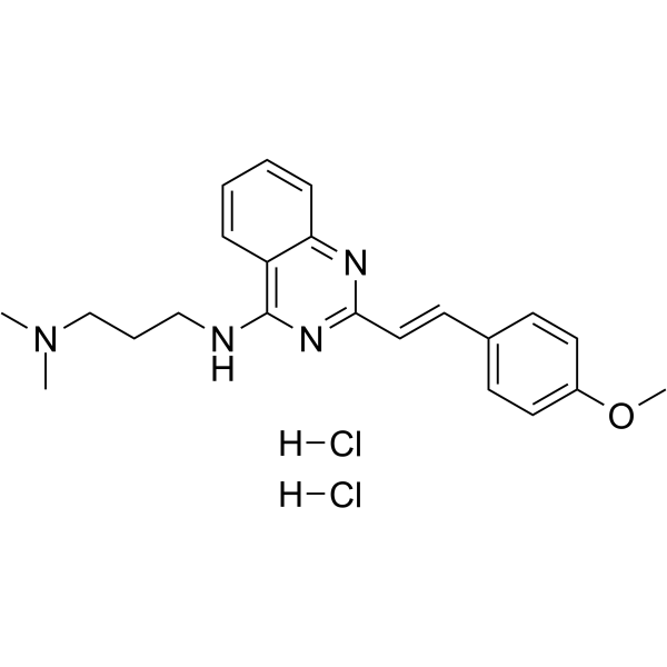
CP-31398 dihydrochloridep53 Stabilizer -
HY-18634
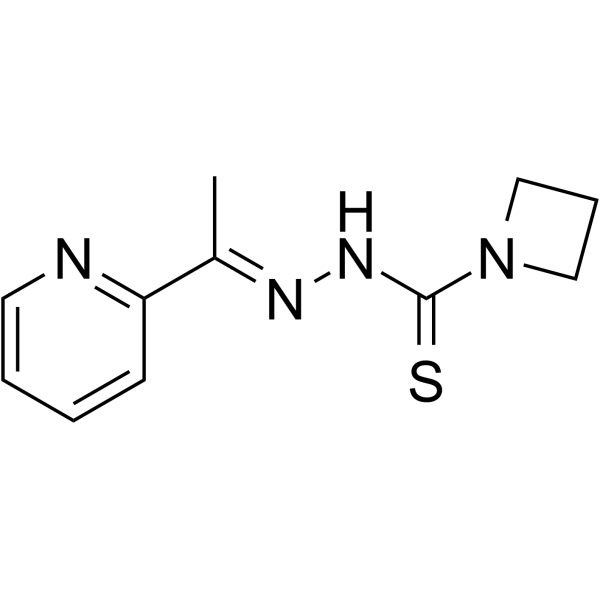
NSC319726MDM-2/p53 p53 Activator -
HY-18935A
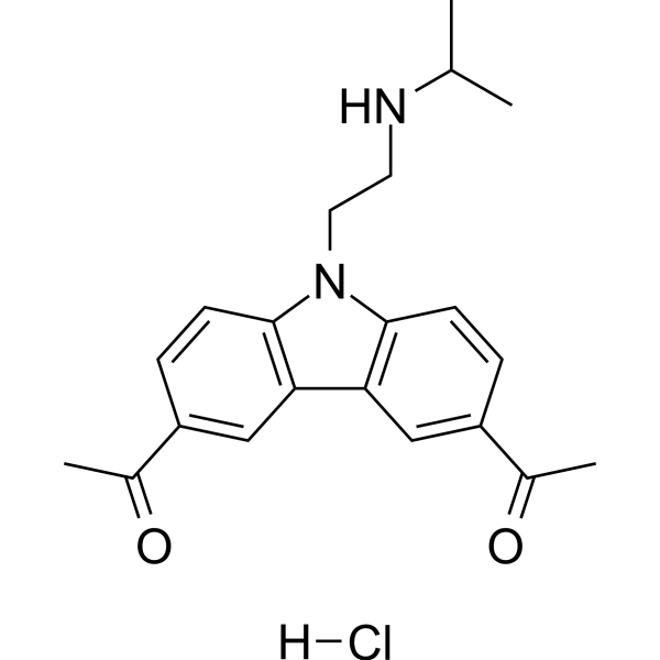
CBL0137 hydrochlorideNF-κB Inhibitor -
HY-18935
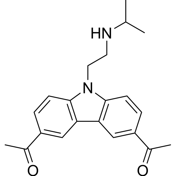
CBL0137FACT Inhibitor -
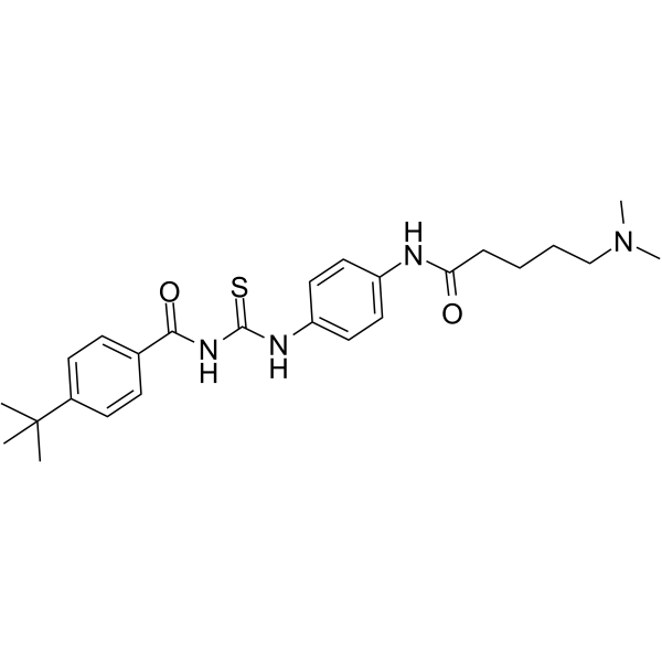
HY-15510
Tenovin-6p53 Activator -
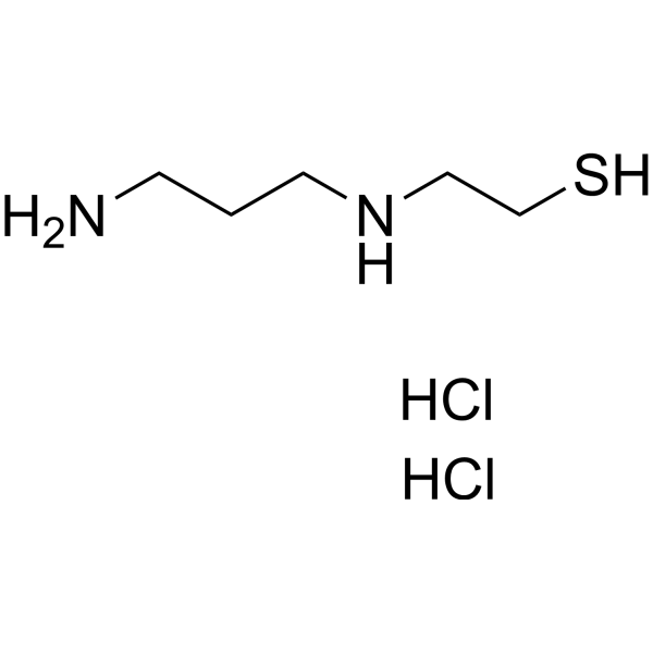
HY-103640
Amifostine thiol dihydrochloridep53 Activator -
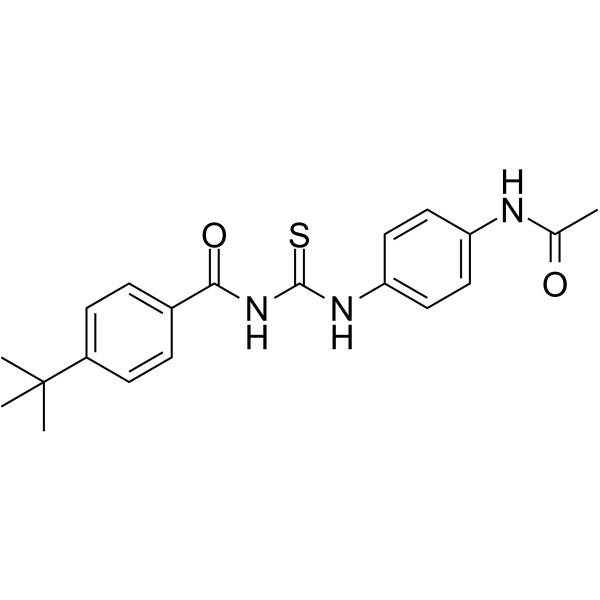
HY-13423
Tenovin-1p53 Activator -

HY-108637R
PhiKan 083 (Standard)Reference Standard


Biologische Aktivität
Beschreibung
Cellular Effect
|
Cell Line

|
Type | Value | Description | References |
|---|---|---|---|---|
| A549 | IC50 |
>25 μM
Compound: CP-31398
|
Antiproliferative activity against human A549 cells after 72 hrs by CellTiter 96 aqueous one solution assay
Antiproliferative activity against human A549 cells after 72 hrs by CellTiter 96 aqueous one solution assay
|
[PMID: 30562697] |
| A549 | IC50 |
24.78 μM
Compound: CP-31398
|
Cytotoxicity against human A549 cells expressing wild type p53 assessed as reduction in cell viability preincubated for 48 hrs and measured after 4 hrs by MTT assay
Cytotoxicity against human A549 cells expressing wild type p53 assessed as reduction in cell viability preincubated for 48 hrs and measured after 4 hrs by MTT assay
|
[PMID: 31761381] |
| DU-145 | IC50 |
15.34 μM
Compound: CP-31398
|
Cytotoxicity against human DU145 cells expressing mutant type p53 assessed as reduction in cell viability preincubated for 48 hrs and measured after 4 hrs by MTT assay
Cytotoxicity against human DU145 cells expressing mutant type p53 assessed as reduction in cell viability preincubated for 48 hrs and measured after 4 hrs by MTT assay
|
[PMID: 31761381] |
| HCT-116 | IC50 |
18.63 μM
Compound: CP-31398
|
Antiproliferative activity against p53+/+ human HCT116 cells after 72 hrs by CellTiter 96 aqueous one solution assay
Antiproliferative activity against p53+/+ human HCT116 cells after 72 hrs by CellTiter 96 aqueous one solution assay
|
[PMID: 30562697] |
| HCT-116 | IC50 |
18.63 μM
Compound: CP-31398
|
Anticancer activity against human HCT116 cells assessed as inhibition of cell proliferation
Anticancer activity against human HCT116 cells assessed as inhibition of cell proliferation
|
[PMID: 31158748] |
| HCT-116 | IC50 |
18.63 μM
Compound: CP-31398
|
Cytotoxicity against human HCT116 cells expressing wild type p53 incubated for 72 hrs by MTS assay
Cytotoxicity against human HCT116 cells expressing wild type p53 incubated for 72 hrs by MTS assay
|
[PMID: 31158748] |
| HCT-116 | IC50 |
26.28 μM
Compound: CP-31398
|
Antiproliferative activity against p53-/- human HCT116 cells after 72 hrs by CellTiter 96 aqueous one solution assay
Antiproliferative activity against p53-/- human HCT116 cells after 72 hrs by CellTiter 96 aqueous one solution assay
|
[PMID: 30562697] |
| HCT-116 | IC50 |
26.28 μM
Compound: CP-31398
|
Cytotoxicity against human HCT116 cells deficient in p53 incubated for 72 hrs by MTS assay
Cytotoxicity against human HCT116 cells deficient in p53 incubated for 72 hrs by MTS assay
|
[PMID: 31158748] |
| L02 | IC50 |
25.34 μM
Compound: CP-31398
|
Cytotoxicity against human HL-7702 cells expressing wild type p53 assessed as reduction in cell viability preincubated for 48 hrs and measured after 4 hrs by MTT assay
Cytotoxicity against human HL-7702 cells expressing wild type p53 assessed as reduction in cell viability preincubated for 48 hrs and measured after 4 hrs by MTT assay
|
[PMID: 31761381] |
| MCF7 | IC50 |
26.96 μM
Compound: CP-31398
|
Antiproliferative activity against human MCF7 cells after 72 hrs by CellTiter 96 aqueous one solution assay
Antiproliferative activity against human MCF7 cells after 72 hrs by CellTiter 96 aqueous one solution assay
|
[PMID: 30562697] |
| MGC-803 | IC50 |
18.27 μM
Compound: CP-31398
|
Cytotoxicity against human MGC-803 cells expressing mutant type p53 assessed as reduction in cell viability preincubated for 48 hrs and measured after 4 hrs by MTT assay
Cytotoxicity against human MGC-803 cells expressing mutant type p53 assessed as reduction in cell viability preincubated for 48 hrs and measured after 4 hrs by MTT assay
|
[PMID: 31761381] |
| NHDF | IC50 |
12.26 μM
Compound: CP-31398
|
Antiproliferative activity against human NHDF cells after 72 hrs by CellTiter 96 aqueous one solution assay
Antiproliferative activity against human NHDF cells after 72 hrs by CellTiter 96 aqueous one solution assay
|
[PMID: 30562697] |
| NHDF | IC50 |
12.26 μM
Compound: CP-31398
|
Cytotoxicity against human NHDF cells incubated for 72 hrs by MTS assay
Cytotoxicity against human NHDF cells incubated for 72 hrs by MTS assay
|
[PMID: 31158748] |
| PANC-1 | IC50 |
>25 μM
Compound: CP-31398
|
Cytotoxicity against human PANC1 cells harboring mutant TP53 gene incubated for 72 hrs by MTS assay
Cytotoxicity against human PANC1 cells harboring mutant TP53 gene incubated for 72 hrs by MTS assay
|
[PMID: 31158748] |
| PC-3 | IC50 |
16.34 μM
Compound: CP-31398
|
Cytotoxicity against human PC-3 cells assessed as reduction in cell viability preincubated for 48 hrs and measured after 4 hrs by MTT assay
Cytotoxicity against human PC-3 cells assessed as reduction in cell viability preincubated for 48 hrs and measured after 4 hrs by MTT assay
|
[PMID: 31761381] |
| T-24 | IC50 |
20.34 μM
Compound: CP-31398
|
Cytotoxicity against human T-24 cells expressing wild type p53 assessed as reduction in cell viability preincubated for 48 hrs and measured after 4 hrs by MTT assay
Cytotoxicity against human T-24 cells expressing wild type p53 assessed as reduction in cell viability preincubated for 48 hrs and measured after 4 hrs by MTT assay
|
[PMID: 31761381] |
| U-251 | IC50 |
18.77 μM
Compound: CP-31398
|
Cytotoxicity against human U251 cells harboring mutant TP53 gene incubated for 72 hrs by MTS assay
Cytotoxicity against human U251 cells harboring mutant TP53 gene incubated for 72 hrs by MTS assay
|
[PMID: 31158748] |
In Vitro
CP-31398 (36.75 μM, 16 h) induces p21 in p53-mutant cells[1]. CP-31398 (15 μg/mL, 20 hours) could induce apoptosis and cell cycle arrest in SW480 cells[2].
MedChemExpress (MCE) has not independently confirmed the accuracy of these methods. They are for reference only.
-
Cell Line:Saos-2 cells expressing transfected mutant p53
-
Concentration:36.75 μM
-
Incubation Time:16 h
-
Result:Induction of p21 in cells expressing only mutant p53.
-
Cell Line:LN-18 and U87MG cells
-
Concentration:36 μM
-
Incubation Time:16 h
-
Result:Decreased the levels of procaspase 3 and induced cleavage of caspase 7.
In Vivo
MedChemExpress (MCE) has not independently confirmed the accuracy of these methods. They are for reference only.
-
Animal Model:Small human tumor xenografts in mice[1]
-
Dosage:100 mg/kg
-
Administration:Orally twice daily for 7 days
-
Result:Suppressed A375.S2 tumor growth by -50%.


Chemical Information
-
CAS. Nr. 259199-65-0
-
Molecular Weight 362.47
-
Formel C22H26N4O
-
SMILES
CN(C)CCCNC1=C2C=CC=CC2=NC(/C=C/C3=CC=C(OC)C=C3)=N1
-
Versand
Room temperature in continental US; may vary elsewhere.
-
Speicherung
Please store the product under the recommended conditions in the Certificate of Analysis.


Reinheit & Dokumentation
Verweise
[1]. B A Foster, et al. Pharmacological rescue of mutant p53 conformation and function. Science. 1999 Dec 24;286(5449):2507-10. [Content Brief]
[2]. Rishu Takimoto, et al. The Mutant p53-Conformation Modifying Drug, CP-31398, Can Induce Apoptosis of Human Cancer Cells and Can Stabilize Wild-Type p53 Protein. Cancer Biol Ther. Jan-Feb 2002;1(1):47-55. [Content Brief]
[3]. J Wischhusen, et al. CP-31398, a novel p53-stabilizing agent, induces p53-dependent and p53-independent glioma cell death. Oncogene. 2003 Nov 13;22(51):8233-45. [Content Brief]

Calculators
Konzentration (Stammlösung) × Volumen (Stammlösung) = Konzentration (Ziellösung) × Volumen (Ziellösung)


CP-31398 Related Classifications
HY-18343 Related Classifications
 Powered by Bioz