



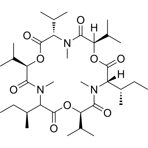
Enniatin A1
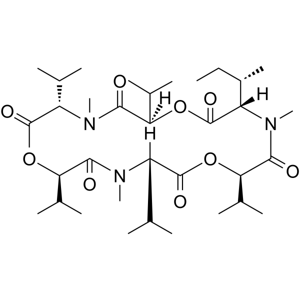
Enniatin A1 isolated from Fusarium mycotoxins is a cyclic hexadepsipeptide consisting of alternating D-α-hydroxyisovaleric acids and N-methyl-L-amino acids. Enniatin A1 possesses anticarcinogenic properties by induction of apoptosis and disruption of ERK signalling pathway. Enniatin A1 inhibits ACAT with an IC50 of 49 μM in rat liver microsomes.
For research use only. We do not sell to patients.
- CAS No.: 4530-21-6
- Formula: C35H61N3O9
- Molecular Weight:667.87
-
Storage:
Please store the product under the recommended conditions in the Certificate of Analysis.

To place orders, for customer services and technical support, please contact: MedChemExpress USA
Tel: 609-228-6898 E-mail: [email protected] [email protected]


-
 Biological Activity
-
Chemical Information
- Protocols
- Purity & Documentation
- References
-
 Help & FAQs


-
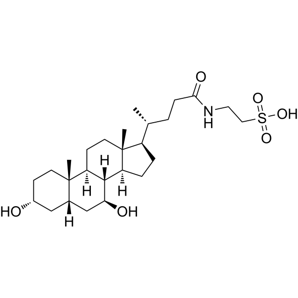
HY-19696
TauroursodeoxycholateEndoplasmic Reticulum Stress Inhibitor -
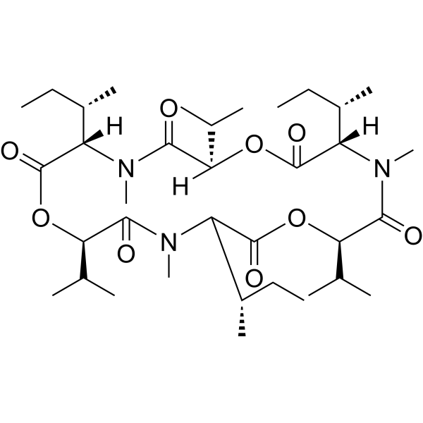
HY-N6702
Enniatin AAcyltransferase Inhibitor -
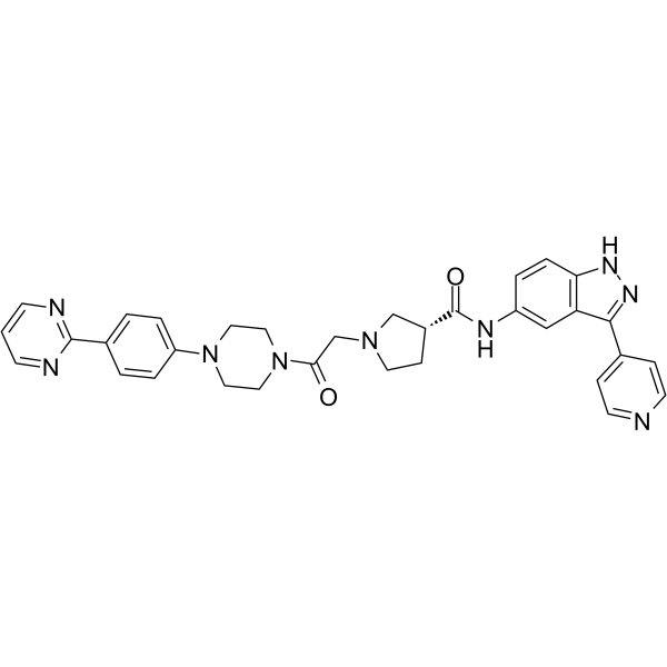
HY-50846
SCH772984ERK1/2 Inhibitor -
HY-108330
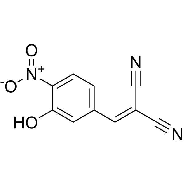
AG126Tyrosine Kinase Inhibitor -
HY-N3807
Enniatin B1ACAT Inhibitor -
HY-12275
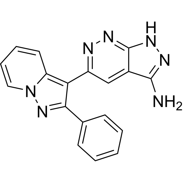
FR 180204ERK Inhibitor -

HY-19696R
Tauroursodeoxycholate (Standard)Endoplasmic Reticulum Stress Inhibitor -
HY-B1864A
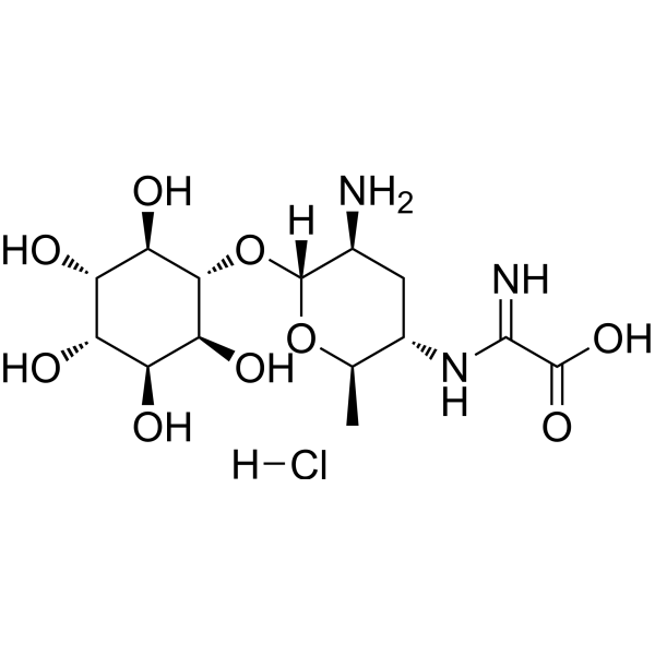
Kasugamycin hydrochlorideAntibiotic -
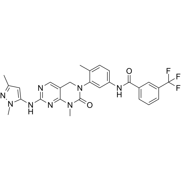
HY-10579
PluripotinERK1 and RasGAP Dual Inhibitor -
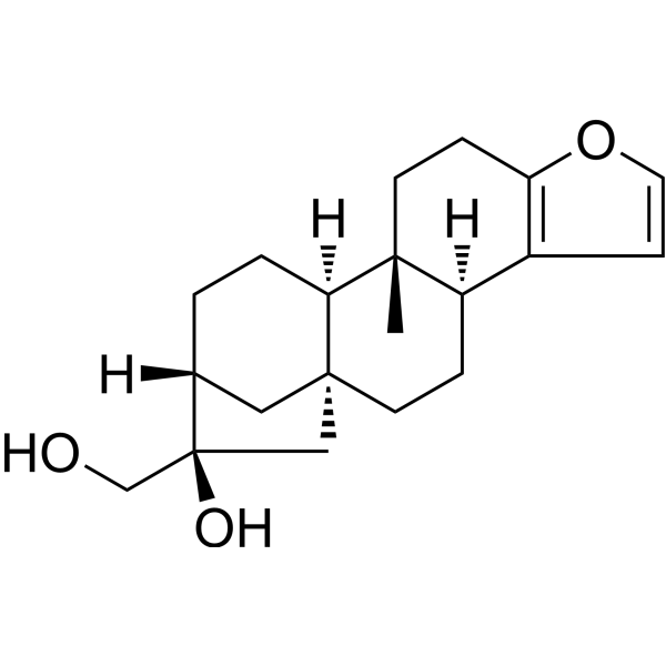
HY-N6257
CafestolERK2 Inhibitor -
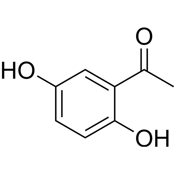
HY-W001174
2,5-Dihydroxyacetophenone -
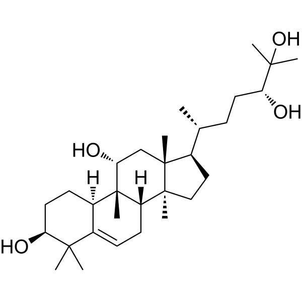
HY-N2312
MogrolSTAT Inhibitor


Biological Activity
Description
IC50 & Target
[1]|
ERK |


Chemical Information
-
CAS No. 4530-21-6
-
Molecular Weight 667.87
-
Formula C35H61N3O9
-
SMILES
O=C([C@@]([C@@H](C)CC)([H])N(C)C([C@@H](C(C)C)OC(C([C@@H](C)CC)N(C)C([C@@H](C(C)C)OC([C@H](C(C)C)N1C)=O)=O)=O)=O)O[C@@](C(C)C)([H])C1=O
-
Structure Classification
-
Initial Source
Fusarium tricinctum
-
Shipping
Room temperature in continental US; may vary elsewhere.
-
Storage
Please store the product under the recommended conditions in the Certificate of Analysis.


Purity & Documentation
References

Calculators
Concentration (start) × Volume (start) = Concentration (final) × Volume (final)


Enniatin A1 Related Classifications
HY-N6704 Related Classifications
 Powered by Bioz