



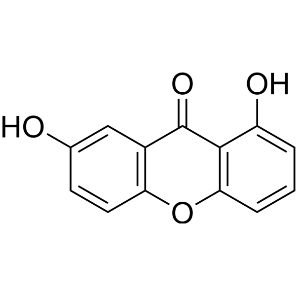
Euxanthone

Based on 1 Customer Validation
Euxanthone, a xanthone derivative, attenuates Aβ1-42-induced oxidative stress and apoptosis by triggering autophagy. Euxanthone exhibits anti-neoplastic and neuroprotective activities.
For research use only. We do not sell to patients.
- Purity : 99.96%
- CAS No.: 529-61-3
- Formula: C13H8O4
- Molecular Weight:228.20
-
Storage:
-20°C, protect from light
* In solvent : -80°C, 6 months; -20°C, 1 month (protect from light)

To place orders, for customer services and technical support, please contact: MedChemExpress USA
Tel: 609-228-6898 E-mail: [email protected] [email protected]


-
 Biological Activity
-
Chemical Information
- Protocols
- Purity & Documentation
- References
-
 Help & FAQs




Biological Activity
Description
Cellular Effect
|
Cell Line

|
Type | Value | Description | References |
|---|---|---|---|---|
| A549 | IC50 |
>200 μM
Compound: 8
|
Cytotoxicity against Taxol-resistant human A549 cells measured after 48 hrs by MTT assay
Cytotoxicity against Taxol-resistant human A549 cells measured after 48 hrs by MTT assay
|
[PMID: 28065566] |
| A549 | IC50 |
14.5 mg/mL
Compound: 13
|
Cytotoxicity against human A549 cells after 3 days by SRB assay
Cytotoxicity against human A549 cells after 3 days by SRB assay
|
[PMID: 15568778] |
| HeLa S3 | IC50 |
>10 μM
Compound: 19
|
Cytotoxicity in human HeLaS3 cells incubated for 72 hrs by MTT assay
Cytotoxicity in human HeLaS3 cells incubated for 72 hrs by MTT assay
|
[PMID: 30978023] |
| HepG2 | IC50 |
>10 μM
Compound: 19
|
Cytotoxicity in human HepG2 cells incubated for 72 hrs by MTT assay
Cytotoxicity in human HepG2 cells incubated for 72 hrs by MTT assay
|
[PMID: 30978023] |
| HT-29 | IC50 |
>10 μM
Compound: 19
|
Cytotoxicity in human HT-29 cells incubated for 72 hrs by MTT assay
Cytotoxicity in human HT-29 cells incubated for 72 hrs by MTT assay
|
[PMID: 30978023] |
| KB | IC50 |
>10 μM
Compound: 19
|
Cytotoxicity in human KB cells incubated for 72 hrs by MTT assay
Cytotoxicity in human KB cells incubated for 72 hrs by MTT assay
|
[PMID: 30978023] |
| MCF7 | IC50 |
>10 μM
Compound: 19
|
Cytotoxicity in human MCF7 cells incubated for 72 hrs by MTT assay
Cytotoxicity in human MCF7 cells incubated for 72 hrs by MTT assay
|
[PMID: 30978023] |
| MCF7 | IC50 |
19.5 mg/mL
Compound: 13
|
Cytotoxicity against human MCF7 cells after 3 days by SRB assay
Cytotoxicity against human MCF7 cells after 3 days by SRB assay
|
[PMID: 15568778] |
| NCI/ADR-RES | IC50 |
>200 μM
Compound: 8
|
Cytotoxicity against human MCF7/ADR cells measured after 48 hrs by MTT assay
Cytotoxicity against human MCF7/ADR cells measured after 48 hrs by MTT assay
|
[PMID: 28065566] |
| OVCAR-3 | IC50 |
>10 μM
Compound: 27
|
Antiproliferative activity against human OVCAR3 cells assessed as reduction in cell viability after 48 hrs by CCK8 assay
Antiproliferative activity against human OVCAR3 cells assessed as reduction in cell viability after 48 hrs by CCK8 assay
|
[PMID: 30830783] |
| SK-OV-3 | IC50 |
>10 μM
Compound: 27
|
Antiproliferative activity against human SKOV3 cells assessed as reduction in cell viability after 48 hrs by CCK8 assay
Antiproliferative activity against human SKOV3 cells assessed as reduction in cell viability after 48 hrs by CCK8 assay
|
[PMID: 30830783] |
| SMMC-7721 | IC50 |
60.35 μM
Compound: 8
|
Cytotoxicity against Taxol-resistant human SMMC7721 cells measured after 48 hrs by MTT assay
Cytotoxicity against Taxol-resistant human SMMC7721 cells measured after 48 hrs by MTT assay
|
[PMID: 28065566] |
In Vitro
Euxanthone (10-20 μM; 24 h) compromises the capability of OS cells to migrate in a dose-dependent fashion, and significantly suppresses cell invasion. Euxanthone presents a significant decrease in adhesion to fibronectin[1].
Euxanthone (10-20 μM; 24 h) modulates the COX-2 expression through the miR-21/PDCD4/c-jun signaling pathway. The repression of COX-2 by Euxanthone mediated its anti-metastatic activities[1].
MedChemExpress (MCE) has not independently confirmed the accuracy of these methods. They are for reference only.
-
Cell Line:Osteosarcoma (OS) cells
-
Concentration:10 μM, 20 μM
-
Incubation Time:24 h
-
Result:Inhibited cell migration at 24 hr.
-
Cell Line:Osteosarcoma (OS) cells
-
Concentration:10 μM, 20 μM
-
Incubation Time:24 h
-
Result:Repressed both the mRNA and protein level of COX-2 in OS cells in a dose-dependent fashion.
In Vivo
Euxanthone (30-60 mg/kg; p.o.;once a day; for 7 days) treatment normalized Bnip3, Beclin1, Pink1, Parkin, p53, Bax, caspase-3, and LC3 II/I in bearing bilateral common carotid artery occlusion (BCCAO). Euxanthone modulates mitophagy and apoptosis induces by mitochondrial stress mediated by mitochondrial fragmentation[2].
MedChemExpress (MCE) has not independently confirmed the accuracy of these methods. They are for reference only.
-
Animal Model:Forty male ICR mice (20 g) induced cerebral ischemia and reperfusion[2]
-
Dosage:30 mg/kg, 60 mg/kg
-
Administration:p.o.;once a day; for 7 days
-
Result:Markedly attenuated BCCAO triggered mitochondrial stress and related breakdown.


Chemical Information
-
CAS No. 529-61-3
-
Appearance Solid
-
Molecular Weight 228.20
-
Formula C13H8O4
-
Color Light yellow to yellow
-
SMILES
O=C1C2=C(OC3=C1C=C(O)C=C3)C=CC=C2O
-
Structure Classification
-
Shipping
Room temperature in continental US; may vary elsewhere.
-
Storage
-20°C, protect from light
* In solvent : -80°C, 6 months; -20°C, 1 month (protect from light)


Purity & Documentation
-
Data Sheet (274 KB)
-
SDS (393 KB)
- English - EN (393 KB)
- Français - FR (393 KB)
- Deutsch - DE (393 KB)
- Norwegian - NO (393 KB)
- Español - ES (393 KB)
- Swedish - SV (393 KB)
- Italian - IT (393 KB)
- Korean - KR (393 KB)
- Portuguese - PT (393 KB)
-
Handling Instructions (2659 KB)
References
[1]. Xiaodong Chen, et al. Euxanthone Impairs the Metastatic Potential of Osteosarcoma by Reducing COX-2 Expression. Anat Rec (Hoboken). 2019 Aug;302(8):1399-1408. [Content Brief]
[2]. Wei Sun, et al. Euxanthone improves cognitive impairment by attenuating mitochondrial fragmentation and suppressing oxidative stress. Cent Eur J Immunol. 2021;46(4):446-455. [Content Brief]
[3]. aicheng Yuan, et al. Euxanthone Attenuates Aβ1-42-Induced Oxidative Stress and Apoptosis by Triggering Autophagy. J Mol Neurosci. 2018 Dec;66(4):512-523. [Content Brief]

Calculators
Concentration (start) × Volume (start) = Concentration (final) × Volume (final)


Euxanthone Related Classifications
HY-N3883 Related Classifications
 Powered by Bioz