



Fidasimtamab

Based on 1 Customer Validation
Fidasimtamab is a bispecific antibody targeting human epidermal growth factor receptor 2 (Her2) and programmed death protein 1 (PD-1), with a Ka of 3.55e-10 M for human Her2 and a Ka of 1.17e-9 M for human PD-1. Fidasimtamab cross-links Her2-positive tumor cells with PD-1-positive T cells to form immune synapses, blocks PD-1-ligand interactions, preserves antibody-dependent cellular cytotoxicity, induces gasdermin B (GSDMB)-mediated pyroptosis, and activates T cells. Fidasimtamab is applicable to relevant research on Her2-positive gastric cancer.
For research use only. We do not sell to patients.
- Purity : ≥95.0%
- CAS No.: 2377419-89-9
- Molecular Weight:144.62 kDa
-
Storage:
Please store the product under the recommended conditions in the Certificate of Analysis.


To place orders, for customer services and technical support, please contact: MedChemExpress USA
Tel: 609-228-6898 E-mail: [email protected] [email protected]


-
 Biological Activity
-
Chemical Information
- Protocols
- Purity & Documentation
- References
-
 Help & FAQs


All EGFR Isoforms
More

Biological Activity
Description
Isotype
Human IgG1 kappa
Recommend Isotype Controls
Species Reactivity
Human
IC50 & Target
ERBB2 & PDCD1
In Vitro
Fidasimtamab binds to purified human PD-1 and Her2 proteins with high affinity, with KD values of 1.17 × 10⁻⁹ M and 3.55 × 10⁻¹⁰ M, respectively[1].
Fidasimtamab (600 nM; 30 min at 4 °C) induces a 24.10% association rate between N87 gastric cancer cells and activated human T cells[1].
Fidasimtamab (serial dilutions; 3 d) dose-dependently activates human CD4+ T cells in an MLR assay, inducing secretion of IFNγ and IL-2 with potency comparable to the parental anti-PD-1 antibody[1].
Fidasimtamab (600 nM; 24 h) significantly enhances T cell-mediated cytotoxicity against N87 and SNU-216 Her2-positive gastric cancer cells, as measured by increased LDH release[1].
Fidasimtamab (600 nM; 6 h) induces pyroptosis in N87 and SNU-216 Her2-positive gastric cancer cells in the presence of activated T cells, and increases IL-18 secretion by 24 h, via cleavage of GSDMB to its active N-terminal fragment[1].
Fidasimtamab (serial dilutions; 4 h at 37 °C) exhibits potent ADCC activity against N87 Her2-positive gastric cancer cells when incubated with human NK cells[1].
Fidasimtamab (600 nM; 24 h) induces a 2-3 fold increase in cell death in PDXO-1 Her2-positive gastric cancer organoids in the presence of T cells, and promotes association between T cells and organoids[1].
Conditioned media from Her2-positive gastric cancer cell-T cell cocultures treated with Fidasimtamab (600 nM; 24 h) activates human CD8+ T cells, increasing CD25 expression and enhancing IFNγ secretion[1].
MedChemExpress (MCE) has not independently confirmed the accuracy of these methods. They are for reference only. Further protocols information, click here.
-
Cell Line:N87 (Her2-positive gastric cancer) cells, SNU-216 (Her2-positive gastric cancer) cells, activated human T cells
-
Concentration:600 nM
-
Incubation Time:24 h
-
Result:Significantly increased LDH release from N87 and SNU-216 cells compared to control groups, indicating enhanced T cell-mediated tumor cell killing.
-
Cell Line:N87 (Her2-positive gastric cancer) cells, human NK cells
-
Concentration:Serial dilutions
-
Incubation Time:4 h at 37 °C
-
Result:Demonstrated potent ADCC activity against N87 cells, with stronger tumor-killing activity than its parental trastuzumab.
In Vivo
Fidasimtamab (IBI-315/BH2950) (5 mg/kg; i.p.; every 3 days; 2 weeks) achieves a 77% tumor volume reduction in human PBMC-reconstituted NOG mice bearing PDX-1 Her2-positive gastric cancer, with enhanced intratumoral T cell infiltration, activation, and proliferation[1].
Fidasimtamab (IBI-315/BH2950) (5 mg/kg; i.p.; every 3 days; 2 weeks) loses its significant tumor inhibitory activity in human PBMC-reconstituted NOG mice bearing GSDMB-knockdown N87 Her2-positive gastric cancer, confirming dependency on GSDMB-mediated pyroptosis[1].
MedChemExpress (MCE) has not independently confirmed the accuracy of these methods. They are for reference only.
-
Animal Model:NOG (NOD/ShiLtj-scid IL2rg
) (female, 4 weeks old, subcutaneously implanted with N87 human Her2-positive gastric cancer cells, reconstituted with human PBMCs)[1] -
Dosage:5 mg/kg
-
Administration:i.p.; every 3 days; 2 weeks
-
Result:Achieved a tumor volume of 54.1 mm3 (day 31), corresponding to a 116.8% tumor inhibition rate relative to control.
Induced the highest infiltration of CD3+, CD8+, and CD4+ lymphocytes in N87 tumors compared to control, parental antibodies, and parental antibody combination groups.
-
Animal Model:NOG (NOD/ShiLtj-scid IL2rg
) (female, 4 weeks old, subcutaneously implanted with PDX-1 human Her2-positive gastric cancer tissue, reconstituted with human PBMCs)[1] -
Dosage:5 mg/kg
-
Administration:i.p.; every 3 days; 2 weeks
-
Result:Achieved a tumor volume of 182.2 mm3 (day 39), corresponding to a 77% reduction relative to control.
Induced the highest infiltration of CD3+, CD8+, and CD4+ lymphocytes in PDX-1 tumors compared to control, parental antibodies, and parental antibody combination groups.
Increased the proportion of granzyme A-positive CD8+ T cells in PDX-1 tumors relative to control.
Boosted the frequency of Ki67+ proliferating CD8+ and CD4+ T cells, CD107a+ activated CD8+ and CD4+ T cells, CD25+ activated CD8+ and CD4+ T cells, and Tbet+IFNγ+ CD3+ T cells in PDX-1 tumors relative to control.
-
Animal Model:NOG (NOD/ShiLtj-scid IL2rg
) (female, 4 weeks old, subcutaneously implanted with GSDMB-knockdown N87 human Her2-positive gastric cancer cells, reconstituted with human PBMCs)[1] -
Dosage:5 mg/kg
-
Administration:i.p.; every 3 days; 2 weeks
-
Result:Showed almost completely blocked tumor inhibitory effect in GSDMB-knockdown N87 tumors, with no significant reduction in tumor volume relative to control.
Reduced infiltration of CD3+ and CD8+ T cells in GSDMB-knockdown N87 tumors compared to its effect in wild-type N87 tumors.
Gene ID
Accession
Conjugated
Unconjugated
Reconsititution
The product can be reconstituted/diluted with sterile PBS or saline.
Format
-
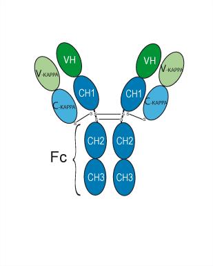
Human IgG1 kappa
Application
ELISA, FACS, Functional assay


Chemical Information
-
CAS No. 2377419-89-9
-
Appearance Liquid
-
Molecular Weight 144.62 kDa
-
Color Colorless to light yellow
-
SMILES
[Fidasimtamab]
-
Synonyms
IBI-315; BH2950
-
Shipping
Shipping with dry ice.
-
Formulation
Please refer to the lot-specific COA for specific buffer information.
-
Storage
Please store the product under the recommended conditions in the Certificate of Analysis.

Protocols
-
Cell Cytotoxicity Assay
Cytotoxicity assays are usually based on the assessment of cell membrane damage, which can also be indirectly detected by measuring cell viability. Detection methods include MTT assay, CKK-8 assay, LDH assay and ATP assay, etc.
-
Mammalian live/dead viability and cytotoxicity staining
Live/dead viability and cytotoxicity staining assays are based on the simultaneous detection of intracellular esterase activity in metabolically active (viable) cells and membrane integrity loss in non-viable cells. In commonly used dual-staining approaches, membrane-permeant fluorogenic substrates are converted by intracellular esterases into fluorescent products in live cells, while impermeant DNA-binding dyes selectively enter cells with compromised plasma membranes and label nucleic acids in dead or dying cells, enabling discrimination between viable and non-viable populations by fluorescence microscopy or flow cytometry.
-
Cell Viability Determination by MTT Colorimetric Assay
The following protocol uses the MTT colorimetric assay as a classic literature-established method for assessing cell viability/metabolic activity in cultured mammalian cells. MTT[3-(4,5-dimethylthiazol-2-yl)-2,5-diphenyltetrazolium bromide] is reduced by metabolically active cells to a colored formazan product; the amount of formazan is quantified spectrophotometrically and provides an indirect measure of metabolically active viable cells. Importantly, MTT reduction reflects cellular oxidoreductase/metabolic activity rather than an absolute direct count of living cells, so changes in cellular metabolism can alter the signal independently of cell number.
-
Pyroptosis Solutions
Pyroptosis is a lytic inflammatory cell-death pathway executed by gasdermin pores, most classically through inflammasome-mediated activation of caspase-1, cleavage of gasdermin D, membrane pore formation, LDH release, and secretion of IL-1β and IL-18. The canonical pathway is commonly modeled by priming cells with an inflammatory signal such as LPS to induce pro-IL-1β and inflammasome components, followed by an activation signal such as ATP or nigericin to activate NLRP3, ASC speck formation, caspase-1 cleavage, GSDMD cleavage, cytokine release, and pyroptotic membrane rupture. The non-canonical pathway is triggered when cytosolic LPS activates mouse caspase-11 or human caspase-4/5, leading to GSDMD cleavage and pyroptosis, and this can secondarily activate NLRP3-dependent IL-1β release. Pyroptosis is linked to inflammatory injury, infection, cancer, liver disease, ocular disease, placental inflammation, and other disease phenotypes, but unresolved questions include which gasdermin fam


Purity & Documentation
-
Data Sheet (267 KB)
-
SDS (251 KB)
- English - EN (251 KB)
- Français - FR (251 KB)
- Deutsch - DE (251 KB)
- Norwegian - NO (251 KB)
- Español - ES (251 KB)
- Swedish - SV (251 KB)
- Italian - IT (251 KB)
- Korean - KR (251 KB)
- Portuguese - PT (251 KB)
-
Inhibitory Antibodies User Guide (603 KB)
References

Calculators
Concentration (start) × Volume (start) = Concentration (final) × Volume (final)


Fidasimtamab Related Classifications
HY-P99618 Related Classifications
 Powered by Bioz
Keywords
- Fidasimtamab
- 2377419-89-9
- IBI-315
- BH2950
- IBI315
- IBI 315
- BH2950
- BH 2950
- BH-2950
- EGFR
- PD-1/PD-L1
- Her2-positive tumor cells
- programmed cell death protein 1
- interferon-gamma
- pyroptosis
- gasdermin B
- Her2-positive breast cancer cells
- PD-1-positive T cells
- human epidermal growth factor receptor 2
- NOG mice
- Her2-positive gastric cancer cells
- Inhibitor
- inhibitor
- inhibit