



DFHBI-1T (solution)

Based on 1 Customer Validation
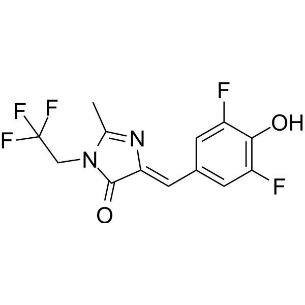
DFHBI-1T (solution) is a membrane-permeable RNA aptamers-activated fluorescence probe (ex/em=472 nm/507 nm). DFHBI-1T binds to RNA aptamers (Spinach, Spinach2, iSpinach, and Broccoli) and causes specific fluorescence and lower background fluorescence. DFHBI-1T is used to image RNA in live cells.
Solvent and concentration: DMSO: 20 mM
Para uso exclusivo en investigación. No vendemos a pacientes.
- No. CAS: 1539318-36-9
- Fòrmula: C13H9F5N2O2
- Peso molecular:320.21
-
Almacenamiento:
Please store the product under the recommended conditions in the Certificate of Analysis.


To place orders, for customer services and technical support, please contact: MedChemExpress USA
Tel: 609-228-6898 E-mail: [email protected] [email protected]


-
 Actividad biológica
-
Chemical Information
- Pureza y Documentación
- Referencias
-
 Help & FAQs


-
HY-110251
DFHBI-1T
-
HY-101894
Dihydrorhodamine 123
-
HY-110250
DFHBI
-
HY-100041
Bromobimane
-
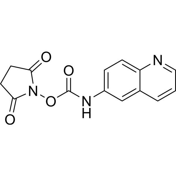
HY-117695
AQC
-
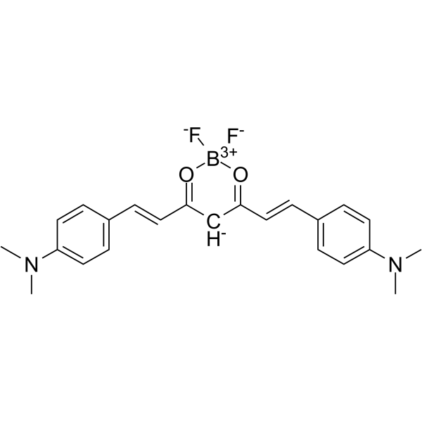
HY-103242
CRANAD-2
-

HY-DY1037
Dihydrorhodamine 123 (solution)
-
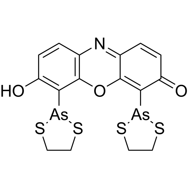
HY-130533
ReAsH-EDT2
-
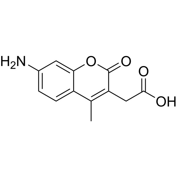
HY-D0025
7-Amino-4-methylcoumarin-3-acetic acid
-
HY-D1303
Marina blue
-

HY-100041R
Bromobimane (Standard)
-

HY-101894R
Dihydrorhodamine 123 (Standard)


Actividad biológica
Solvent and concentration: DMSO: 20 mM
Guide (The following is our recommended protocol. This protocol is only a guide and should be modified according to your specific needs).
1. Preparation of stock solution[1]
It is recommended that the DFHBI-1T stock solution be stored at -20°C or -80°C in the dark after aliquoting.
2. Preparation of working solution
Dilute the stock solution with serum-free medium. The corresponding stock solution can be diluted according to the actual situation. Note that if the solvent is DMSO, the cytotoxicity of DMSO must be considered, and a solvent control should be prepared; if the solvent is pure water, the working solution needs to be filtered and sterilized before adding cells.
Note: Please adjust the concentration of DFHBI-1T working solution according to actual conditions and prepare it before use.
3. Specific staining steps
3.1 Inoculate cells expressing Spinach2-tagged RNA in a suitable culture dish or culture plate and culture under standard cell culture conditions.
3.2 Aspirate the old medium and gently wash the cells twice with pre-warmed PBS to remove residual serum and cell debris.
3.3 Add 20 μM DFHBI-1T working solution to the cells, ensuring complete coverage of the cell monolayer.
3.4 Incubate the cells with the dye for 10 min in a 37°C, 5% CO2 humidified incubator.
3.5 After incubation, aspirate the dye-containing medium and wash the cells three times with pre-warmed PBS to remove unbound DFHBI-1T.
3.6 Replace PBS with fresh cell culture medium or imaging buffer.
3.7 Immediately image the cells using a fluorescence microscope equipped with a GFP filter set to detect fluorescence of the Spinach2-DFHBI-1T complex.
MedChemExpress (MCE) has not independently confirmed the accuracy of these methods. They are for reference only.


Chemical Information
-
No. CAS 1539318-36-9
-
Appearance Liquid
-
Peso molecular 320.21
-
Fòrmula C13H9F5N2O2
-
Color Light yellow to yellow
-
SMILES
O=C1N(C(C)=N/C1=C\C2=CC(F)=C(C(F)=C2)O)CC(F)(F)F
-
Envío
Room temperature in continental US; may vary elsewhere.
-
Almacenamiento
Please store the product under the recommended conditions in the Certificate of Analysis.


Pureza y Documentación
-
Ficha de datos (271 KB)
-
SDS (252 KB)
- English - EN (252 KB)
- Français - FR (252 KB)
- Deutsch - DE (252 KB)
- Norwegian - NO (252 KB)
- Español - ES (252 KB)
- Swedish - SV (252 KB)
- Italian - IT (252 KB)
- Korean - KR (252 KB)
- Portuguese - PT (252 KB)
-
Instrucciones de manejo (2659 KB)
Referencias
[1]. Wenjiao Song, et al. Plug-and-play fluorophores extend the spectral properties of Spinach. J Am Chem Soc. 2014 Jan 29;136(4):1198-201. [Content Brief]
[2]. Grigory S Filonov, et al. Broccoli: rapid selection of an RNA mimic of green fluorescent protein by fluorescence-based selection and directed evolution. J Am Chem Soc. 2014 Nov 19;136(46):16299-308. [Content Brief]

Calculators
Concentration (start) × Volume (start) = Concentration (final) × Volume (final)


DFHBI-1T (solution) Related Classifications
HY-DY1047 Related Classifications
 Powered by Bioz